Skip to main page content
U.S. flag

An official website of the United States government

Dot gov

The VSports app下载. gov means it’s official. Federal government websites often end in . gov or . mil. Before sharing sensitive information, make sure you’re on a federal government site. .

Https

The site is secure V体育官网. The https:// ensures that you are connecting to the official website and that any information you provide is encrypted and transmitted securely. .

. 2017 Jan;76(1):218-226.
doi: 10.1136/annrheumdis-2015-208577. Epub 2016 May 4.

VSports - WNT16 antagonises excessive canonical WNT activation and protects cartilage in osteoarthritis

Affiliations

"V体育官网入口" WNT16 antagonises excessive canonical WNT activation and protects cartilage in osteoarthritis

Giovanna Nalesso et al. Ann Rheum Dis. 2017 Jan.

Abstract

Objective: Both excessive and insufficient activation of WNT signalling results in cartilage breakdown and osteoarthritis. WNT16 is upregulated in the articular cartilage following injury and in osteoarthritis. Here, we investigate the function of WNT16 in osteoarthritis and the downstream molecular mechanisms VSports手机版. .

Methods: Osteoarthritis was induced by destabilisation of the medial meniscus in wild-type and WNT16-deficient mice. Molecular mechanisms and downstream effects were studied in vitro and in vivo in primary cartilage progenitor cells and primary chondrocytes. The pathway downstream of WNT16 was studied in primary chondrocytes and using the axis duplication assay in Xenopus. V体育安卓版.

Results: WNT16-deficient mice developed more severe osteoarthritis with reduced expression of lubricin and increased chondrocyte apoptosis. WNT16 supported the phenotype of cartilage superficial-zone progenitor cells and lubricin expression. Increased osteoarthritis in WNT16-deficient mice was associated with excessive activation of canonical WNT signalling. In vitro, high doses of WNT16 weakly activated canonical WNT signalling, but, in co-stimulation experiments, WNT16 reduced the capacity of WNT3a to activate the canonical WNT pathway V体育ios版. In vivo, WNT16 rescued the WNT8-induced primary axis duplication in Xenopus embryos. .

Conclusions: In osteoarthritis, WNT16 maintains a balanced canonical WNT signalling and prevents detrimental excessive activation, thereby supporting the homeostasis of progenitor cells. VSports最新版本.

Keywords: Chondrocytes; Knee Osteoarthritis; Osteoarthritis. V体育平台登录.


Conflict of interest statement

Conflicts of Interest: None declared.

Figures (V体育官网入口)

Figure 1
Figure 1
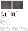
WNT16 is upregulated in experimental osteoarthritis induced by destabilisation of the medial meniscus (DMM). Wild-type mice received DMM of the left knee and sham operation on the right and were killed at 2 days (C and D), 7 days (E and F) and 8 weeks (G) post surgery. (A) WNT16 mRNA was upregulated in the whole joints of DMM-operated mice compared with sham-operated controls, reaching statistical significance after 7 days. WNT16 protein was undetectable in non-operated knees (B), but was upregulated at 2 days following DMM (C) and, to a lesser extent, following sham operation (D). WNT16 expression was further increased in DMM-operated mice at 7 days (E) and to less of an extent following sham operation (F) (joint tissues labelled as follows: C, cartilage; B, subchondral bone; M, meniscus). After 8 weeks, (G) WNT16 was no longer detected in DMM-operated knees. (H) Isotype negative control at the 2-day time point. N=3 per condition.
Figure 2
Figure 2
Wnt16−/− mice are more susceptible to instability-induced osteoarthritis. The 10-week-old mice (7 wild-type (WT) and 10 wnt16−/− (KO)) were subjected to destabilisation of the medial meniscus and the outcome was compared 8 weeks following surgery. (A) Safranin O staining of the medial compartment of representative sections. (B) OARSI histological score. (C) Intensity of the safranin O in the medial tibial plateau. LF, lateral femur; LT, lateral tibia; MF, medial femur; MT, medial tibia.
Figure 3
Figure 3
Lubricin was upregulated following destabilisation of the medial meniscus (DMM). Wild-type (WT) and wnt16−/− mice were subjected to DMM and killed 2 days thereafter. (A) The joint surface, including the articular cartilage and the subchondral bone, was dissected and processed for gene expression analysis by real-time PCR. Lubricin was upregulated in WT mice but not in wnt16−/− mutants at mRNA level 2 days following DMM (N≥10). (B) At protein level, lubricin staining following DMM was detected in the superficial layer of the articular cartilage and in a region between the intermediate and the deep layer in WT mice. (C) The area of the articular cartilage positive for lubricin staining 2 days following DMM was significantly higher in WT mice (N=4) than in wnt16−/− mice (N=6). (D) WNT16 recombinant protein upregulated lubricin mRNA expression in bovine articular chondrocyte (BAC) at 24 h (N=3). KO, knockout. *p<0.05; **p<0.01; ***p<0.001.
Figure 4
Figure 4
Disruption of WNT16 results in apoptosis of superficial cartilage cells and in the loss of superficial zone molecular markers. Terminal deoxynucleotidyl transferase dUTP nick end labeling (TUNEL) staining of representative sections from the medial compartment of the knee of wild-type (WT) and wnt16−/− mice 2 days after destabilisation of the medial meniscus (DMM) (N=6) (A). The TUNEL staining (shown in red) was superimposed on the Nomarski image for anatomical orientation. Quantification of TUNEL-positive cells in the superficial and deep layers of the articular cartilage within the medial compartment (most affected in DMM) and lateral compartment (mildly or unaffected) (B). Gene expression analysis of superficial zone cells from wnt16−/− mice with or without rescue with exogenous 200 ng/mL recombinant WNT16 (N=4) (C). KO, knockout. *p<0.05.
Figure 5
Figure 5
WNT16 is a weak activator of the β-catenin-dependent pathway but also limits its activation by other more potent canonical WNTs. WNT16 caused a weak upregulation of the canonical WNT reporter gene Axin2 compared with WNT3a, in human articular chondrocytes (HAC) (N=3 patients, 4 replicates each) (A). Lubricin expression caused by WNT16 stimulation (ng/mL) in HAC (B), and mouse superficial zone cell (SZC) (C), was higher than that caused by WNT3a stimulation. Lubricin expression decreased in the presence of canonical inhibitor DKK1 (100 ng/mL) in mouse SZC (N=4) (C). All at 24 h. Following destabilisation of the medial meniscus (DMM) surgery, wnt16−/− mice had a higher expression of Axin2 mRNA in their joints (D). WNT16 reduced the capacity of WNT3a to activate the SUPER8XTOPFlash reporter assay in HEK293 cells treated with WNT3a, alone and in combination with WNT16 (ng/mL) (N=4) (E and F). wnt16 inhibited axis duplication caused by wnt8 in Xenopus laevis (G and H). Embryos were injected ventrally with 0.02 ng/µL wnt16 or 0.002 ng/µL wnt8 at the four-cell stage and left for 2 days to develop (n=36–47).
Figure 6
Figure 6
Cartilage injury causes activation of WNT signalling through downregulation of antagonists such as FRZB and upregulation of several agonists such as WNT16 and WNT8. WNT16 buffers the canonical WNT activation to homeostatic levels through its capacity to directly support a weak activation and preventing excessive activation induced by other ligands. Excessive canonical WNT activation causes cartilage breakdown by driving inappropriate maturation particularly within the superficial zone progenitor cells, whereas homeostatic levels of activation are necessary for supporting the superficial progenitor population and lubricin expression (this work and refs 5, 7).

References

    1. Dieppe P, Lim K, Lohmander S. Who should have knee joint replacement surgery for osteoarthritis? Int J Rheum Dis 2011;14:175–80. 10.1111/j.1756-185X.2011.01611.x - DOI - PubMed
    1. Hartmann C, Tabin CJ. Wnt-14 plays a pivotal role in inducing synovial joint formation in the developing appendicular skeleton. Cell 2001;104:341–51. 10.1016/S0092-8674(01)00222-7 - DOI - PubMed
    1. Guo X, Day TF, Jiang X, et al. . Wnt/beta-catenin signaling is sufficient and necessary for synovial joint formation. Genes Dev 2004;18:2404–17. 10.1101/gad.1230704 - DOI - PMC - PubMed
    1. Yuasa T, Kondo N, Yasuhara R, et al. . Transient activation of Wnt/{beta}-catenin signaling induces abnormal growth plate closure and articular cartilage thickening in postnatal mice. Am J Pathol 2009;175:1993–2003. 10.2353/ajpath.2009.081173 - "VSports手机版" DOI - PMC - PubMed
    1. Yasuhara R, Ohta Y, Yuasa T, et al. . Roles of β-catenin signaling in phenotypic expression and proliferation of articular cartilage superficial zone cells. Lab Invest 2011;91:1739–52. 10.1038/labinvest.2011.144 - DOI - PMC - PubMed

VSports最新版本 - MeSH terms