Skip to main page content
U.S. flag

An official website of the United States government

Dot gov

The . gov means it’s official. Federal government websites often end in . gov or . mil VSports app下载. Before sharing sensitive information, make sure you’re on a federal government site. .

Https

The site is secure. The https:// ensures that you are connecting to the official website and that any information you provide is encrypted and transmitted securely V体育官网. .

. 2013 May 31;8(5):e64950.
doi: 10.1371/journal.pone.0064950. Print 2013.

Identification of diverse lipid droplet targeting motifs in the PNPLA family of triglyceride lipases

Affiliations

V体育安卓版 - Identification of diverse lipid droplet targeting motifs in the PNPLA family of triglyceride lipases

V体育平台登录 - Sricharan Murugesan et al. PLoS One. .

Abstract

Members of the Patatin-like Phospholipase Domain containing Protein A (PNPLA) family play key roles in triglyceride hydrolysis, energy metabolism, and lipid droplet (LD) homoeostasis. Here we report the identification of two distinct LD targeting motifs (LTM) for PNPLA family members. Transient transfection of truncated versions of human adipose triglyceride lipase (ATGL, also known as PNPLA2), PNPLA3/adiponutrin, or PNPLA5 (GS2-like) fused to GFP revealed that the C-terminal third of these proteins contains sequences that are sufficient for targeting to LDs. Furthermore, fusing the C-termini of PNPLA3 or PNPLA5 confers LD localization to PNPLA4, which is otherwise cytoplasmic. Analyses of additional mutants in ATGL, PNPLA5, and Brummer Lipase, the Drosophila homolog of mammalian ATGL, identified two different types of LTMs. The first type, in PNPLA5 and Brummer lipase, is a set of loosely conserved basic residues, while the second type, in ATGL, is contained within a stretch of hydrophobic residues. These results show that even closely related members of the PNPLA family employ different molecular motifs to associate with LDs. VSports手机版.


Conflict of interest statement

Competing Interests: The authors have declared that no competing interests exist.

Figures

Figure 1
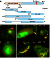
Figure 1. GFP-tagged proteins containing the C-terminal third of human PNPLA5 localize to LDs.
(A) Schematic of the domain structure of full length PNPLA5 highlighting the N-terminal catalytic dyad within the Patatin domain, the C-terminal basic patch region, and all truncation mutants generated. HeLa cells were treated overnight with OA, transfected with the indicated constructs for 24 h, fixed and stained with LipidTox Red, and then analyzed by fluorescence microscopy. N-terminal GFP-tagged PNPLA5 constructs missing the C-terminal third of the protein were found in the cytoplasm or nucleus (B, C, and E), whereas constructs containing the C-terminal third of PNPLA5 (residues 286–429), or one lacking a portion of the patatin domain localized to LipidTox stained LDs (D, F, and G). Bars, 5 μm.
Figure 2
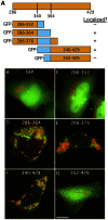
Figure 2. Additional truncation mutants of PNPLA5 identify a 25 amino acid domain necessary for LD localization.
(A) Schematic of the C-terminal constructs fused to GFP. HeLa cells were treated overnight with OA, transfected with the indicated constructs for 24 h, fixed and stained with LipidTOX Red. Cells were then analyzed by fluorescence microscopy to observe LD localization as in Figure 1. Constructs containing residues 340–364 of PNPLA5 localized to LipidTox stained LDs (D, E, and F), whereas those missing a portion of this sequence or a control expressing GFP alone, remained in the cytoplasm (B, C, and G). Bars, 5 μm.
Figure 3
Figure 3. The C-terminal domains of other PNPLA family members are important for LD localization.
(A, B) Schematic depicting PNPLA family C-terminal domains N-terminally fused to GFP or PNPLA4. HeLa cells were treated overnight with OA, transfected with the indicated constructs for 24 h, fixed and stained with LipidTOX Red, and analyzed by fluorescence microscopy to observe LD localization. (C, top panels) PNPLA family C-terminal domains fused to GFP localize to LDs. (C, bottom panels) Fusing C-terminal domains of PNPLA3 and PNPLA5, but not ATGL, confers LD localization to PNPLA4. The number of cells with LD surface localization in a given population was quantified as a percentage of GFP-transfected cells by scoring cells that displayed ‘rings’ of GFP signal surrounding Lipidtox stained LDs. Cells lacking LDs were not scored. To account for inherent variations in LD size/number per cell, ≥300 cells were scored per condition in three independent experiments. These results, quantified in D and E, are plotted as means ± SE (≥3 experiments/condition, ≥300 cells counted/experiment, ***, p<0.0001). (F) Quantitation of fluorescence intensity on LD surface:cytoplasm ratio from line plots (n≥15 LDs and cells/condition). FL = full length, Bar, 5 μm.
Figure 4
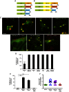
Figure 4. Alterations in the basic charge LTM of PNPLA5 abolish LD localization.
(A) Comparison of amino acid sequences 340–364 of PNPLA5 between several species. Boxed in red are the conserved proline-rich and arginine-rich domains. (B) HeLa cells were treated overnight with OA, transfected with the indicated constructs for 24 h, fixed and stained with LipidTOX Red. Cells were then analyzed by fluorescence microscopy and scored for LD localization. GFP-PNPLA5(A347AAAA352) and GFP-PNPLA5(K360KLV363) localized to LDs, whereas GFP-PNPLA5(A360AEE363) was cytoplasmic (quantified in D). Bar, 5 μm. (C) Quantitation of LD diameters showed that overexpession of wildtype GFP-PNPLA5 reduced LD size, whereas LDs in cells over-expressing GFP-PNPLA5(A360AEE363) were similar to those in control cells. (D) Different combinations of arginines in the LTM were mutated within full length GFP-tagged PNPLA5 and LD localization was observed. No single arginine was critical for LD association but removal of each one incrementally reduced LD binding. Data are plotted as means ± SEM; ≥3 experiments/condition; ≥300 cells counted/experiment; ***indicates p<0.0001, **indicates p<0.001, *indicates p<0.05 compared to wildtype (RSRRLV). (E) Quantitation of fluorescence intensity on LD surface:cytoplasm ratio from line plots (n≥15 LDs and cells/condition). (F) HeLa cells were treated as in ‘B’ except transfections were followed by cell fractionation. Wildtype PNPLA5 (RSRRLV), but not the mutant lacking all three arginines in the basic patch (ASAALV), was enriched on LDs as shown by western blotting of isolated LD fractions in comparison to pooled cytoplasmic fractions (cyto); quantitation in (G). Data are plotted as means± SEM, n = 3.
Figure 5
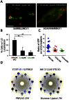
Figure 5. The basic patch LTM is conserved in Drosophila Brummer Lipase
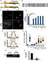
. HeLa cells were treated overnight with OA, transfected with the indicated constructs for 24 h, fixed, and stained with LipidTOX Red. Cells were then analyzed by fluorescence microscopy and scored for LD localization. (A) Wildtype Drosophila Brummer Lipase, the homolog of human ATGL, localized to LDs (A’) whereas mutating the three basic residues downstream of a proline knot-like motif abolished LD localization (B’). Bar, 5 μm. (B) Quantitation of LD localization. Data are plotted as means ± SEM, ≥3 experiments/condition, ≥300 cells counted/experiment; **indicates p<0.001 compared to wildtype (RIRLNK). (C) Quantitation of fluorescence intensity on LD surface:cytoplasm ratio from line plots. Wildtype Brummer lipase had a high LD:cytoplasm ratio, which was lost upon mutating the three basic residues, while mutating two residues had an intermediate effect (n≥15 LDs and cells/condition). (D) The LTM sequences of PNPLA5 (354–367) and Brummer Lipase (319–332) when projected as a helical wheel form potential amphipathic helices as determined by DNAstar.
Figure 6
Figure 6. ATGL has a hydrophobic LTM.
(A) A schematic diagram of ATGL depicting the putative hydrophobic LTM region within the long variable C-terminal domain. (B) Species comparison of sequences in the hydrophobic domain (residues 320–360) of ATGL orthologs. Black amino acids = identical, Red = similar, Green = unique. (C) HeLa cells were treated overnight with OA, transfected with the indicated constructs for 24 h, fixed, and stained with LipidTOX Red. Cells were then analyzed by fluorescence microscopy and scored for LD localization. Bar, 5 μm. Expression of full-length ATGL lacking the hydrophobic region (residues 320–360) in HeLa cells resulted in a decreased LD surface signal and an increased cytoplasmic signal; (D) GFP-tagged N-terminal truncations lacking residues 1–319 or a C-terminal truncation lacking residues 361–504 (but containing the hydrophobic region) were able to localize to LDs at wildtype levels; an N-terminal truncation lacking residues 1–360 (missing hydrophobic region) did not localize to LDs at all. Expressing either full-length ATGL lacking the hydrophobic residues 320–360 or a C-terminal truncation lacking residues 320–504 (found in NLSDM patients) resulted in reduced LD localization. Data are plotted as means ± SEM; ≥3 experiments/condition, ≥300 cells counted/experiment. ***indicates p<0.0001. (E) Fluorescence intensity line plot profiles from line plots in C (see arrowheads for lines). (F) Quantitation of LD surface:cytoplasm intensity ratio from line intensity plots for all ATGL constructs (plotted as mean±SEM, ≥20 LDs and cells/condition). (G) HeLa cells were treated as in ‘A’ except transfections were followed by cell fractionation. Wildtype ATGL, but not ATGL lacking the hydrophobic residues 320–360, was enriched on LDs as shown by western blotting of isolated LD fractions in comparison to pooled cytoplasmic fractions (cyto); quantitation in (H). Data are plotted as means ± SEM, n = 3.

References

    1. Brasaemle DL, Wolins NE (2012) Packaging of fat: an evolving model of lipid droplet assembly and expansion. J Biol Chem 287: 2273–2279. - VSports最新版本 - PMC - PubMed
    1. Fujimoto T, Parton RG (2011) Not just fat: the structure and function of the lipid droplet. Cold Spring Harb Perspect Biol 3. - PMC - PubMed
    1. Reue K (2011) A thematic review series: lipid droplet storage and metabolism: from yeast to man. J Lipid Res 52: 1865–1868. - PMC - PubMed
    1. Farese RV Jr, Walther TC (2009) Lipid droplets finally get a little R-E-S-P-E-C-T. Cell 139: 855–860. - PMC - PubMed
    1. Lass A, Zimmermann R, Oberer M, Zechner R (2011) Lipolysis - a highly regulated multi-enzyme complex mediates the catabolism of cellular fat stores. Prog Lipid Res 50: 14–27. - PMC - PubMed

Publication types

MeSH terms