<kbd dir="t0TITV"></kbd>
Skip to main page content
U.S. flag

An official website of the United States government

Dot gov

The . gov means it’s official. Federal government websites often end in . gov or . mil. Before sharing sensitive information, make sure you’re on a federal government site. VSports app下载.

Https

The site is secure. The https:// ensures that you are connecting to the official website and that any information you provide is encrypted and transmitted securely. V体育官网.

. 2020 May:32:101483.
doi: 10.1016/j.redox.2020.101483. Epub 2020 Mar 2.

Identification of Frataxin as a regulator of ferroptosis

Affiliations

Identification of Frataxin as a regulator of ferroptosis

Jing Du et al. Redox Biol. 2020 May.

Erratum in (VSports)

"VSports在线直播" Abstract

Ferroptosis is a newly discovered form of non-apoptotic regulated cell death and is characterized by iron-dependent and lipid peroxidation. Due to the enhanced dependence on iron in cancer cells, induction of ferroptosis is becoming a promising therapeutic strategy VSports手机版. However, the precise underlying molecular mechanism and regulation process of ferroptosis remains largely unknown. In the present study, we demonstrate that the protein Frataxin (FXN) is a key regulator of ferroptosis by modulating iron homeostasis and mitochondrial function. Suppression of FXN expression specifically repressed the proliferation, destroyed mitochondrial morphology, impeded Fe-S cluster assembly and activated iron starvation stress. Moreover, suppression of FXN expression significantly enhanced erastin-induced cell death through accelerating free iron accumulation, lipid peroxidation and resulted in dramatic mitochondria morphological damage including enhanced fragmentation and vanished cristae. In addition, this type of cell death was confirmed to be ferroptosis, since it could be pharmacologically restored by ferroptotic inhibitor Fer-1 or GSH, but not by inhibitors of apoptosis, necrosis. Vice versa, enforced expression of FXN blocked iron starvation response and erastin-induced ferroptosis. More importantly, pharmacological or genetic blocking the signal of iron starvation could completely restore the resistance to ferroptosis in FXN knockdown cells and xenograft graft in vivo. This paper suggests that FXN is a novel ferroptosis modulator, as well as a potential provided target to improve the antitumor activity based on ferroptosis. .

Keywords: Ferroptosis; Frataxin; Iron-sulfur cluster; Mitochondria V体育安卓版. .


Conflict of interest statement

Declaration of competing interest The authors declare that there is no conflict of interest.

"V体育安卓版" Figures

Image 1
Graphical abstract
Fig. 1
Fig. 1
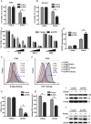
FXN knockdown repressed the proliferation of HT-1080 cells. (A) Western blot analysis of FXN expression in indicated FXN knockdown and control HT-1080 cells. Tom20 was served as internal loading control. (B) Quantitative real-time PCR analysis of the mRNA of FXN in the three indicated HT-1080 cells. (C) The ability of proliferation was detected by CCK8 assay between control and sh-FXN cells. Induction of cycle arrest in HT-1080 cells was detected by flow cytometry (D-E). Effects of FXN suppression in HT-1080 cells on the proliferation capacity were measured by plate clone formation assay (F-G) and soft agar colony formation assay (H-I), corresponding histograms were shown on the right . Migration of HT-1080 cells was evaluated by transwell assay; corresponding histograms were shown on the right (J-K). All histograms were represented as mean ± SD. P < 0.05, ★★P < 0.01 versus control.
Fig. 2
Fig. 2
Suppression of FXN induced the dysfunction of mitochondria and accumulation of free iron. (A) To evaluate mitochondrial morphology, HT-1080 cells were stained with MitoTracker probe (100 nM) and DAPI (10 mg/ml), and then photographed by confocal laser microscope. Scale bars: 75 μm. (B) Flow cytometry was performed to measure mitochondrial membrane potential (MMP) by TMRE probe (100 nM) in indicated FXN knockdown and control HT-1080 cells. (C) OCR rate was carried out by extracellular flux analyzer after the additions of oligomycin, FCCP and antimycin A. (D-G) The basal respiration, maximal respiration, spare respiration and ATP production were calculated. (H) Mitochondrial and cytoplasmic aconitase activity was analyzed by the in-gel activity assay. (I) The protein levels of mitochondrial and cytoplasmic aconitase were detected by western blot assay. β-actin and Tom20 were used as a loading control. (J-K) Western blot analysis of iron-starvation stress and ISC turnover in iron-sulfur protein. (L) HT-1080 cells were stained with Rhodamine B-[(1,10-phenanthroline-5-yl)-aminocarbonyl]benzyl ester (RPA) probe (4 μM) and DAPI (10 mg/ml) to detect the cellular labile iron by confocal laser microscope. Scale bars: 75 μm. (M) The quantitative PCR analysis was performed to measure mitochondrial DNA copy number. Primer specific for HGB1 genes were used for the determination of nuclear DNA (nDNA) and another primer (ND-1) for the detection of mtDNA. P < 0.05,★★P < 0.01 versus control.
Fig. 3
Fig. 3
Suppression of FXN accelerated erastin induced ferroptosis. (A) HT-1080 cells were exposed to various concentrations of erastin for 12 h respectively and followed by CCK8 assay. (B) Propidium iodide positive cells were stained and observed by fluorescent microscopy after erastin exposure for 12 h. Scale bars: 30 μm. (C) CFDA-SE probe (5 μM) stained HT-1080 cells were exposed to 2.5 μM erastin and cultured for 3 days, then subjected to flow cytometry. (D) The morphological changes of mitochondria were detected by transmission electron microscopy (TEM) in the absence or presence of erastin. Flow cytometry was performed to measure lipid peroxides (E), cytoplasm ROS (F), mitochondrial membrane potential(G) after erastin treatment for 12 h in HT-1080 cells. (H) Representative images of BODIPY staining in erastin treated HT-1080 cells which observed by confocal laser microscope. Scale bars: 75 μm. (I) HT-1080 cells were treated with erastin (10 μM) with or without small molecule inhibitor for 12 h and respective cell viability was detected by CCK8. (ferroptosis inhibitor ferrostatin-1,0.5 μM; GSH, 1 mM; apoptosis inhibitor Z-VAD-FMK, 4 μM; necroptosis inhibitor Necrosulfonamide, 0.5 μM; autophagy inhibitor BafA1, 20 nM ). All histograms were represented as mean ± SD. ★★P < 0.01 versus control.
Fig. 4
Fig. 4
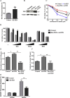
Enforced expression of FXN contributed to ferroptosis resistance. (A) The proliferation rate was analyzed by the CFDA-SE probe between the indicated cells. (B) Quantitative PCR analysis was performed to measure mtDNA copy number. Primers specific for the HGB1 gene were used for the determination of nuclear DNA (nDNA) and primers specific for ND-1 were used to detect mtDNA. (C) Indicated HT-1080 cells were incubated with different concentrations of erastin for 12 h and cell viability was assayed by CCK8 assay. (D) Intracellular Fe2+ was measured by the staining of RPA and photographed by the confocal microscope. Scale bars: 75 μm. (E) MitoTracker Red labeled HT-1080 cells were subjected to the confocal microscope for observing the changes of mitochondrial morphology. Scale bars: 75 μm. (F) The morphological changes of mitochondria were detected by transmission electron microscopy (TEM) in the absence or presence of erastin. Lower scale bars: 0.2 μm. (G-H) To assess lipid ROS production, HT-1080 cells were treated with 5 μM erastin and loaded with BODIPY C11 probe for 30 min followed by flow cytometry measurement (G) and confocal laser microscope (H). Scale bars: 75 μm. (I) Indicated HT-1080 cells were treated with erastin (10 μM) with or without small molecule inhibitor and respective cell viability was detected by CCK8. (ferrostatin-1,0.5 μM; GSH, 1 mM; iron chelator DFO, 100 μM; Z-VAD-FMK, 4 μM; Necrosulfonamide, 0.5 μM). All histograms were represented as mean ± SD. (For interpretation of the references to colour in this figure legend, the reader is referred to the Web version of this article.) P < 0.05,★★P < 0.01 versus control.
Fig. 5
Fig. 5
Overexpress of FTH eliminated the proliferation arrest and mitochondria dysfunction induced by FXN suppression. (A-B) The ability of cell proliferation was measured by CCK8 assay (A) and plate clone formation assay(B) between the three indicated cells. (C-D) To assess intracellular Fe2+, cells were stained with RPA probe for 30 min and photographed by a confocal laser microscope(C) or subjected to flow cytometry (D). (E) Cells were stained by MitoTracker Red and mitochondrial morphology changes were observed under the confocal microscope. Scale bars: 75 μm. (F) The quantitative PCR analysis was performed to measure mtDNA copy number. (For interpretation of the references to colour in this figure legend, the reader is referred to the Web version of this article.) P < 0.05,★★P < 0.01 versus control.
Fig. 6
Fig. 6
FTH expression restored the resistance to ferroptosis in FXN knockdown cells. (A) Indicated cells were treated with erastin (0–10 μM) for 12 h and cell viability was assayed by CCK8. (B) HT-1080 cells were treated with erastin (10 μM) for 12 h with or without small molecule inhibitor and respective cell viability was detected by CCK8. (C) The morphological changes of mitochondria were detected by transmission electron microscopy (TEM) in the absence or presence of erastin. Lower scale bars: 0.2 μm. (D) Cells were treated with 5 μM erastin and loaded with DCFH-DA (4 μM) probe for 30 min followed by flow cytometry to assess cytoplasm ROS formation. (E) Indicated HT-1080 cells were treated with erastin (5 μM) for 12 h. Levels of lipid ROS production were detected with BODIPY C11 (2.5 μM) probe by flow cytometry measurement. ★★P < 0.01 versus control.
Fig. 7
Fig. 7
Knockdown of FXN inhibited the growth of xenograft in vivo. (A) The tumor volumes were measured every 2 days and significant inhibition of tumor growth was observed after FXN knockdown. Representative photographs showed the tumor size in mice after the mice were sacrificed. (B) H&E and IHC analysis for Ki67, FXN and FTH in indicated tumor specimens. Scale bars: 50 μm. (C-E) The Specimens were scored by relative integrated optical density (IOD) value. P < 0.05,★★P < 0.01 versus control.
Fig. S1
Fig. S1
Kaplan-meier analysis was performed base on TCGA database to evaluate the association between FXN expression level and patients' overall survival.
Fig. S2
Fig. S2
Suppression of FXN induced the decrease of NRF2-Keap1 axis. Related to Fig. 2. (A) The quantitation analysis of the in-gel aconitase activity. (B) Western blot analysis of NRF2 signal pathway associated protein. (C) Quantitative real-time polymerase chain reaction analysis of the mRNA of NRF2 in the three indicated cells. (D) The quantitation analysis of the fluorescence of RPA biosensor. ★P < 0.05, ★★P < 0.01 versus control. ns means no significant statistical difference.
None
Fig. S3 Suppression of FXN accelerated erastin-induced ferroptosis but not RSL3 induced ferroptosis. Related to Fig. 3. Indicated FXN suppression U266 (A) and Kasumi-1 (B) cells were treated with or without erastin (20 μM) for 12 h and cell viability was assayed by CCK8. (C) Indicated FXN suppression cells were exposed to various concentrations of IKE (0 to 5 μM), RSL3 (0 to 1 μM) for 12 hours and BSO (0 to 100 mM) for 48 hours, cell viability was measured by CCK8. Flow cytometry was performed to measure lipid peroxides (E), cytoplasm ROS (F) after erastin treatment for 12h in U266 cells. (G) GSH level was monitored in the indicated HT-1080 cells treated with or without erastin for 12 h. (H) Suppression of FXN accelerated erastin-induced accumulation of free iron level. Western blot analysis of iron-starvation stress (I) and LC3(J) conversion in the indicated cells under the treatment of erastin. ★P < 0.05, ★★P < 0.01 versus control.
Fig. S
Fig. S
Ectopic expression of an shRNA-resistant FXN cDNA reversed the erastin-induced ferroptosis. Related to Fig. 4. (A) The quantitation analysis of RPA biosensor fluorescence. (B)An shRNA-resistant FXN cDNA (Res-FXN) was generated and co-transduced with shFXN, and western blot was used to confirm the expression of FXN. (C, D) Cell death in HT-1080 stably transduced with Res-FXN, shFXN or vector control was detected through CCK8 assay after the exposure of various concentrations of erastin(0 to 10μM), IKE (0 to 5 μM), RSL3 (0 to 1 μM) for 12 hours and BSO (0 to 100 mM) for 48 hours. (E, F) Mitochondrial and cytoplasmic iron was measured by the ICP-MS. (G) The quantitation analysis of Bodipy biosensor fluorescence. ★P < 0.05, ★★P < 0.01 versus control.
Fig. s5
Fig. s5
The quantitation analysis of RPA biosensor fluorescence. Related to Fig. 5. ★★P < 0.01 versus control.

References

    1. Puig S., Ramos-Alonso L., Romero A.M., Martinez-Pastor M.T. The elemental role of iron in DNA synthesis and repair. Metallomics. 2017;9:1483–1500. - PubMed
    1. Furuyama K., Kaneko K. Iron metabolism in erythroid cells and patients with congenital sideroblastic anemia. Int. J. Hematol. 2018;107:44–54. - PubMed (VSports)
    1. Manz D.H., Blanchette N.L., Paul B.T., Torti F.M., Torti S.V. Iron and cancer: recent insights. Ann. N. Y. Acad. Sci. 2016;1368:149–161. - PMC - PubMed
    1. Dixon Scott J., Lemberg Kathryn M., Lamprecht Michael R., Skouta R., Zaitsev Eleina M., Gleason Caroline E., Patel Darpan N., Bauer Andras J., Cantley Alexandra M., Yang Wan S. Ferroptosis: an iron-dependent form of nonapoptotic cell death. Cell. 2012;149:1060–1072. - V体育ios版 - PMC - PubMed
    1. A P., Fl I., Jm M., Pj C. Regulated necrosis in kidney ischemia-reperfusion injury. Kidney Int. 2019;96:291–301. - PubMed

Publication types

LinkOut - more resources