"VSports注册入口" Skip to main page content
U.S. flag

An official website of the United States government

Dot gov

The . gov means it’s official. Federal government websites often end in VSports app下载. gov or . mil. Before sharing sensitive information, make sure you’re on a federal government site. .

Https

The site is secure. The https:// ensures that you are connecting to the official website and that any information you provide is encrypted and transmitted securely V体育官网. .

. 2017 Oct 17;16(1):163.
doi: 10.1186/s12943-017-0732-6.

"VSports注册入口" Reprogramming of stromal fibroblasts by SNAI2 contributes to tumor desmoplasia and ovarian cancer progression

Affiliations

"VSports" Reprogramming of stromal fibroblasts by SNAI2 contributes to tumor desmoplasia and ovarian cancer progression

Zongyuan Yang et al. Mol Cancer. .

Abstract

Background: Molecular profiling in ovarian cancer (OC) revealed that the desmoplasia subtype presented the poorest prognosis, highlighting the contribution of stromal fibroblasts in tumor progression. This study aimed to investigate the molecular characteristics of SNAI2 driving the transcriptional reprogramming of fibroblasts within tumors. VSports手机版.

Methods: SNAI2 expression was evaluated in microdissected profiles of various cancers and in various molecular subtypes of OC. Gene set enrichment analysis (GSEA) and single sample GSEA (ssGSEA) were performed to explore the correlation between SNAI2 and stromal fibroblast activation. The SNAI2 defined signature in the mesenchymal OC subtype was identified through an integrative analysis of the TCGA and the Tothill datasets. The predictive value of this signature was validated in independent datasets V体育安卓版. SNAI2 expression alteration influence of tumor growth in primary CAFs was evaluated in 3D organotypic and murine xenograft models. .

Results: We demonstrated that SNAI2 was frequently activated in the tumor stroma, correlated with fibroblast activation and worse patient outcome in OC. SNAI2 transformed normal fibroblasts to a CAF-like state and boosted their tumor-supporting role in 3D organotypic culture and in OC xenograft model V体育ios版. SNAI2 drove a transcriptional signature in the mesenchymal subtype of OC that contributed to tumor desmoplasia, which fed back to increase SNAI2 expression and sustain fibroblast activation. .

Conclusions: Our results address the role of SNAI2 in reprogramming stromal fibroblasts VSports最新版本. The identified SNAI2 mesenchymal signature has both a predictive value and biological relevance and might be a therapeutic target for stroma-oriented therapy against the desmoplasia OC subtype. .

Keywords: Ovarian cancer; Reprogram; SNAI2; Stiffness; Stromal fibroblast V体育平台登录. .


VSports在线直播 - Conflict of interest statement

Ethics approval and consent to participate

All participants provided written informed consent, and the study was approved by the Institutional Research Ethic Committee of Tongji hospital, Huazhong University of Science and Technology.

Consent for publication

Not applicable.

Competing interests

The authors declare that they have no competing interests.

Publisher’s Note

Springer Nature remains neutral with regard to jurisdictional claims in published maps and institutional affiliations.

Figures
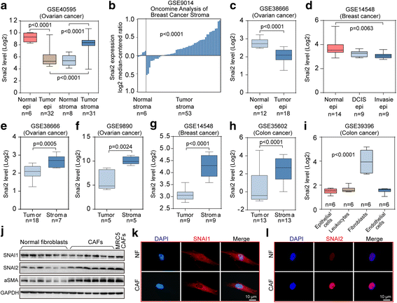
Fig. 1
Fig. 1
The specific upregulation of SNAI2 in the ovarian tumor stroma. a Boxplots showing the expression level of SNAI2 in microdissected normal epithelium, normal stroma, tumor epithelium and tumor stroma included in the ovarian dataset GSE40595. b Oncomine analysis of SNAI2 in the microdissected normal and tumor stroma in the breast profile GSE9014. c Boxplots showing the expression level of SNAI2 in the microdissected normal and tumor epithelium of GSE38666. d Boxplots showing the expression level of SNAI2 in the normal epithelium, the DCIS epithelium and the invasive epithelium of GSE14548. e–h Boxplots showing the expression level of SNAI2 in the tumor and stroma compartment in the ovarian dataset GSE38666 (e). and GSE9890 (f)., the breast dataset GSE14548 (g). and the colon dataset GSE35602 (h). i Illustration of the relative gene expression of SNAI2 in the epithelial cells, leukocytes, endothelial cells, and fibroblasts as deposited in the colon cancer profile GSE39396. j Immunoblot showing the expression of SNAI1, SNAI2 and aSMA in the microdissected normal ovarian fibroblasts and the cancer associated fibroblasts from OC patients. MRC5-CAFs served as the positive control for activated fibroblasts. GAPDH served as the loading control. k–l Immunofluorescence staining showing the relative expression of SNAI1 (k). and SNAI2 (l). in the normal ovarian fibroblasts and in the cancer associated fibroblasts from OC patients. The nuclei were counterstained with DAPI. The P value is indicated
Fig. 2
Fig. 2
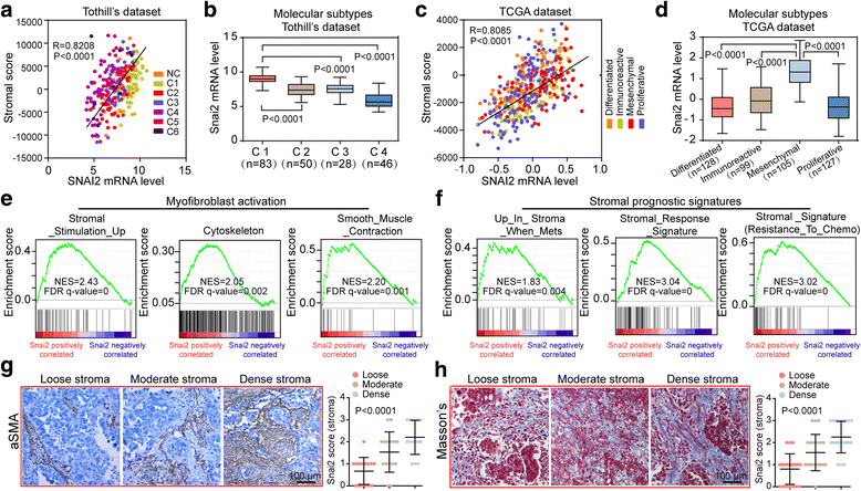
SNAI2 expression is associated with a reactive and desmoplastic stroma in OC. a Scatter plots and Spearman’s correlation showing the expression level of SNAI2 and that of the calculated stroma score in the Tothill’s dataset GSE9891. b Boxplots showing the relative SNAI2 expression in the major molecular subtypes (C1-C4) of GSE9891. c Scatter plots and Spearman’s correlation showing the expression level of SNAI2 and that of the calculated stroma score in the ovarian TCGA dataset. d Boxplots showing the relative SNAI2 expression in various subtypes of the ovarian TCGA dataset. e and f GSEA plot of the association between gene sets positively correlated with SNAI2 in the stroma profile of GSE40595 and the signatures representative of myofibroblast activation (e) or the signatures predictive of patient prognosis in the tumor stroma (f). g and h Scoring of SNAI2 in the stromal compartment of OC tissues with variant tumor stroma matrix (Loose stroma, moderate stroma and dense stroma) stratified by the aSMA staining (g) and Masson’s staining (h). The P value is indicated
Fig. 3
Fig. 3
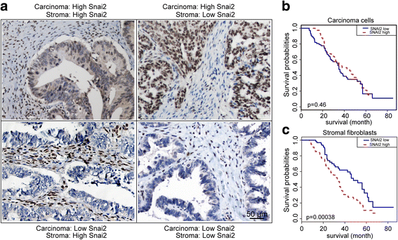
The expression pattern and clinical relevance of tumoral and stromal SNAI2 expression in OC patients. a Representative IHC images of the SNAI2 protein expression in the carcinoma and stromal compartments of the OC tumor tissues. b and c Kaplan–Meier survival curves depicting the overall survival (OS) of patients with serous OC stratified by SNAI2 protein expression levels (relative nuclear staining intensity) in carcinoma cells (b) or in tumor stromal fibroblasts (c). The P value is indicated
Fig. 4
Fig. 4
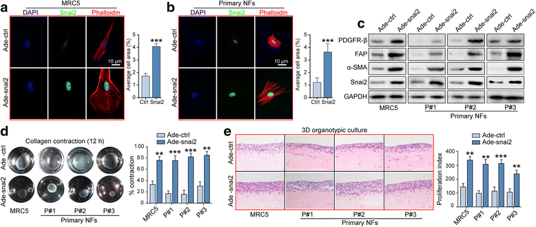
SNAI2 transforms normal fibroblasts to a CAF-like state. a and b Representative images and quantification of the cellular cytoskeleton by F-actin staining and SNAI2 immunofluorescence in the MRC5 cells (a). and the ovarian normal fibroblasts (NFs) (b). after transfection with Ade-ctrl or Ade-SNAI2. The nuclei were counterstained with DAPI. c Western blot analysis of PDGFRB, FAP, ACTA2 and SNAI2 in the MRC5 cells and the primary ovarian NFs after a 72 h transfection with Adeno-ctrl or Adeno-SNAI2. GAPDH served as the loading control. d Representative images and quantification of the collagen contraction capacity of the MRC5 cells and the primary ovarian NFs in the Adeno-ctrl and Adeno-SNAI2 transfection group. e Representative images and the quantification of the proliferation index of SKOV3 cells cocultured with MRC5 cells and primary ovarian NFs in the Adeno-ctrl and Adeno-SNAI2 transfection group (**P < 0.01, ***P < 0.001)
Fig. 5
Fig. 5
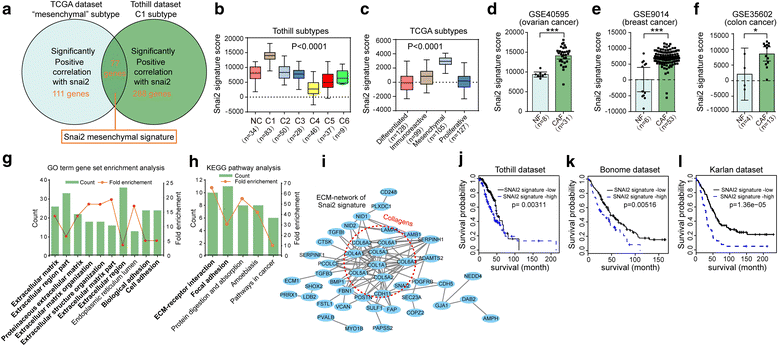
SNAI2 Drives a Transcriptional Program in the mesenchymal OC subtype that predicts clinical outcome. a Graphical depiction of the designation of the SNAI2 mesenchymal signature through overlapping genes significantly correlated with snai2 expression in the TCGA mesenchymal and Tothill C1 subtype. b and c Single sample GSEA (ssGSEA) analysis showing the relative expression of the SNAI2 mesenchymal signature score in various subtypes included in the Tothill’s dataset (b) and the ovarian TCGA dataset (c). d–f ssGSEA analysis showing the relative expression of the SNAI2 mesenchymal signature in NFs and CAFs in the stroma profile of OC (GSE0595) (d), breast cancer (GSE9014) (e) and colon cancer (GSE35602) (f). g and h Top ten most enriched GO annotation (g) and five most enriched KEGG pathways (h) in the set of the 77 genes included in SNAI2 mesenchymal signature. Count: number of genes that are involved in a given pathway. The fold enrichment shows how many fold more a given term was overrepresented in the 77 genes compared with a background of the total human genome. i STRING (Search Tool for the Retrieval of Interacting Genes) analysis of the proteins included in the SNAI2 mesenchymal signature, with the ECM network central in the network. Lines represent associations between proteins, and the line thickness reflects the number lines of evidence for each association. The dashed circles indicate the proteins included in the collagen family. j–l Kaplan-Meier analysis of the SNAI2 mesenchymal signature score expression in the patients included in three independent sets of the OC as a Tothill’s dataset (GSE9891) (j), Bonome’s dataset (GSE26712) (k) and Karlan’s dataset (GSE51088) (l). Kaplan-Meier curves were performed using the log-rank test. (*P < 0.05, ***P < 0.001)
Fig. 6
Fig. 6
Matrix stiffening modulates fibroblast activation involving upregulating SNAI2 expression. a Schematic illustration of the generation of normal-like stiffness (~1kpa) and tumor-like stiffness (~100 kpa) using the PAA gel substrate. b Representative images of the MRC5 cells growing on substrate with variant matrix stiffness (1~100 kpa), with a plastic plate being a positive control for cell spreading. c–e Representative images and quantification of the cellular cytoskeleton by F-actin staining in the MRC5 cells (c), the primary NFs (d) and the CAFs (e) grown on a soft or stiff matrix. f Western blot showing the protein expression of ACTA2, YAP1, SNAI2 and SNAI1 in the MRC5 cells and the primary CAFs grown on a soft or stiff matrix. g Immunoblotting showing the protein expression of ACTA2, YAP1, SNAI2 and SNAI1 in the MRC5 cells and the primary CAFs grown on soft or stiff matrix, in the presence or absence of a scramble siRNA or an siRNA specifically targeting YAP1, SNAI2 or SNAI1. h Representative images of the MRC5 cells and the primary CAFs grown on a soft or stiff substrate and in the presence or absence of an siRNA specifically targeting SNAI2. i Representative images and quantification of the cellular cytoskeleton by F-actin staining in MRC5 cells and in primary CAFs grown on a soft or stiff substrate and in the presence or absence of an siRNA specifically targeting SNAI2
Fig. 7
Fig. 7
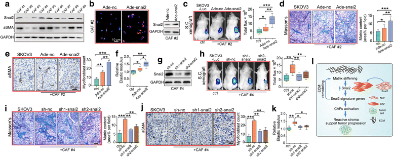
SNAI2 is required for the procarcinogenic role of fibroblasts in an OC xenograft model. a Western blot showing the protein expression of SNAI2 and aSMA in a panel of primary CAFs isolated from metastatic tumor tissues of serous OC patients. b Left: Immunofluorescence staining of SNAI2 to verify the acquisition of SNAI2 expression in primary CAFs after Ade-SNAI2 transfection (left panel). Right: Western blot analysis confirmed the effect of the SNAI2 expressing adenovirus in increasing SNAI2 expression. c Representative bioluminescence images of mice (n = 8 each group) bearing SKOV3-Luc alone or co-injection with primary CAFs transfected with either Ade-NC or Ade-SNAI2 at 4 weeks after tumor implantation. Bar graph showing the quantification of the normalized total photon counts of the subcutaneous xenografts in each group. d Masson trichrome staining and quantification of the matrix content in tumor sections from mice in groups described in (c). e Representative aSMA staining images and quantification of the amount of myofibroblasts in xenograft tissues of the above groups. f Xenograft tissue stiffness was measured as Young’s Modulus and presented as the relative elastic modulus compared with that of the SKOV3 alone group. g Western blot analysis confirmed the silencing efficiency of sh-SNAI2 in reducing SNAI2 expression in the primary CAFs. h Representative bioluminescence images of mice (n = 8 each group) bearing SKOV3-Luc alone or co-injection with primary CAFs transfected with either sh-NC or sh-SNAI2 at 4 weeks after tumor implantation. Bar graph showing the quantification of the normalized total photon counts of the subcutaneous xenografts of each group. i Masson trichrome staining and quantification of the matrix content in tumor sections from mice in groups described in (h). j Representative aSMA staining images and quantification of the amount of myofibroblasts in xenograft tissues of the above groups. k Xenograft tissue stiffness was measured as Young’s Modulus and presented as the relative elastic modulus compared with that of the SKOV3 alone group. l Graphical illustration of the feed-forward regulation mechanism between matrix stiffness and SNAI2 to drive CAF activation, desmoplasia and tumor progression. Matrix stiffness promotes SNAI2 expression in stromal CAFs, which feeds back to maintain a stiff matrix and the subsequent SNAI2 mesenchymal signature that drives a reactive tumor stroma and facilitates tumor promotion. (*P < 0.05;**P < 0.01; ***P < 0.001)

V体育2025版 - References

    1. Bowtell DD. The genesis and evolution of high-grade serous ovarian cancer. Nat Rev Cancer. 2010;10:803–808. doi: 10.1038/nrc2946. - DOI - PubMed
    1. Vang R, Shih Ie M, Kurman RJ. Ovarian low-grade and high-grade serous carcinoma: pathogenesis, clinicopathologic and molecular biologic features, and diagnostic problems. Adv Anat Pathol. 2009;16:267–282. doi: 10.1097/PAP.0b013e3181b4fffa. - DOI - PMC - PubMed
    1. De Sousa E, Melo F, Wang X, Jansen M, Fessler E, Trinh A, de Rooij LP, de Jong JH, de Boer OJ, van Leersum R, Bijlsma MF, et al. Poor-prognosis colon cancer is defined by a molecularly distinct subtype and develops from serrated precursor lesions. Nat Med. 2013;19:614–618. doi: 10.1038/nm.3174. - DOI (V体育官网) - PubMed
    1. Tothill RW, Tinker AV, George J, Brown R, Fox SB, Lade S, Johnson DS, Trivett MK, Etemadmoghadam D, Locandro B, et al. Novel molecular subtypes of serous and endometrioid ovarian cancer linked to clinical outcome. Clin Cancer Res. 2008;14:5198–5208. doi: 10.1158/1078-0432.CCR-08-0196. - DOI - PubMed
    1. Bell D, Berchuck A, Birrer M, Chien J, Cramer D, Dao F, Dhir R, DiSaia P, Gabra H, Glenn P, et al. Integrated genomic analyses of ovarian carcinoma. Nature. 2011;474:609–615. doi: 10.1038/nature10166. - DOI - PMC - PubMed

Publication types

MeSH terms

Substances

LinkOut - more resources