Skip to main page content
U.S. flag

An official website of the United States government

Dot gov

The . gov means it’s official VSports app下载. Federal government websites often end in . gov or . mil. Before sharing sensitive information, make sure you’re on a federal government site. .

Https

The site is secure V体育官网. The https:// ensures that you are connecting to the official website and that any information you provide is encrypted and transmitted securely. .

. 2016 Oct 19;8(361):361ra139.
doi: 10.1126/scitranslmed.aaf5504.

NAD+ repletion improves muscle function in muscular dystrophy and counters global PARylation

Affiliations

NAD+ repletion improves muscle function in muscular dystrophy and counters global PARylation

"VSports手机版" Dongryeol Ryu et al. Sci Transl Med. .

Abstract (VSports在线直播)

Neuromuscular diseases are often caused by inherited mutations that lead to progressive skeletal muscle weakness and degeneration. In diverse populations of normal healthy mice, we observed correlations between the abundance of mRNA transcripts related to mitochondrial biogenesis, the dystrophin-sarcoglycan complex, and nicotinamide adenine dinucleotide (NAD+) synthesis, consistent with a potential role for the essential cofactor NAD+ in protecting muscle from metabolic and structural degeneration. Furthermore, the skeletal muscle transcriptomes of patients with Duchene's muscular dystrophy (DMD) and other muscle diseases were enriched for various poly[adenosine 5'-diphosphate (ADP)-ribose] polymerases (PARPs) and for nicotinamide N-methyltransferase (NNMT), enzymes that are major consumers of NAD+ and are involved in pleiotropic events, including inflammation. In the mdx mouse model of DMD, we observed significant reductions in muscle NAD+ levels, concurrent increases in PARP activity, and reduced expression of nicotinamide phosphoribosyltransferase (NAMPT), the rate-limiting enzyme for NAD+ biosynthesis VSports手机版. Replenishing NAD+ stores with dietary nicotinamide riboside supplementation improved muscle function and heart pathology in mdx and mdx/Utr-/- mice and reversed pathology in Caenorhabditis elegans models of DMD. The effects of NAD+ repletion in mdx mice relied on the improvement in mitochondrial function and structural protein expression (α-dystrobrevin and δ-sarcoglycan) and on the reductions in general poly(ADP)-ribosylation, inflammation, and fibrosis. In combination, these studies suggest that the replenishment of NAD+ may benefit patients with muscular dystrophies or other neuromuscular degenerative conditions characterized by the PARP/NNMT gene expression signatures. .


Figures

Fig. 1
Fig. 1. The relationship between transcripts involved in NAD+ biosynthesis and consumption with muscular dystrophy in mice and DMD patients
(A) Schematic illustrating NAD+ consumption by sirtuins and PARPs and the salvage of NAD+ from NAM via NAMPT and NMNAT enzymes. (B and C) Correlation of Nampt (B) and Nmnat3 (C) transcripts from 42 strains of genetically diverse BXD mice with various measurements of muscle mass, as a percentage of body weight. CD, chow diet. (D) Correlation of Nampt transcript expression in the BXD strains with transcript expression for regulators of mitochondrial transcription and for genes encoding the components of the mitochondria. (E) A factor loading plot (biplot) showing the correlation of transcripts for mitochondrial-related genes (Tfam, Hspd1, Atp5h, and Tomm70a), utrophin (Utr), dystrophin-associated glycoproteins (Dag1, Sgcb, and Sgcd), muscle growth–related genes (Maged1 and Il6), and genes involved in the pathology of mdx mice (Tgfb1, Tnf, Mstn, Il1b, and Redd2) with NAD+ synthesis transcripts (Nampt, Nmnat1, and Nrk1) in the BXD mouse strains. Angles more than 90° between gene vectors represent negative correlation (an angle of 180° indicates perfect negative correlation). (F) These transcripts were then plotted in a circular schematic using Pearson’s r ≥ |0.2| in BXD strains showing negative (red) and positive (green) correlations. The expression of transcripts related to NAD+ (G) consumption or (H) biosynthesis shows an enrichment signature of PARP genes and of the NNMT gene in human DMD skeletal muscle data sets (n = 5 per group) (24, 25). CON, control.
Fig. 2
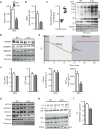
Fig. 2. NAD+ as a marker and limiting factor for in vivo energetics and mitochondrial function in mdx mice
Measurements on gastrocnemius from 16-week-old male mdx or control mice. As a new marker for muscular dystrophy, (A) NAD+ levels were reduced, as measured by 31P MRS [wild type (WT), n = 7; mdx, n = 12]. (B) These values paralleled total intracellular NAD+ levels measured by mass spectrometry in tissue extracts (n = 5). Reductions in NAD+ levels may be due to augmented NAD+ consumption, evidenced by enhanced (C) PARP activity (n = 4) and total PARylation content (WT, 1.00 ± 0.25; mdx, 2.19 ± 0.31; P = 0.042), in addition to reduced NAD+ salvage demonstrated by lower (D) NAMPT protein (WT, 1.00 ± 0.03; mdx, 0.69 ± 0.09; P = 0.036), with NMNAT1 (WT, 1.00 ± 0.09; mdx, 0.88 ± 0.03) and NMNAT3 (WT, 1.00 ± 0.05; mdx, 0.89±0.01; P = 0.009) unchanged, using heat shock protein 90 (HSP90) or Ponceau staining as loading controls. prot, protein; IB, immunoblot. (E) Schematic of in vivo 31P MRS of mdx skeletal muscle mitochondrial energetics. Dynamic magnetic resonance (MR) spectra were acquired during the following periods: rest (2 min), ischemia (9 min), and recovery (9 min). (F) High-performance liquid chromatography (HPLC) and 31P MRS measurements of ATP and PCr, respectively, showing unchanged ATP levels (n = 7) and a reduction in the PCr/ATP ratio (WT, n = 7; mdx, n = 5). Because the resting ATPase activity did not change (indicating unaltered ATP demand) (WT, n = 7; mdx, n = 5), mdx animals must function at an increased fraction of their now reduced maximal capacity (ATPmax) (WT, n = 7; mdx, n = 5) to meet the unchanged ATP demand, when MRS data are compared to control animals. mdx muscle exhibited corresponding reductions in mitochondrial proteins and function in mdx muscle, as represented by (G) ATP5A (WT, 1.00 ± 0.06; mdx, 0.51 ± 0.04; P = 0.002), UQCRC2 (WT, 1.00 ± 0.06; mdx, 0.36 ± 0.09; P = 0.004), MTCO1 (WT, 1.00 ± 0.20; mdx, 0.59 ± 0.04), SDHB (WT, 1.00 ± 0.27; mdx, 0.36 ± 0.16), and TOM20 (WT, 1.00 ± 0.07; mdx, 0.35 ± 0.12; P = 0.012) compared to HSP90 (D) as loading control, (H) blue native polyacrylamide gel electrophoresis (BN PAGE) of isolated mitochondria, and (I) CS activity (n = 9). VDAC, voltage-dependent anion channel; OD, optical density.
Fig. 3
Fig. 3. NR enhances mitochondrial function in C2C12 myotubes and increases SIRT1-dependent muscle integrity and function in dys-1;hlh-1 double-mutant C. elegans
C2C12 myotubes were treated with 0.5 to 1 mM NR. Twelve hours of NR increases (A) intracellular NAD+ levels (n = 6) and (B) increased oxygen consumption rate (OCR) in myotubes exposed to the uncoupler FCCP (n = 6). Veh, vehicle. (C) Total protein acetyl-lysine levels in myotubes were reduced after 6 hours of exposure to 0.5 and 1 mM NR. (D) SIRT1 inhibition with EX527 (10 µM) in myotubes attenuated increases in mitochondrial proteins [ATP5A (Ctrl, 1.00 ± 0.08; NR, 2.28 ± 0.14; NR + EX527, 0.80 ± 0.24; Ctrl versus NR, P = 0.001; NR versus NR + EX527, P = 0.006), UQCRC2 (Ctrl, 1.00 ± 0.15; NR, 3.54 ± 0.44; NR + EX527, 1.04 ± 0.17; Ctrl versus NR, P = 0.006; NR versus NR + EX527, P = 0.006), MTCO1 (Ctrl, 1.00 ± 0.18; NR, 2.25 ± 0.61; NR + EX527, 1.04 ± 0.07), and SDHB (Ctrl, 1.00 ± 0.19; NR, 3.54 ± 0.15; NR + EX527, 1.84 ± 0.11; Ctrl versus NR, P = 0.001; NR versus NR + EX527, P = 0.001)], reductions in FOXO1 acetylation (Ctrl, 1.00 ± 0.14; NR, 0.46 ± 0.26; NR + EX527, 1.50 ± 0.25; NR versus NR + EX527, P = 0.044), and (E) inductions of mitochondrial-related transcripts after 12 and 6 hours of NR treatment, respectively. EX, EX527. (F) Improvements in dys-1;hlh-1 mutant worm fitness on days 1 to 6 of adulthood after NR and its sir-2.1 dependence shown in animals fed RNA interference (RNAi) for sir-2.1 (R11A8.4), as measured by examining worm motility using the worm tracker (n = 3 or 60 worms per experiment) (10). a.u., arbitrary units. Reduced worm paralysis (n = 3 or 60 worms per experiment) (G) and muscle degeneration, measured by rhodamine-coupled phalloidin staining (n = 6 or 60 worms per experiment) (H) in dys-1;hlh-1 mutant worms after NR, were not observed in dys-1;hlh-1 worms fed RNAi for sir-2.1. Arrows indicate degenerated muscle fibers.
Fig. 4
Fig. 4. In vivo monitoring of NAD+ provides an efficacy marker for improvements in the phenotype of NR-treated mdx mice
Sixteen-week-old mdx mice received a dietary supplement with NR (400 mg/kg per day) for 12 weeks. (A) 31P MRS measurements of increasing NAD+ levels upon NR treatment in mdx mice (mdx, n = 5; mdx NR, n = 10). (B) The recovery of mdx muscle NAD+ levels was verified by the analysis of gastrocnemius extracts by mass spectrometry (n = 5). (C) PARP activity (n = 4) and total protein PARylation was reduced (CD, 1.00 ± 0.31; NR, 0.23 ± 0.07; P = 0.035), whereas (D) NAMPT (CD, 1.00 ± 0.06; NR, 1.20 ± 0.23) and NMNAT3 (CD, 1.00 ± 0.23; NR, 2.64 ± 0.30; P = 0.012) content increased, after NR treatment of mdx mice. (E) Unchanged ATP levels were measured by HPLC in gastrocnemius (mdx, n = 7; mdx NR, n = 10). (F) In vivo 31P MRS measurements of mitochondrial energetics revealed an improvement in the PCr/ATP ratio (mdx, n = 5; mdx NR, n = 10), unchanged resting ATPase activity (mdx, n = 5; mdx NR, n = 10), and a recovery of ATPmax (mdx, n = 5; mdx NR, n = 10) in NR-treated mdx mice. NR-treated mdx animals hence function at a reduced fraction of their capacity (ATPmax) to meet ATP demand. The enhancement of NAD+ metabolism with NR prevented improved (G) ATP5A (CD, 1.00 ± 0.15; NR, 1.18 ± 0.15), UQCRC2 (CD, 1.00 ± 0.24; NR, 2.21 ± 0.57), MTCO1 (CD, 1.00 ± 0.53; NR, 1.62 ± 0.16), SDHB (CD, 1.00 ± 0.38; NR, 2.96 ± 0.57; P = 0.023), and TOM20 (CD, 1.00 ± 0.15; NR, 2.63 ± 0.22; P = 0.002) protein levels, using HSP90 (D) or Ponceau loading control, (H) mitochondrial complexes as evidenced by blue native PAGE of isolated gastrocnemius mitochondria, using a Coomassie loading control, (I) CS activity in frozen gastrocnemius extracts (mdx, n = 9; mdx NR, n = 6), and (J) cytochrome c oxidase staining of soleus (arrow) and gastrocnemius (asterisks) muscle fibers. (K) NR increased the running distance and time during an endurance treadmill test in mdx mice (mdx, n = 9; mdx NR, n = 12). (L and M) Hematoxylin and eosin (H&E) and Masson’s trichrome staining of hearts from control C57BL/10 and mdx male mice at 16 months of age with and without NR treatment. Representative images of the (L) whole heart and (M) magnified sections showing necrosis and fibrosis (n = 4).
Fig. 5
Fig. 5. NAD+ repletion causes compensatory increases in the expression of structural proteins and protects muscle from damage and fibrosis
Sixteen-week-old mdx mice received a dietary supplement with NR (400 mg/kg per day) for 12 weeks. (A) NR-treated mdx mice exhibited increased α-dystrobrevin (α-DB) (CD, 1.00 ± 0.35; NR, 1.61 ± 0.09) and δ-sarcoglycan (δ-SG) (CD, 1.00 ± 0.15; NR, 2.24 ± 0.43; P = 0.026) protein expression compared to HSP90 (Fig. 4D) as a loading control in gastrocnemius extracts. NR attenuated mdx muscle damage as evidenced through (B) Evans Blue staining of both TA muscle and (C) sections of gastrocnemius and soleus muscles [red, Evans Blue (EB); white, 4′,6-diamidino-2-phenylindole (DAPI)], (D) quantified using ImageJ software (mdx, n = 6; mdx NR, n = 12). (E) NR also reduced basal plasma creatine kinase levels (mdx, n = 5; mdx NR, n = 6). (F) mdx mice treated with NR demonstrated an attenuation in the percent loss of torque in quadriceps, after muscle damage induced by lengthening contractions (mdx, n = 7; mdx NR, n = 6). (G) Increases in the average minimal Feret’s diameter (in micrometers) and cross-sectional area (CSA) (in square micrometers) of tibialis anterior muscle fibers, indicating increases in fiber size with NR treatment in mdx mice. These data were acquired from images stained with DAPI and laminin [(H) shows a representative image]. Quantification of images was performed with ImageJ software (8 weeks of NR treatment, n = 5). (I) Similarly, reduced CD45 staining was seen in diaphragms of mdx mice treated with NR. (J) Reduced acetylation of p65 (NF-κB) protein after NR in quadriceps (CD, 1.00 ± 0.15; NR, 0.38 ± 0.16; P = 0.023; n = 3). (K) FAPs expressing mesenchymal PDGFRα were decreased in diaphragms of mdx mice treated with NR (n = 3). Also, reductions in the fibrosis of mdx diaphragms were observed with less Picrosirius red staining in transverse muscle sections of NR-treated mice. (L) PARylation intensity in skeletal muscle nuclei of mdx mice is reduced with NR, as shown with immunohistological staining of tibialis anterior muscle with PAR, CD45, and laminin antibodies.
Fig. 6
Fig. 6. mdx/Utr−/− mice recover skeletal muscle function and exhibit improved heart pathology with NR treatment
Three-week-old mdx/Utr−/− mice received a dietary supplement with NR (400 mg/kg per day) for 5 to 7 weeks. Images stained with DAPI and laminin were used to quantify increases in the (A) average and (B) distribution of mdx/Utr−/− mouse muscle fiber cross-sectional areas (in µm2) of the tibialis anterior muscle (mdx/Utr−/−n = 3; 7 weeks of NR treatment). (C) NR-treated mdx/Utr−/− mice showed an increase in the minimal Feret’s diameter (in micrometers) (mdx/Utr−/−n = 3; 7 weeks of NR treatment). Quantification of images was performed with ImageJ software. (D) As evidence for the therapeutic effectiveness of NR treatment, mdx/Utr−/− mice grip strength was improved from 8 weeks (mdx/Utr−/−n = 4; mdx/Utr−/− NR, n = 7; 5 weeks of NR treatment) to 10 weeks of age (mdx/Utr−/−n = 3; mdx/Utr−/− NR, n = 5; 7 weeks of NR treatment). BW, body weight. (E) Sections of heart tissue were (immuno) histochemically stained with Masson’s trichrome, hematoxylin and eosin, and CD45, showing a reduction of cardiac fibrosis, necrosis, and macrophage infiltration, respectively, in the ventricles of mdx/Utr−/− mice at 4 weeks of age and treated with NR for 6 weeks (representative images from n = 3 mdx/Utr−/− and n = 3 mdx/Utr−/− NR). (F) Scheme summarizing the SIRT1-dependent effects of NR on mdx mouse muscle.

VSports最新版本 - References

    1. Moat SJ, Bradley DM, Salmon R, Clarke A, Hartley L. Newborn bloodspot screening for Duchenne muscular dystrophy: 21 years experience in Wales (UK) Eur. J. Hum. Genet. 2013;21:1049–1053. - PMC - PubMed
    1. Mendell JR, Shilling C, Leslie ND, Flanigan KM, al-Dahhak R, Gastier-Foster J, Kneile K, Dunn DM, Duval B, Aoyagi A, Hamil C, Mahmoud M, Roush K, Bird L, Rankin C, Lilly H, Street N, Chandrasekar R, Weiss RB. Evidence-based path to newborn screening for Duchenne muscular dystrophy. Ann. Neurol. 2012;71:304–313. - PubMed
    1. Fairclough RJ, Wood MJ, Davies KE. Therapy for Duchenne muscular dystrophy: Renewed optimism from genetic approaches. Nat. Rev. Genet. 2013;14:373–378. - "VSports手机版" PubMed
    1. Chalkiadaki A, Igarashi M, Nasamu AS, Knezevic J, Guarente L. Muscle-specific SIRT1 gain-of-function increases slow-twitch fibers and ameliorates pathophysiology in a mouse model of Duchenne muscular dystrophy. PLOS Genet. 2014;10:e1004490. - PMC - PubMed
    1. Handschin C, Kobayashi YM, Chin S, Seale P, Campbell KP, Spiegelman BM. PGC-1α regulates the neuromuscular junction program and ameliorates Duchenne muscular dystrophy. Genes Dev. 2007;21:770–783. - PMC - PubMed

Publication types (V体育官网入口)

MeSH terms