Skip to main page content
U.S. flag

An official website of the United States government

Dot gov

The VSports app下载. gov means it’s official. Federal government websites often end in . gov or . mil. Before sharing sensitive information, make sure you’re on a federal government site. .

Https

The site is secure. The https:// ensures that you are connecting to the official website and that any information you provide is encrypted and transmitted securely. V体育官网.

. 2014 Sep 11;9(9):e106601.
doi: 10.1371/journal.pone.0106601. eCollection 2014.

DJ-1 interacts with and regulates paraoxonase-2, an enzyme critical for neuronal survival in response to oxidative stress

Affiliations

"VSports app下载" DJ-1 interacts with and regulates paraoxonase-2, an enzyme critical for neuronal survival in response to oxidative stress

Mohammad Parsanejad et al. PLoS One. .

Abstract (VSports app下载)

Loss-of-function mutations in DJ-1 (PARK7) gene account for about 1% of all familial Parkinson's disease (PD). While its physiological function(s) are not completely clear, DJ-1 protects neurons against oxidative stress in both in vitro and in vivo models of PD. The molecular mechanism(s) through which DJ-1 alleviates oxidative stress-mediated damage remains elusive. In this study, we identified Paraoxonase-2 (PON2) as an interacting target of DJ-1. PON2 activity is elevated in response to oxidative stress and DJ-1 is crucial for this response. Importantly, we showed that PON2 deficiency hypersensitizes neurons to oxidative stress induced by MPP+ (1-methyl-4-phenylpyridinium). Conversely, over-expression of PON2 protects neurons in this death paradigm VSports手机版. Interestingly, PON2 effectively rescues DJ-1 deficiency-mediated hypersensitivity to oxidative stress. Taken together, our data suggest a model by which DJ-1 exerts its antioxidant activities, at least partly through regulation of PON2. .


Conflict of interest statement (V体育安卓版)

Competing Interests: The authors have declared that no competing interests exist.

Figures

Figure 1
Figure 1. DJ-1 and PON2 interact.
(A) DJ-1 full length protein sequence. Peptide observed from DJ-1 after using PON2 as bait is highlighted. Mascot peptide score is 30.2. (B) HEK293 cells expressing GST-DJ-1 or GST as control were lysed and GST-DJ-1 was precipitated by glutathione sepharose beads and analyzed with Western blotting using PON2 antibody. (C) HEK293 cells were transfected with plasmid expressing Myc-PON2 (M-PON2). Cells were lysed and Myc-PON2 was precipitated with Myc antibody. Isolated complexes were analyzed with Western blotting using DJ-1 antibody. (D) DJ-1 was pulled down by DJ-1 antibody from cell lysate extracted from cultured cortical neurons. Immune complexes were analyzed with Western blotting using PON2 antibody.
Figure 2
Figure 2. DJ-1 and oxidative stress modulate PON2 activity.
(A) Cultured WT and DJ-1 KO cortical neurons were treated with MPP+ (20 µM) for 12 hours and cells were washed and membrane was extracted. Crude membrane was exposed to the substrate C12 for 60 minutes and the percentage of remaining C12 was measured. (B) Cultured WT and DJ-1 KO cortical neurons were treated with MPP+ (20 µM) for 24 hours. Neurons were then exposed to DHC for 10 minutes and the amount of hydrolysis of DHC was assessed with measuring UV absorbance. One unit of PON2 activity is equal to 1 µmol DHC hydrolyzed/ml/min. (C) WT and DJ-1 KO MEFs were treated with hydrogen peroxide (100 µM) for 24 hours and PON2 activity was measured as described in B. (D) WT and DJ-1 KO MEFs were infected with adenovirus expressing DJ-1 or GFP alone as control. After 48 hours of expression, cells were lysed and exposed to C12 as the substrate for 60 minutes. Percentage of C12 remaining in activity buffer was measured. Statistical significance was assessed by Anova and post-hoc test Tukey on data obtained from three independent experiments (n = 3). * denotes p<0.05, ** denotes p<0.01, and *** denotes p<0.001.
Figure 3
Figure 3. DJ-1 has no lactonase activity and no effects on PON2 protein level.
(A) WT and PON2 deficient MEFs were infected with adenovirus expressing DJ-1 or GFP. PON2 activity was then measured using C12 as described before. (B) Samples used in panel A was exposed to SDS-PAGE analysis to assess their levels of DJ-1, PON2 and GFP. (C) Cultured cortical neurons extracted from DJ-1 WT and DJ-1 KO were treated with MPP+ (20 µM) for different durations. Cells were lysed and PON2 protein level was assessed by western blotting. Statistical significance was assessed by Anova and post-hoc test Tukey on data obtained from three independent experiments (n = 3). * denotes p<0.05, **denotes p<0.01 and *** denotes p<0.001.
Figure 4
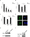
Figure 4. PON2 protects neurons against MPP+.
(A) Primary cortical neurons obtained from PON2 deficient or wild type mice were subjected to 10, 20 and 40 µM MPP+ treatment for 48 hours. Cells were lysed and viability was assessed by direct microscopy and counting intact nuclei. (B) WT and PON2 def cortical neurons were transfected with plasmid expressing Myc-PON2 and GFP (under independent promoters), or GFP as control, and subjected to 20 µM MPP+ for 48 hours. Cells were fixed and the nuclei were stained with Hoechst. Survival percentage represents the ratio of GFP-expressing cells with morphologically intact nuclei (D, a and b) to the total number of GFP positive cells. (C) WT and DJ-1 KO cortical neurons over-expressing PON2 and GFP or GFP alone as control (using adenovirus expressing PON2 or GFP) were subjected to 20 µM MPP+ for 48 hours. The survival assay was performed as described in part B. (D) Representative image of GFP positive neurons (a and c), and Hoechst-stained surviving (b) and dead (d) nuclei. (E) Western blot analysis of PON2 levels in PON2 deficient (PON2 def) and WT MEFs and also in WT MEFs infected with PON2-expressing adenovirus (WT+PON2 AV). The membrane was probed with PON2 antibody. (F) Western blot analysis for Myc in WT MEFs expressing control (Ctr) or Myc-PON2 plasmids. The Western blot was analyzed by Myc antibody. Statistical significance was assessed by Anova and post-hoc test Tukey on data obtained from three independent experiments (n = 3). * denotes p<0.05, **denotes p<0.01 and *** denotes p<0.001.

References

    1. Hirsch E, Graybiel AM, Agid YA (1988) Melanized dopaminergic neurons are differentially susceptible to degeneration in Parkinson's disease. Nature 334: 345–348. - PubMed
    1. Tanner CM, Ottman R, Goldman SM, Ellenberg J, Chan P, et al. (1999) Parkinson disease in twins: an etiologic study. JAMA 281: 341–346. - PubMed
    1. Polymeropoulos MH, Lavedan C, Leroy E, Ide SE, Dehejia A, et al. (1997) Mutation in the alpha-synuclein gene identified in families with Parkinson's disease. Science 276: 2045–2047. - "VSports app下载" PubMed
    1. Kitada T, Asakawa S, Hattori N, Matsumine H, Yamamura Y, et al. (1998) Mutations in the parkin gene cause autosomal recessive juvenile parkinsonism. Nature 392: 605–608. - "VSports最新版本" PubMed
    1. Leroy E, Boyer R, Auburger G, Leube B, Ulm G, et al. (1998) The ubiquitin pathway in Parkinson's disease. Nature 395: 451–452. - PubMed

Publication types

MeSH terms