Skip to main page content
U.S. flag

An official website of the United States government

Dot gov

The . gov means it’s official. Federal government websites often end in VSports app下载. gov or . mil. Before sharing sensitive information, make sure you’re on a federal government site. .

Https

The site is secure. The https:// ensures that you are connecting to the official website and that any information you provide is encrypted and transmitted securely V体育官网. .

. 2020 Dec 16;13(12):468.
doi: 10.3390/ph13120468.

Eudebeiolide B Inhibits Osteoclastogenesis and Prevents Ovariectomy-Induced Bone Loss by Regulating RANKL-Induced NF-κB, c-Fos and Calcium Signaling

Affiliations

V体育安卓版 - Eudebeiolide B Inhibits Osteoclastogenesis and Prevents Ovariectomy-Induced Bone Loss by Regulating RANKL-Induced NF-κB, c-Fos and Calcium Signaling

Mi-Hwa Kim et al. Pharmaceuticals (Basel). .

Abstract

Eudebeiolide B is a eudesmane-type sesquiterpenoid compound isolated from Salvia plebeia R. Br. , and little is known about its biological activity. In this study, we investigated the effects of eudebeiolide B on osteoblast differentiation, receptor activator nuclear factor-κB ligand (RANKL)-induced osteoclastogenesis in vitro and ovariectomy-induced bone loss in vivo. Eudebeiolide B induced the expression of alkaline phosphatase (ALP) and calcium accumulation during MC3T3-E1 osteoblast differentiation. In mouse bone marrow macrophages (BMMs), eudebeiolide B suppressed RANKL-induced osteoclast differentiation of BMMs and bone resorption. Eudebeiolide B downregulated the expression of nuclear factor of activated T-cells 1 (NFATc1) and c-fos, transcription factors induced by RANKL. Moreover, eudebeiolide B attenuated the RANKL-induced expression of osteoclastogenesis-related genes, including cathepsin K (Ctsk), matrix metalloproteinase 9 (MMP9) and dendrocyte expressed seven transmembrane protein (DC-STAMP). Regarding the molecular mechanism, eudebeiolide B inhibited the phosphorylation of Akt and NF-κB p65. In addition, it downregulated the expression of cAMP response element-binding protein (CREB), Bruton's tyrosine kinase (Btk) and phospholipase Cγ2 (PLCγ2) in RANKL-induced calcium signaling. In an ovariectomized (OVX) mouse model, intragastric injection of eudebeiolide B prevented OVX-induced bone loss, as shown by bone mineral density and contents, microarchitecture parameters and serum levels of bone turnover markers. Eudebeiolide B not only promoted osteoblast differentiation but inhibited RANKL-induced osteoclastogenesis through calcium signaling and prevented OVX-induced bone loss. Therefore, eudebeiolide B may be a new therapeutic agent for osteoclast-related diseases, including osteoporosis, rheumatoid arthritis and periodontitis. VSports手机版.

Keywords: OVX mouse model; RANKL; calcium signal; eudebeiolide B; osteoporosis V体育安卓版. .


Conflict of interest statement

The authors declare no conflict of interest.

Figures

Figure 1
Figure 1
Eudebeiolide B inhibits RANKL-induced osteoclast differentiation and promotes osteoblast differentiation. (A) Structure of eudebeiolide B (n = 3). (B) Cytotoxicity of eudebiolide B. Bone marrow macrophages (BMMs) were seeded and treated with indicated concentrations of eudebeiolide B in the presence of M-CSF (30 ng/mL) for 72 h. Cell viability was analyzed using the XTT assay (n = 3). (C) BMMs were treated with RANKL (100 ng/mL) and M-CSF (30 ng/mL) after pretreatment with 1, 5, 10 or 30 μM of eudebeiolide B for 1 h. Cells were fixed and stained with TRAP staining solution. TRAP-positive multinucleated cells (TRAP + MNCs) with more than three nuclei were defined as osteoclasts and counted (n = 3). (D) BMMs were seeded and treated with RANKL (50 ng/mL) and M-CSF (30 ng/mL) for 72 h. Cells were then treated with eudebeiolide B for 48 h. Bone resorption areas were measured using ImageJ (n = 3). (E) MC3T3-E1 cells were seeded and incubated with differentiation media and eudebeiolide B for 7 days. Osteoblast differentiation was determined by ALP staining, and ALP activity was measured using the cell lysate (n = 3). (F) MC3T3-E1 cells were seeded and incubated with differentiation media and eudebeiolide B for 21 days. Osteoblast differentiation was assessed by Alizarin red staining, and calcium accumulation was quantified with cetylpyridinium chloride solution (n = 3). (G) MC3T3-E1 cells were treated with or without eudebeiolide B (10 μM) and cultured for 24, 48 and 72 h. The mRNA expression of Runx2, Osterix, OPG and RANKL was assessed by quantitative PCR (n = 3). Values are expressed as the means ± S.D. of three individual experiments. * p < 0.05 and ** p < 0.01 versus control (CE) obtained through one-way ANOVA followed by Dunnett’s test; * p < 0.05 and ** p < 0.01 versus the only-DM-treated group (G) obtained through the unpaired Student’s t-test.
Figure 2
Figure 2
Attenuation of RANKL-induced osteoclastogenesis-related transcription factor and gene expression by eudebeiolide B. (AC) BMMs were pretreated with or without eudebeiolide B and stimulated with RANKL (100 ng/mL) for the indicated time. (A) Western blot analysis of c-Fos and NFATc1 protein was performed, and band densities of (B) c-Fos and (C) NFATc1 were quantified by ImageJ (n = 3). (DG) BMMs were cultured in the presence of RANKL (100 ng/mL) with or without eudebeiolide B for 48 h. (D) NFATc1, (E) cathepsin K, (F) MMP9 and (G) DC-STAMP mRNAs were analyzed by quantitative RT-PCR (n = 3). All data are expressed as the means ± S.D. of three individual experiments. * p < 0.05 and ** p < 0.01 versus the only-RANKL-treated group (B,C) obtained through the unpaired Student’s t-test; * p < 0.05 and ** p < 0.01 versus the only-RANKL-treated group (DG) obtained through one-way ANOVA followed by Dunnett’s test.
Figure 3
Figure 3
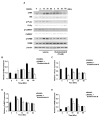
Effect of eudebeiolide B on RANKL-induced cellular signaling. BMM cells were pretreated with or without eudebeiolide B (10 μM) for 1 h and then stimulated with RANKL (100 ng/mL) for the indicated times. (A) Expression of signaling molecules determined by Western blot analysis. Band optical densities of (B) ERK, (C) P38, (D) JNK, I AKT and (F) NF-κB p65 were evaluated by ImageJ software (n = 3). All data are expressed as the means ± S.D. * p < 0.05 and ** p < 0.01 versus the only-RANKL-treated group obtained through the unpaired Student’s t-test.
Figure 4
Figure 4
Eudebeiolide B inhibits RANKL-induced calcium signaling. (A) Phosphorylation was evaluated by Western blot analysis. Eudebeiolide B inhibits the phosphorylation of (B) Btk, (C) PLCγ2, (D) CaMKIV and (E) CREB. BMMs were pretreated with or without eudebeiolide B (10 μM) for 1 h and then stimulated with RANKL (100 ng/mL) for the indicated times. Western blot analyses were performed with the indicated antibodies (n = 3). All data are expressed as the means ± S.D. * p < 0.05 versus the only-RANKL-treated group obtained through the unpaired Student’s t-test.
Figure 5
Figure 5
Bone mineral density BMD, bone mineral content BMC and biochemical serum analysis of the sham-operated, OVX + vehicle, OVX + eudebeiolide B-treated and OVX + alendronate-treated mice. (A) BMD and (B) BMC of the whole body were analyzed using DEXA (n = 6). The serum levels of (C) ALP, (D) c-terminal telopeptide of type 1 collagen (CTX), (E) osteoprotegerin (OPG) and (F) RANKL were determined by ELISA (n = 6). (G) RANKL/OPG ratio was calculated (n = 6). Data are expressed as the means ± S.D. # p < 0.05 versus the sham group obtained through the unpaired Student’s t-test; * p < 0.05, ** p < 0.01 versus the only-OVX-treated group obtained through one-way ANOVA followed by Dunnett’s test.
Figure 6
Figure 6
Micro-CT analysis of the proximal femurs from the eudebeiolide B- or alendronate-treated OVX mice. (A) The micro-CT images of longitudinal and transverse of the proximal femurs from the sham-operated, OVX + vehicle, OVX + eudebeiolide B-treated and OVX + alendronate-treated mice were obtained. (B) The bone volume/total volume (BV/TV), (C) trabecular thickness (Tb. Th.) and (D) trabecular number (Tb. N.) of the femurs were analyzed using micro-CT (n = 6). Data are expressed as the means ± S.D. # p < 0.05 versus the sham group obtained through the unpaired Student’s t-test; * p < 0.05, ** p < 0.01 versus the only-OVX-treated group obtained through one-way ANOVA followed by Dunnett’s test.
Figure 7
Figure 7
Histological analysis of the proximal tibia tissue sections from the eudebeiolide B- or alendronate-treated OVX mice. (A) H&E stains, (B) histochemical stains for TRAP and (C) immunohistochemical stains for ALP of the sham-operated, OVX + vehicle, OVX + eudebeiolide B-treated and OVX + alendronate-treated mice were obtained (n = 6). (D) Osteoclast surface/bone surface (Oc.S/BS) and (E) Osteoblast surface/bone surface (Ob.S/BS) were evaluated by HistoMorph software (n = 6). Data are expressed as the means ± S.D. # p < 0.05 versus the sham group obtained through the unpaired Student’s t-test; * p < 0.05 versus the only-OVX-treated group obtained through one-way ANOVA followed by Dunnett’s test.

References (VSports最新版本)

    1. Rodan G.A., Martin T.J. Therapeutic approaches to bone diseases. Science. 2000;289:1508–1514. doi: 10.1126/science.289.5484.1508. - "V体育官网入口" DOI - PubMed
    1. Khosla S., Oursler M.J., Monroe D.G. Estrogen and the skeleton. Trends Endocrinol. Metab. 2012;23:576–581. doi: 10.1016/j.tem.2012.03.008. - DOI - PMC - PubMed
    1. Rodan G.A. Bone homeostasis. Proc. Natl. Acad. Sci. USA. 1998;95:13361–13362. doi: 10.1073/pnas.95.23.13361. - VSports在线直播 - DOI - PMC - PubMed
    1. Boyle W.J., Simonet W.S., Lacey D.L. Osteoclast differentiation and activation. Nature. 2003;423:337–342. doi: 10.1038/nature01658. - DOI - PubMed
    1. Heino T.J., Hentunen T.A. Differentiation of osteoblasts and osteocytes from mesenchymal stem cells. Curr. Stem Cell Res. Ther. 2008;3:131–145. doi: 10.2174/157488808784223032. - DOI - PubMed