Skip to main page content
U.S. flag

An official website of the United States government

Dot gov

The . gov means it’s official. Federal government websites often end in VSports app下载. gov or . mil. Before sharing sensitive information, make sure you’re on a federal government site. .

Https

The site is secure. The https:// ensures that you are connecting to the official website and that any information you provide is encrypted and transmitted securely. V体育官网.

. 2025 Sep 15;23(9):358.
doi: 10.3390/md23090358.

"VSports在线直播" Single-Cell Transcriptomic Analysis Reveals Cell Heterogeneity and Altered Signaling Pathways in Jellyfish Sting Patients

Affiliations

Single-Cell Transcriptomic Analysis Reveals Cell Heterogeneity and Altered Signaling Pathways in Jellyfish Sting Patients

Zhen Qin et al. Mar Drugs. .

Abstract

Jellyfish stings induce a range of symptoms, from localized irritation to life-threatening systemic reactions, yet the underlying immune mechanisms remain poorly understood. This study employed single-cell RNA sequencing (scRNA-seq) to analyze peripheral blood mononuclear cells (PBMCs) from a severely affected patient and healthy controls, uncovering the immune landscape at single-cell resolution and identifying the signaling pathways. We identified 11 major immune cell types, with a marked increase in CD14+ monocytes (81 VSports手机版. 86% of total cells) and significant reductions in T cells, B cells, and CD16+ monocytes in the envenomated patient. Subclustering revealed six monocyte and four neutrophil subsets, each displaying distinct functional profiles. Patient monocytes were enriched for MMP9+ and RETN+ subsets, associated with leukocyte migration and inflammation, whereas healthy controls exhibited CD74+ monocytes linked to oxidative phosphorylation. Neutrophils in the patient were predominantly LTF+ and S100A12+, implicating inflammatory and immune regulatory pathways. These findings provide a detailed single-cell atlas of immune dysregulation post-jellyfish sting, highlighting the pivotal roles of MMP9+ monocytes and S100A12+ neutrophils in driving inflammation. This study offers potential therapeutic targets for mitigating severe immune responses in jellyfish envenomation. .

Keywords: CD74+ monocytes; MMP9+ monocytes; S100A12+ neutrophils; cell heterogeneity; jellyfish stings; scRNA-seq V体育安卓版. .


Conflict of interest statement (V体育安卓版)

There are no conflicts of interest.

Figures

Figure 1
Figure 1
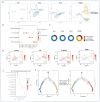
Overview and cell type analysis of PBMC single-cell transcriptomics. (A) Schematic representation of the study design and workflow. The figure outlines the steps from patient sample processing to single-cell RNA sequencing and downstream analysis. (B) UMAP projection of sampled cell types. A UMAP plot illustrating the dimensionality reduction of different cell types identified within the PBMC samples. Each dot represents a single cell, colored by its annotated cell type to visualize the distribution and clustering of cellular diversity. (C) Dot plot of canonical marker genes. A dot plot displaying the expression of classical marker genes across various cell types. The size of each dot indicates the percentage of cells expressing the gene, while the color intensity indicates the average expression level. (D) Pie charts illustrating the proportion of 11 major cell types across the four samples. Each bar represents a sample, and the colors correspond to different cell types. Pie charts showing the relative proportions of eleven major cell types identified in the PBMCs of the four individual samples. (E) Feature plots for selected canonical marker genes providing a spatial representation of gene expression levels on the UMAP plot. These plots help to identify the specific cell types associated with the expression of these marker genes.
Figure 2
Figure 2
Subclustering and functional analysis of monocytes. (A) UMAP plot depicting the distribution of monocyte (Mono) subtypes across the four samples. Each color represents a distinct monocyte subtype. (B) Dot plot showing the expression of marker genes for each monocyte subtype. The dot size and color intensity represent the percentage of cells expressing the gene and the average expression level, respectively. (C) Pie charts display the proportion of six monocyte subtypes across the four samples. Each bar represents a sample, and the colors correspond to different monocyte subtypes. (D) Feature plot illustrating the spatial distribution of marker genes for each monocyte subtype on the UMAP plot. UMAP_1 and UMAP_2 represent the two primary dimensions resulting from dimensionality reduction. (E) Volcano plot highlighting differentially expressed genes between MMP9+ monocytes and CD74+ monocytes. Significantly upregulated genes are marked in red. (F) Pathway enrichment analysis of upregulated genes in MMP9+ monocytes. The bar plot shows the top enriched pathways. (G) Pathway enrichment analysis of upregulated genes in CD74+ monocytes. The bar plot shows the top enriched pathways.
Figure 3
Figure 3
Subclustering and functional analysis of neutrophils. (A) UMAP plot showing the distribution of neutrophil subtypes across the four samples. Each color represents a distinct neutrophil subtype. (B) Dot plot displaying the expression of marker genes for each neutrophil subtype. The dot size and color intensity represent the percentage of cells expressing the gene and the average expression level, respectively. (C) Pie charts illustrate the proportion of four neutrophil subtypes across the four samples. Each bar represents a sample, and the colors correspond to different neutrophil subtypes. (D) Feature plot showing the spatial distribution of marker genes for each neutrophil subtype on the UMAP plot. UMAP_1 and UMAP_2 represent the two primary dimensions resulting from dimensionality reduction. (E) Dot plot summarizing the functional pathways associated with each neutrophil subtype. The dot size represents the significance of the pathway, and the color indicates the enrichment score. (F) Monocle pseudotime analysis of neutrophil subtypes. A trajectory analysis using Monocle to infer the potential differentiation paths or states of neutrophil subtypes over pseudotime. Component 1 and Component 2 in the pseudotime analysis reflect the dominant trajectory components.
Figure 4
Figure 4
Cell–cell communication analysis in PBMC samples. (A) Heatmap showing the interaction strength between cell types in healthy controls (HCs, left) and the patient (right). The color intensity represents the interaction strength. (B) Dot plot summarizing the key interaction pairs in healthy controls (HCs). The dot size represents the interaction strength, and the color indicates the significance. (C) Dot plot summarizing the key interaction pairs in the patient. The dot size represents the interaction strength, and the color indicates the significance.

References

    1. Naveed M., Chan M.W.H., Aslam S., Wang F., Sajjad A., Ullah A., Saleem N., Haider M.S., Arija V. Nutritional composition assessment and antimicrobial activity of Catostylus perezi, jellyfish blooms along the coast of Pakistan: An awareness to avoid food neophobia in Pakistan. Nat. Prod. Res. 2024;38:3957–3963. doi: 10.1080/14786419.2023.2269597. - DOI - PubMed
    1. Killi N., Mariottini G.L. Cnidarian Jellyfish: Ecological Aspects, Nematocyst Isolation, and Treatment Methods of Sting. Results Probl. Cell Differ. 2018;65:477–513. - PubMed
    1. Malacarne P.F., Menezes T.N., Martins C.W., Naumann G.B., Gomes H.L., Pires R.G.W., Figueiredo S.G., Campos F.V. Advances in the characterization of the Scorpaena plumieri cytolytic toxin (Sp-CTx) Toxicon. 2018;150:220–227. doi: 10.1016/j.toxicon.2018.06.065. - DOI - PubMed
    1. Baysoy A., Bai Z., Satija R., Fan R. The technological landscape and applications of single-cell multi-omics. Nat. Rev. Mol. Cell Biol. 2023;24:695–713. doi: 10.1038/s41580-023-00615-w. - "VSports注册入口" DOI - PMC - PubMed
    1. Ruwanpathirana P., Priyankara D. Clinical manifestations of wasp stings: A case report and a review of literature. Trop. Med. Health. 2022;50:82. doi: 10.1186/s41182-022-00475-8. - DOI - PMC - PubMed

MeSH terms (V体育官网入口)

Substances