Skip to main page content
U.S. flag

An official website of the United States government

Dot gov

The . gov means it’s official. Federal government websites often end in . gov or . mil VSports app下载. Before sharing sensitive information, make sure you’re on a federal government site. .

Https

The site is secure V体育官网. The https:// ensures that you are connecting to the official website and that any information you provide is encrypted and transmitted securely. .

. 2024 Aug 17;25(1):313.
doi: 10.1186/s12931-024-02932-w.

The role and mechanism of thrombospondin-4 in pulmonary arterial hypertension associated with congenital heart disease

Affiliations

The role and mechanism of thrombospondin-4 in pulmonary arterial hypertension associated with congenital heart disease

Haowei Zeng et al. Respir Res. .

Abstract

Background: Due to a special hemodynamic feature, pulmonary vascular disease in pulmonary arterial hypertension associated with congenital heart disease (PAH-CHD) has two stages: reversible and irreversible. So far, the mechanism involved in the transition from reversible to irreversible stage is elusive. Moreover, no recognized and reliable assessments to distinguish these two stages are available. Furthermore, we found that compared with control and reversible PAH, thrombospondin-4 (THBS4) was significantly upregulated in irreversible group by bioinformatic analysis VSports手机版. Hence, we further verify and investigate the expression and role of THBS4 in PAH-CHD. .

Methods: We established the monocrotaline plus aorto-cava shunt-induced (MCT-AV) rat model. We measured the expression of THBS4 in lung tissues from MCT-AV rats. Double immunofluorescence staining of lung tissue for THBS4 and α-SMA (biomarker of smooth muscle cells) or vWF (biomarker of endothelial cells) to identify the location of THBS4 in the pulmonary artery. Primary pulmonary artery smooth muscle cells (PASMCs) were cultivated, identified, and used in this study V体育安卓版. THBS4 was inhibited and overexpressed by siRNA and plasmid, respectively, to explore the effect of THBS4 on phenotype transformation, proliferation, apoptosis, and migration of PASMCs. The effect of THBS4 on pulmonary vascular remodeling was evaluated in vivo by adeno-associated virus which suppressed THBS4 expression. Circulating level of THBS4 in patients with PAH-CHD was measured by ELISA. .

Results: THBS4 was upregulated in the lung tissues of MCT-AV rats, and was further upregulated in severe pulmonary vascular lesions. And THBS4 was expressed mainly in PASMCs V体育ios版. When THBS4 was inhibited, contractile markers α-SMA and MYH11 were upregulated, while the proliferative marker PCNA was decreased, the endothelial-mensenchymal transition marker N-cad was downregulated, proapototic marker BAX was increased. Additionally, proliferation and migration of PASMCs was inhibited and apoptosis was increased. Conversely, THBS4 overexpression resulted in opposite effects. And the impact of THBS4 on PASMCs was probably achieved through the regulation of the PI3K/AKT pathway. THBS4 suppression attenuated pulmonary vascular remodeling. Furthermore, compared with patients with simple congenital heart disease and mild PAH-CHD, the circulating level of THBS4 was higher in patients with severe PAH-CHD. .

Conclusions: THBS4 is a promising biomarker to distinguish reversible from irreversible PAH-CHD before repairing the shunt. THBS4 is a potential treatment target in PAH-CHD, especially in irreversible stage. VSports最新版本.

Keywords: Congenital heart disease; Pulmonary arterial hypertension; Pulmonary vascular remodeling; Reversibility; Thrombospondin-4. V体育平台登录.


Conflict of interest statement

The authors declare no competing interests.

Figures
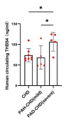
Fig. 1
Fig. 1
Circulating level of THBS4 in patients with PAH-CHD. The resultant data are represented as mean ± SD. *P < 0.05
Fig. 2
Fig. 2
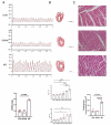
Hemodynamics and right ventricular hypertrophy in the MCT plus aorto-caval shunt induced PAH rats model. (A) Right ventricular systolic pressure (RVSP). (B)Time course of the right ventricular hypertrophy index (RVHI) and weight ratio of the right heart/total weight (R/w). R: weight of the right ventricular, L + I: weight of the left ventricle and interventricular septum. Scare bars,100 μm. (C) Cross-sectional area of the right ventricular cardiomyocyte. Scare bars,100 μm. CON: control group; SHAM: sham group; MS: MCT plus aorto-caval shunt (MS) induced PAH rats from 1 week (1w) to 4 weeks (4w). The resultant data are represented as mean ± SD. *P < 0.05; **P < 0.01; ***P < 0.001; ****P < 0.0001
Fig. 3
Fig. 3
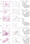
Time course of morphological changes in PAH rats induced by MCT plus aorto-caval shunt. (A) Hematoxylin and Eosin staining of lung tissues. Scare bars,100 μm. (B) Immunofluorescence staining of α-SMA in lung tissues. Scare bars,100 μm. (C) The percentage of non-muscularized pulmonary arterioles (D) The percentage of partially-muscularized pulmonary arterioles. (E) The percentage of fully-muscularized pulmonary arterioles. (F) The percentage of wall thickness. (G) The percentage of wall area. CON: control group; SHAM: sham group; MS: MCT plus aorto-caval shunt (MS) induced PAH rats from 1 week (1w) to 4 weeks (4w). The resultant data are represented as mean ± SD. *P < 0.05; **P < 0.01
Fig. 4
Fig. 4
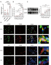
Expression of THBS4 in PAH rats induced by MCT plus aorto-caval shunt. (A) THBS4 expression in GSE149899. qRT-PCR (B) and Western blot (C) showed the time course of THBS4 expression in PAH rats induced by MCT plus aorto-caval shunt. (D) Double immunofluorescence staining of THBS4 with α-SMA or VWF. Scare bars: 50 μm. CON: the control group; SHAM: the sham group; MS: PAH rats induced by MCT plus aorto-caval shunt (MS) from 1 week (1w) to 4 weeks (4w). The resultant data are represented as mean ± SD. *P < 0.05; **P < 0.01; ***P < 0.001; ****P < 0.0001
Fig. 5
Fig. 5
qRT-PCR (A) and Western blot (B) showed the transfection efficiency of THBS4 in PASMCs. (C) The influence of THBS4 on the transformation of phenotype of PASMCs. The resultant data are represented as mean ± SD. *: si-THBS4 vs.si-CON; #: pEX3-THBS4 vs. pEX3-CON.*P < 0.05; ***P < 0.001; #P < 0.05; ##P < 0.01; ###P < 0.001
Fig. 6
Fig. 6
Effect of THBS4 on the proliferation, apoptosis, and migration of PASMCs. (A) Effect of THBS4 on proliferation of PASMCs, as determined by the EdU assay. Scare bars, 100 μm. (B) Effect of THBS4 on apoptosis of PASMCs, as determined by the Annexin-V flow cytometry. (C) Effect of THBS4 on migration of PASMCs, as determined by the Wound healing assay. Scare bars, 200 μm. (D) Effect of THBS4 on migration of PASMCs, as determined by the Transwell assay. Scare bars, 100 μm. The resultant data are represented as mean ± SD. *: si-THBS4 vs.si-CON; #: pEX3-THBS4 vs. pEX3-CON. *P < 0.05; **P < 0.01; ****P <  0.0001; #P < 0.05; ##P < 0.01
Fig. 7
Fig. 7
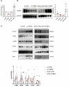
Phosphorylation level of PI3K/AKT in PASMCs after inhibition and overexpression of THBS4 in PASMCs. The resultant data are represented as mean ± SD. *: si-THBS4 vs. si-CON; #: pEX3-THBS4 vs. pEX3-CON.*P < 0.5; #P < 0.5
Fig. 8
Fig. 8
Effect of THBS4 suppression on pulmonary vascular remodeling in PAH rats induced by MCT plus aorto-caval shunt. (A) The AAV transfection efficiency was determined by detecting green fluorescence protein which is encoded by AAV in lung tissues. Scare bars, 100 μm. (B) THBS4 suppression efficiency determined by immunofluorescence staining of THBS4. Scare bars, 50 μm. (C) Hematoxylin and Eosin staining of lung tissues. Scare bars,100 μm. (D) Immunofluorescence staining of α-SMA in lung tissues. Scare bars,100 μm. The percentage of non-muscularized (E), partially-muscularized (F), and fully-muscularized pulmonary arterioles (G). (H) The percentage of wall thickness. (I) The percentage of wall area. (G) Cross-sectional area of the right ventricular cardiomyocyte. Scare bars,100 μm. The resultant data are represented as mean ± SD. CON: control group; SHAM: sham group; MS-AAV-CON: administration of control AAV + MCT plus aorto-caval shunt induced PAH rats; MS-AAV-siTHBS4: administration of THBS4 suppression AAV + MCT plus aorto-caval shunt induced PAH rats. PA: pulmonary arteriole. *P < 0.05; **P < 0.01; ***P < 0.001; ****P < 0.0001

References

    1. Humbert M, Kovacs G, Hoeper MM, Badagliacca R, Berger RMF, Brida M, Carlsen J, Coats AJS, Escribano-Subias P, Ferrari P, et al. 2022 ESC/ERS guidelines for the diagnosis and treatment of pulmonary hypertension. Eur Heart J. 2022;43:3618–731. 10.1093/eurheartj/ehac237 - DOI - PubMed
    1. van Riel AC, Schuuring MJ, van Hessen ID, Zwinderman AH, Cozijnsen L, Reichert CL, Hoorntje JC, Wagenaar LJ, Post MC, van Dijk AP, et al. Contemporary prevalence of pulmonary arterial hypertension in adult congenital heart disease following the updated clinical classification. Int J Cardiol. 2014;174:299–305. 10.1016/j.ijcard.2014.04.072 - DOI - PubMed
    1. Lammers AE, Bauer LJ, Diller GP, Helm PC, Abdul-Khaliq H, Bauer UMM, Baumgartner H. Pulmonary hypertension after shunt closure in patients with simple congenital heart defects. Int J Cardiol. 2020;308:28–32. 10.1016/j.ijcard.2019.12.070 - V体育官网入口 - DOI - PubMed
    1. van Loon RL, Roofthooft MT, Hillege HL, ten Harkel AD, van Osch-Gevers M, Delhaas T, Kapusta L, Strengers JL, Rammeloo L, Clur SA, et al. Pediatric pulmonary hypertension in the Netherlands: epidemiology and characterization during the period 1991 to 2005. Circulation. 2011;124:1755–64. 10.1161/CIRCULATIONAHA.110.969584 - DOI - PubMed
    1. Wagenvoort CA, Wagenvoort N, Draulans-Noë Y. Reversibility of plexogenic pulmonary arteriopathy following banding of the pulmonary artery. J Thorac Cardiovasc Surg. 1984;87:876–86. 10.1016/S0022-5223(19)38415-6 - DOI - PubMed

MeSH terms (V体育安卓版)

VSports app下载 - Substances

LinkOut - more resources