Skip to main page content
U.S. flag

An official website of the United States government

Dot gov

The . gov means it’s official. Federal government websites often end in . gov or VSports app下载. mil. Before sharing sensitive information, make sure you’re on a federal government site. .

Https

The site is secure. The https:// ensures that you are connecting to the official website and that any information you provide is encrypted and transmitted securely V体育官网. .

. 2022 Aug 19;20(1):182.
doi: 10.1186/s12915-022-01380-6.

Modulation of macrophage inflammatory function through selective inhibition of the epigenetic reader protein SP140 (VSports最新版本)

Affiliations

V体育官网 - Modulation of macrophage inflammatory function through selective inhibition of the epigenetic reader protein SP140

Mohammed Ghiboub et al. BMC Biol. .

Abstract

Background: SP140 is a bromodomain-containing protein expressed predominantly in immune cells. Genetic polymorphisms and epigenetic modifications in the SP140 locus have been linked to Crohn's disease (CD), suggesting a role in inflammation VSports手机版. .

Results: We report the development of the first small molecule SP140 inhibitor (GSK761) and utilize this to elucidate SP140 function in macrophages. We show that SP140 is highly expressed in CD mucosal macrophages and in in vitro-generated inflammatory macrophages V体育安卓版. SP140 inhibition through GSK761 reduced monocyte-to-inflammatory macrophage differentiation and lipopolysaccharide (LPS)-induced inflammatory activation, while inducing the generation of CD206+ regulatory macrophages that were shown to associate with a therapeutic response to anti-TNF in CD patients. SP140 preferentially occupies transcriptional start sites in inflammatory macrophages, with enrichment at gene loci encoding pro-inflammatory cytokines/chemokines and inflammatory pathways. GSK761 specifically reduces SP140 chromatin binding and thereby expression of SP140-regulated genes. GSK761 inhibits the expression of cytokines, including TNF, by CD14+ macrophages isolated from CD intestinal mucosa. .

Conclusions: This study identifies SP140 as a druggable epigenetic therapeutic target for CD V体育ios版. .

Keywords: Crohn's disease; Macrophage; SP140 VSports最新版本. .


"VSports注册入口" Conflict of interest statement

PDC, AYFLY, AS, SH, RPB, KG, GY, HPK, AC, AP, LAS, DJM, LL, TGH, RJW, CEB, LAH, GB, UK, NH, JRP, RKP, NRH, and DFT were employed by GSK at the time of conducting this study. MG, JK, ILH, DAZ, OW, TBMH, AATV, JVL, PH, MPJW, and WJDJ were employed by Amsterdam University Medical Centers at the time of conducting this study V体育平台登录. GSK has a patent EP2643462B1 related to the therapeutic use of SP140 inhibitors.

Figures
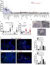
Fig. 1
Fig. 1
SP140 expression associates with inflammatory diseases and mucosal macrophages of CD patients. SP140 gene expression in human tissues and cell types as indicated. Expression is given as count normalized with MAS5.0. SP140 gene expression in white blood cells of normal healthy controls (N) (n=33), Crohn’s disease (CD) (n=6), ulcerative colitis (UC) (n=6), systemic lupus erythematosus (SLE) (n=64), chronic lymphocytic leukemia (CCL) (n=21), acute myeloid leukemia (AML) (n=7), and rheumatoid arthritis (RA) (n=32) patients. SP140 gene expression in human colon tissue obtained from N (n=18), non-inflamed and inflamed CD (n=5 and 13, respectively), and UC (n=3 and 17, respectively) colonic tissues. Data was collected from in-house GSK microarray profiler. Expression is given as count normalized with MAS5.0. d Immunohistochemistry of SP140 protein in colon tissue obtained from N and inflamed CD and inflamed UC tissue, scale bar: 100 μm. e Immunofluorescence staining of DAPI (blue), CD68 (green), and SP140 (red) in N or inflamed CD colon tissue, scale bar: 100 μm. f The average of mucosal cell count per 3 visual fields of total CD68+ macrophages, SP140+ CD68+ macrophages and SP140 CD68+ macrophages in N and inflamed CD tissue (n = 3 patients per group). g Immunofluorescence staining of DAPI (blue), HLA-DR (green), and SP140 (red) in uninflamed or inflamed CD colon tissue, scale bar: 100 μm. h The average of mucosal cell count of 3 visual fields per tissue of total HLA-DR+ macrophages, SP140+ HLA-DR+ macrophages, and SP140 HLA-DR+ macrophages in N and inflamed CD tissue (n=3 patients per group). Statistical significance is indicated as follows: *P < 0.05, **P < 0.01, ****P < 0.0001
Fig. 2
Fig. 2
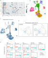
Single-cell analyses of SP140 expression in inflamed and uninflamed CD ileum. Publicly available single-cell RNA sequencing [24] was used to illustrate SP140 expression in intestinal (ileum) macrophages in inflamed (n=11) and uninflamed (n=11) tissue CD patients. a Marker genes expression per cell type where the size of the dots represents the percentage of cells expressing the gene. Darker blue represents more reads per cell. b UMAP of all the cells annotated with their annotated cell type. c Visual illustration of SP140 expression in all cells. Darker blue represents more reads per cell. d Visual illustration of the actual counts of SP140 expression in cluster 6 (MNPs). Darker blue represents more reads per cell. e Comparative analyses of the percentage cells expressing SP140 when comparing inflamed with uninflamed in different immune cells
Fig. 3
Fig. 3
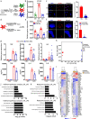
SP140 knockdown reduces the activity of the inflammatory macrophages. a Scheme of polarization protocol of human CD14+ monocytes to “M0,” “M1,” and “M2” macrophage phenotypes, with indicated cytokines as described in the “Methods” section. b Relative gene expression of SP140 in “M0,” “M1,” and “M2” macrophages, n=6. c Immunofluorescence staining of SP140 speckles in “M1” and “M2” macrophages imaged by microscopy (left) and quantified per nuclei as nuclear bodies count (right). Images were counted in 150 cells selected randomly from 3 different staining per condition (Total number of SP140 speckled in 150 cells/150 = average per cell), scale bar: 30 μm. d Scheme of SP140 silencing protocol as described in “Methods” section. e The efficiency of SP140 silencing was assessed by measuring relative gene expression (qPCR) of SP140 (n=6) and f immunofluorescence staining of SP140 speckles nuclear bodies (left) and SP140 speckled nuclear bodies count (counted as described above) (right), scale bar: 3 μm. g Relative gene expression after LPS stimulation: (top, n=6), and LPS-induced protein levels (bottom, n=3) n=3, of TNF, IL-6, and IL-8. h PCA of gene expression dataset of LPS induced genes in scrambled- or SP140 siRNA-treated “M1” macrophages (unstimulated or stimulated with 4 h 100 ng/mL LPS) assessed through microarray; PC1 represents most variance associated with the data (LPS stimulation) and PC2 represents second most variance (siRNA), n=3. i Heatmap of top 50 DEGs from microarray gene expression dataset of LPS induced genes in scrambled- or SP140 siRNA-treated “M1” macrophages (unstimulated or stimulated with 4 h 100 ng/mL LPS) (non-annotated genes were not included). j Hallmark pathways analysis and k Reactome pathway analysis were carried out using ShinyGO v0.60 illustrating the most impacted pathways by SP140 siRNA in unstimulated “M1” macrophages (bottom) or after 4 h of 100 ng/mL LPS stimulation (bottom). In all assays, statistical significance is indicated as follows: *P < 0.05, **P < 0.01, ***P < 0.001, ****P < 0.0001
Fig. 4
Fig. 4
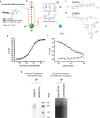
The Development of the first small molecule inhibitor of SP140 (GSK761) and investigating its affinity and selectivity. a Scheme of the three-cycle benzimidazole library and b Spotfire cube view of the SP140 selection output from the benzimidazole library. BB1, cycle 1 building blocks; BB2, cycle 2 building blocks; and BB3, cycle 3 building blocks. Each individual dot in the cube represents discrete small molecule warheads after 3 rounds of affinity selection, while the size of the dot corresponds to the number of unique instances recorded by DNA sequencing (2-24). The dots are colored by the BB3s that compose the library molecules. Library members with a single copy were removed to simplify visualization revealing a prominent line defined by a specific BB1&BB3 combination (disynthon). The most enriched trisynthon (BB1, BB2, and BB3 combination) represents the biggest dot on the line. e Biochemical characterization of the interaction between c GSK761 and recombinant SP140 in a fluorescence polarization (FP) binding assay using d a fluorophore-conjugated version of GSK761: GSK064 (generated by fluorescent labelling of GSK761). The mean binding affinity for this interaction was a Kd = 41.07 ± 1.42 nM (n = 5). f A FP-binding assay was configured using recombinant SP140 and GSK064, which was used to determine the potency of GSK761. Displacement of GSK064 from SP140 by GSK761 (circles) was achieved and determined to have a mean IC50 of 77.79 ± 8.27 nM (n=3). No effect on GSK064 motion was observed in the presence of varying concentrations of GSK761 (triangles). The data presented in d and e are representative data from a single experimental replicate, affinities, and potencies are mean values determined from multiple test occasions. g Endogenous SP140 (HuT78 nuclear extracts) and Halo-tagged SP140 (transfected HEK29 cells) were pulled down using a biotinylated version of GSK761 (GSK675) and visualized by Western blotting and gel electrophoresis. Biotinylated beads only and SP140 untransfected cells were used as control
Fig. 5
Fig. 5
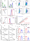
GSK761 affects macrophage polarization and cytokine production in peripheral blood and CD mucosal macrophages. Human primary CD14+ monocytes were differentiated with 20 ng/mL M-CSF for 3 days. The cells where then washed with PBS and treated with 0.1% DMSO or 0.04 μM GSK761 for 1 h prior to 3 days polarization to “M1” and “M2” macrophages phenotypes with 100 ng/mL INF-γ or 40 ng/mL IL-4, respectively (GSK761 and DMSO were not washed and kept in the culture during the 3 days of polarization). a Gene expression of the inflammatory marker CD64 (“M1” marker) and anti-inflammatory marker CD206 (“M2” marker) was measured by qPCR in IFN-γ (“M1”) and IL-4 (“M2”) polarized macrophages and b–e FACS analysis was performed in IFN-γ (“M1”) polarized macrophages. b Scatter plots illustrating the frequency of CD64+ cells. c A graph bar summarizing the frequency of CD64+ macrophages in 3 donors (left) and CD64 protein expression intensity (right). d Scatter plots illustrating the frequency of CD206+ cells. e A graph bar summarizing the frequency of CD206+ macrophages in 3 donors (left) and CD206 protein expression intensity, n=3. f “M1” polarized macrophages were pretreated for 1 h with 1% DMSO (n=4) or with 0.04 μM GSK761 (n=5). The cells were then stimulated for 4 h with 100 ng/mL LPS and Customized RT2 Profiler PCR Arrays was performed, the scatter plot illustrates the differentially expressed genes (2-fold change). g “M1” polarized macrophages were pretreated for 1 h with 1% DMSO or with an increasing concentration of GSK761 (0.01, 0.04, 0.12, 0.37, and 1.11 μM). The cells were then stimulated for 24 h with 100 ng/mL LPS. Protein levels of TNF, IL-6, IL-1β, IL-10, IL-8, and IL-12p70 were measured in the supernatant, n=4–7. The Y axis indicates the fold change in cytokine protein expression relative to DMSO control. h CD14+ cells were isolated from inflamed CD mucosa. The cells were then incubated ex vivo for 4 h with either 0.1% DMSO or 0.04 μM GSK761. Relative gene expression of TNF, IL6, IL10, and CD64 were measured using qPCR, n=4. The Y axis indicates the fold change in mRNA level relative to DMSO control. Statistical significance is indicated as follows: *P < 0.05, **P < 0.01, ***P < 0.001, ****P < 0.0001
Fig. 6
Fig. 6
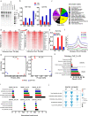
LPS stimulation leads to SP140 protein recruitment to chromatin; GSK761 reduces this recruitment and dampens inflammatory pathways in inflammatory macrophages. a Nuclear extracts from αCD3/αCD28 stimulated HuT78 cells were incubated with unmodified or modified (acetylated and methylated) histone H3 peptides as indicated. SP140 was then pulled down to visualize its interaction with H3 peptides. b ChIP-qPCR of SP140 occupancy at TSS of TNF and IL6 in “M1” macrophages stimulated with 100 ng/mL LPS for 4 h or without LPS stimulation, n=3 donors (DN). c Epigenome roadmap scan illustrating proportions of SP140 genome-wide occupancy after SP140 ChIP-seq. d Heatmap (1000 top SP140-bound genes) of SP140 ChIP-seq reads ranked on 1 h LPS-stimulated (left) or 4 h LPS-stimulated (right) “M1” macrophages rank-ordered from high to low occupancy centered on TSS. Top 20 genes with high SP140 occupancy are listed. e ChIP-qPCR of SP140 occupancy at TSS of TNF in “M1” macrophages pretreated with 0.1% DMSO or 0.04 μM GSK761 for 1 h and then stimulated with 100 ng/mL LPS for 1 or 4 h or kept unstimulated (0 h LPS). f Metagene created from normalized genome-wide average reads for SP140 centered on TSS. g PCA of RNA-seq comparing 0.1% DMSO to 0.04 μM GSK761 treated “M1” macrophages after 4 h of LPS stimulation (left) or after 8 h of LPS stimulation (right). h SP140 ChIP-seq gene ontology analysis of the most enriched molecular function and biological process after 1 h of LPS stimulation, comparing 0.1% DMSO with 0.04 μM GSK761 treated “M1” macrophages. i Hallmarks pathway enrichment analysis at 0, 1, and 4 h of 100 ng/mL LPS stimulation for ChIP-seq and at j 0, 4, and 8 h of 100 ng/mL LPS stimulation for RNA-seq. j The direction and color of the arrow indicate the direction and size of the enrichment score, the size of the arrow is proportional to the −log10 (p-value), and non-transparent arrows represent significantly affected pathways
Fig. 7
Fig. 7
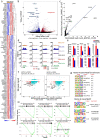
SP140 preferentially controls the expression of specific gene sets involved in the innate immune response. a Heatmap of the top 100 DEGs and b volcano plots of the all genes comparing 0.1% DMSO- with 0.04 μM GSK761-treated “M1” macrophages after 4 h of 100 ng/mL LPS stimulation. c R2 TSS plot comparing a global differentially SP140-bound genes (DBGs) after 1 h of 100 ng/mL LPS stimulation of 0.1% DMSO- and 0.04 μM GSK761-treated “M1” macrophages. d SP140 ChIP-seq genome browser view of some of the most affected DBGs; TNF, TRAF1, IRF1, and TRAFAIP2. Y axis represents a signal score of recovered sequences in 0.1% DMSO and 0.04 μM GSK761 treated macrophages after 0, 1, and 4 h of 100 ng/mL LPS stimulation. e RNA sequencing-derived gene expression of some of the most DBGs. f Comparative analyses of the top 1000 DBGs (signal) with their gene expression (Wald statistic). Gene expression at 4 h and ChIP at 1 h (left) and gene expression at 4 h and ChIP at 4 h (right), Y axis represents the SP140 differential binding signal (BD). g Homer Known Motif Enrichment using TF motifs and their respective p-value scoring (top) and Homer de novo Motif results with best match TFs (bottom) in 0.1% DMSO-treated “M1” macrophages after 1 h of 100 ng/mL LPS stimulation. h An enrichment analysis targeted chemokine activity for SP140 ChIP-seq (top) and RNA-seq (bottom) comparing 0.1% DMSO- with 0.04 μM GSK761-treated “M1” macrophages at 0, 1, and 4 h of 100 ng/mL LPS stimulation

"VSports app下载" References

    1. Rutgeerts P. A critical assessment of new therapies in inflammatory bowel disease. J Gastroenterol Hepatol. 2002;17(s1):S176–SS85. doi: 10.1046/j.1440-1746.17.s1.1.x. - DOI - PubMed
    1. Papadakis KA, Shaye OA, Vasiliauskas EA, Ippoliti A, Dubinsky MC, Birt J, et al. Safety and efficacy of adalimumab (D2E7) in Crohn's disease patients with an attenuated response to infliximab. Am J Gastroenterol. 2005;100(1):75–79. doi: 10.1111/j.1572-0241.2005.40647.x. - "VSports注册入口" DOI - PubMed
    1. Feagan BG, Greenberg GR, Wild G, Fedorak RN, Pare P, McDonald JW, et al. Treatment of active Crohn's disease with MLN0002, a humanized antibody to the alpha4beta7 integrin. Clin Gastroenterol Hepatol. 2008;6(12):1370–1377. doi: 10.1016/j.cgh.2008.06.007. - DOI - PubMed
    1. Sandborn WJ, Feagan BG, Fedorak RN, Scherl E, Fleisher MR, Katz S, et al. A randomized trial of Ustekinumab, a human interleukin-12/23 monoclonal antibody, in patients with moderate-to-severe Crohn's disease. Gastroenterology. 2008;135(4):1130–1141. doi: 10.1053/j.gastro.2008.07.014. - DOI - PubMed
    1. Bain CC, Schridde A. Origin, differentiation, and function of intestinal macrophages. Front Immunol. 2018;9:2733. doi: 10.3389/fimmu.2018.02733. - DOI - PMC - PubMed

Publication types

MeSH terms