Skip to main page content
U.S. flag

An official website of the United States government

Dot gov

The . gov means it’s official. Federal government websites often end in VSports app下载. gov or . mil. Before sharing sensitive information, make sure you’re on a federal government site. .

Https

The site is secure. The https:// ensures that you are connecting to the official website and that any information you provide is encrypted and transmitted securely V体育官网. .

. 2022 Jan 18;6(1):5.
doi: 10.1038/s41698-021-00250-8.

HER3 activation contributes toward the emergence of ALK inhibitor-tolerant cells in ALK-rearranged lung cancer with mesenchymal features (VSports app下载)

Affiliations

HER3 activation contributes toward the emergence of ALK inhibitor-tolerant cells in ALK-rearranged lung cancer with mesenchymal features

Keiko Tanimura (V体育安卓版) et al. NPJ Precis Oncol. .

Abstract

Anaplastic lymphoma kinase-tyrosine kinase inhibitors (ALK-TKIs) have shown dramatic efficacy in patients with ALK-rearranged lung cancer; however, complete response in these patients is rare VSports手机版. Here, we investigated the molecular mechanisms underlying the emergence and maintenance of drug-tolerant cells in ALK-rearranged lung cancer. Cell based-assays demonstrated that HER3 activation and mesenchymal-to-epithelial transition, mediated through ZEB1 proteins, help maintain cell survival and induce the emergence of ALK-TKI-tolerant cells. Compared with ALK-TKIs alone, cotreatment with pan-HER inhibitor afatinib and ALK-TKIs prevented tumor regrowth, leading to the eradication of tumors in ALK-rearranged tumors with mesenchymal features. Moreover, pre-treatment vimentin expression in clinical specimens obtained from patients with ALK-rearranged lung cancer was associated with poor ALK-TKI treatment outcomes. These results demonstrated that HER3 activation plays a pivotal role in the emergence of ALK-TKI-tolerant cells. Furthermore, the inhibition of HER3 signals combined with ALK-TKIs dramatically improves treatment outcomes for ALK-rearranged lung cancer with mesenchymal features. .


"VSports手机版" Conflict of interest statement

T. Y. received commercial research grants from Pfizer, Ono Pharmaceutical, Chugai Pharmaceutical, and Takeda Pharmaceutical Company Limited. J. U. received research grants from Eli Lilly Japan K. K. , AstraZeneca K. K. , and Boehringer Ingelheim Japan. T. S. received research grants from Otsuka Pharmaceutical and Oncolys BioPharma, and a patent fee from JT Pharmaceutical. S. Y. obtained commercial research grants from Chugai Pharmaceutical and Boehringer-Ingelheim and has received speaking honoraria from Chugai Pharmaceutical, Boehringer-Ingelheim, Novartis, and Pfizer. R. K. received research grants from Chugai Pharmaceutical, Takeda Pharmaceutical Company Limited, Toppan Printing, and Daiichi-Sankyo V体育安卓版. K. T. received research grants from Chugai-Roche and Ono Pharmaceutical and personal fees from AstraZeneca, Chugai-Roche, MSD-Merck, Eli Lilly, Boehringer-Ingelheim, and Daiichi-Sankyo. The remaining authors declare no competing interests.

Figures
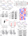
Fig. 1
Fig. 1. HER3 activation plays a pivotal role in the emergence of ALK-TKI-tolerant ALK-rearranged NSCLC cells.
a H2228 and A925L parental and DT cells treated with alectinib (3 µmol/L) or brigatinib (1 µmol/L) for 9 days were incubated with the indicated concentrations of alectinib or brigatinib for 72 h. Cell viability was detected using MTT assays. b Human phospho-RTK array analysis of parental and DT cells treated with ALK-TKIs. Western blotting (c) and with immunoprecipitation (d) of parental and DT cells. e Microarray analysis of H2228 and A925L parental vs. DT cells. Heatmap and Z-score hierarchical clustering of differentially expressed genes associated with KEGG_ERBB_SIGNALING_PATHWAY gene set. Highlighted genes are upregulated in DT cells of both H2228 and A925L. f Scatterplot of genes in the KEGG_ERBB_SIGNALING_PATHWAY gene set with significant differential expression in either H2228 or A925L DT cells vs parent cells. x-Axis: log2FC of the gene between H2228-DT vs. H2228; y-axis: log2FC of the same gene between A925L-DT and A925L cells. Samples are colored according to the significance of differential expression (absolute log2FC > 1) in both groups. g qPCR of neuregulin 1 (NRG1) in parental and DT cells. P-values were calculated by unpaired t tests. h Parental and DT cells were incubated with afatinib for 72 h and i treated with medium only, alectinib, afatinib, or a combination for 72 h. h, i Cell viability MTT assays. *P < 0.05 (two-way ANOVA followed by Tukey’s test). j Western blotting of parental and DT cells treated with 100 nmol/L of alectinib, afatinib, or a combination. Data are represented as mean ± S.D.
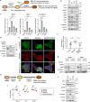
Fig. 2
Fig. 2. HER3-mediated adaptive resistance with alterations in EMT marker expression is reversible under drug-free conditions.
a Plated H2228 cells were untreated (left) or treated with alectinib (3 µmol/L) for 9 days (DT cells; left middle) and DT cells were incubated with drug-free-medium for 9 days (DF9; right middle) or 28 days (DF28; right). b Western blotting of H2228 cells. c qPCR of HER3, NRG1, or E-cadherin in H2228 cells. *P < 0.05 (two-way ANOVA followed by Dunnett’s test). d Western blotting of H2228 cells treated with 3 µmol/L alectinib, 100 nmol/L afatinib, or a combination for 72 h. e Immunocytochemical analysis of H2228 cells treated with or without alectinib (3 µmol/L) and afatinib (100 nmol/L) for 96 h. f Quantification of E-cadherin-positive cells treated with or without alectinib (3 µmol/L) and afatinib (100 nmol/L) for 96 h. *P < 0.05 vs. alectinib-treated cells (Tukey–Kramer method). g Western blotting of cells treated with nonspecific control siRNA or HER3-specific siRNA and incubated with or without alectinib for 48 h. h Plated H2228 cells were untreated (left) or treated with alectinib (3 µmol/L) for 9 days (DT cells; middle), and DT cells were incubated with alectinib (3 µmol/L) for 30 days (drug-tolerant expanded persisters, DTEPs; right). i Cell viability MTT assays of H2228 parental, DT, and DTEP cells treated with medium only, 100 nmol/L of alectinib, afatinib, or a combination for 72 h. *P < 0.05 (two-way ANOVA followed by Tukey’s test). j Western blotting of cells observed in (i). Data are represented as mean ± S.D.
Fig. 3
Fig. 3. ZEB1 facilitates alterations in EMT marker expression and HER3 activation in ALK-TKI treated cells.
ac Western blotting of H2228 and A925L cells incubated with or without alectinib (3 µmol/L) for the indicated time (a); incubated with nonspecific control siRNA or ALK-specific siRNA (b); and incubated with nonspecific control siRNA or ZEB1-specific siRNAs (#1, #2) (c). d Western blotting of H2228 cells transfected with a control vector (Vec) or ZEB1-expressing lentiviral plasmids (ZEB1) and treated with alectinib (100 nmol/L) for 48 h; n ≥ 2 independent experiments. e Cell viability MTT assays of H2228 cells transfected with a control vector (Vec) or ZEB1-expressing lentiviral plasmids (ZEB1) were treated with alectinib (100 nmol/L) for 72 h. Data are represented as mean ± S.D. P values were calculated using two-way ANOVA followed by Tukey’s test. f Schematic diagram of the drug tolerance mechanisms, including mesenchymal-to-epithelial transition (MET) and HER3 activation via ZEB1, in ALK-rearranged NSCLC cells.
Fig. 4
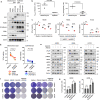
Fig. 4. EMT marker expression is correlated with susceptibility to ALK-TKIs.
a Western blotting of ALK-rearranged NSCLC parental cells. b IC50 values for alectinib and brigatinib in five ALK-rearranged NSCLC cells. c Cell viability MTT assays of H2228 and A925L cells treated with nonspecific control siRNA or HER3-specific siRNAs (#1 and #2) and incubated with or without ALK-TKIs (100 nmol/L) for 72 h. d MTT assay of cells treated with alectinib (100 nmol/L) alone or afatinib (100 nmol/L) for 72 h. e Western blotting of H2228, A925L, or H3122 parental cells incubated with medium only, alectinib (100 nmol/L), afatinib (100 nmol/L), or a combination for 48 h. f Crystal violet staining of cells treated with DMSO, 100 nmol/L of ALK-TKI (alectinib or brigatinib), 100 nmol/L of afatinib, or a combination for 14 days with the drug replenished every 72 h prior to visual examination with crystal violet staining. g Apoptotic cell percentages of H2228 and A925L cells, which are annexin V and propidium iodide double-stained, were detected by flow cytometry following treatment with medium, alectinib (100 nmol/L), afatinib (100 nmol/L), or combination for 48 h. Data are represented as mean ± S.D. *P < 0.05, **P < 0.01, ***P < 0.001 [unpaired t-tests (b, d) and two-way ANOVA followed by Tukey’s test (c, g)].
Fig. 5
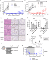
Fig. 5. Afatinib with ALK-TKIs prevents CDX tumor regrowth.
a, b A925L cell line-derived xenograft (CDX) tumors (a) and H2228 CDX tumors (b) were treated with a vehicle (control), ALK-TKI brigatinib (10 mg/kg) or alectinib (6 mg/kg), afatinib (5 mg/kg), or ALK-TKI plus afatinib (5 mg/kg) (n = 6) via oral gavage daily. c Proliferating and apoptotic cells were quantified by their Ki-67-positive proliferation index (percentage of Ki-67-positive cells) and TUNEL assays, respectively. The scale bar indicates 100 μm. d Representative immunohistochemistry images of CDX tumors following staining with specific human Ki-67 and TUNEL antibodies. e Quantification of immunostaining-positive cells, as determined by the H-score for vimentin and E-cadherin calculated from areas randomly selected from CDX tumors treated with or without alectinib (6 mg/kg) for 4 days. f Mice with A925L-CDX tumors were treated with ALK-TKI brigatinib (10 mg/kg) or brigatinib plus afatinib (5 mg/kg) (n = 6) by oral gavage daily for 17 days. g Tumor regrowth was evaluated on day 32. Tumor volumes were measured from the start of treatment. Data are represented as mean ± S.D. *P < 0.05 [two-way ANOVA followed by Bonferroni’s test (a, b, g) and one-way ANOVA followed by Tukey’s post hoc test (d, e)].

References

    1. Miller KD, et al. Cancer statistics for hispanics/latinos, 2018. CA Cancer J. Clin. 2018;68:425–445. - "VSports app下载" PubMed
    1. Govindan R, et al. Changing epidemiology of small-cell lung cancer in the United States over the last 30 years: analysis of the surveillance, epidemiologic, and end results database. J. Clin. Oncol. 2006;24:4539–4544. - VSports - PubMed
    1. Li T, Kung HJ, Mack PC, Gandara DR. Genotyping and genomic profiling of non-small-cell lung cancer: implications for current and future therapies. J. Clin. Oncol. 2013;31:1039–1049. - PMC - PubMed
    1. Recondo G, Facchinetti F, Olaussen KA, Besse B, Friboulet L. Making the first move in EGFR-driven or ALK-driven NSCLC: first-generation or next-generation TKI? Nat. Rev. Clin. Oncol. 2018;15:694–708. - PubMed
    1. Soda M, et al. Identification of the transforming EML4-ALK fusion gene in non-small-cell lung cancer. Nature. 2007;448:561–566. - PubMed