Skip to main page content (V体育平台登录)
U.S. flag

An official website of the United States government

Dot gov

The . gov means it’s official. Federal government websites often end in VSports app下载. gov or . mil. Before sharing sensitive information, make sure you’re on a federal government site. .

Https

The site is secure. The https:// ensures that you are connecting to the official website and that any information you provide is encrypted and transmitted securely. V体育官网.

. 2021 Jun 7;13(11):2850.
doi: 10.3390/cancers13112850.

VDAC1 Silencing in Cancer Cells Leads to Metabolic Reprogramming That Modulates Tumor Microenvironment

Affiliations

VDAC1 Silencing in Cancer Cells Leads to Metabolic Reprogramming That Modulates Tumor Microenvironment (V体育平台登录)

Erez Zerbib (VSports注册入口) et al. Cancers (Basel). .

Abstract

The tumor microenvironment (TME) plays an important role in cell growth, proliferation, migration, immunity, malignant transformation, and apoptosis. Thus, better insight into tumor-host interactions is required VSports手机版. Most of these processes involve the metabolic reprogramming of cells. Here, we focused on this reprogramming in cancerous cells and its effect on the TME. A major limitation in the study of tumor-host interactions is the difficulty in separating cancerous from non-cancerous signaling pathways within a tumor. Our strategy involved specifically silencing the expression of VDAC1 in the mitochondria of human-derived A549 lung cancer xenografts in mice, but not in the mouse-derived cells of the TME. Next-generation sequencing (NGS) analysis allows distinguishing the human or mouse origin of genes, thus enabling the separation of the bidirectional cross-talk between the TME and malignant cells. We demonstrate that depleting VDAC1 in cancer cells led to metabolic reprogramming, tumor regression, and the disruption of tumor-host interactions. This was reflected in the altered expression of a battery of genes associated with TME, including those involved in extracellular matrix organization and structure, matrix-related peptidases, angiogenesis, intercellular interacting proteins, integrins, and growth factors associated with stromal activities. We show that metabolic rewiring upon mitochondrial VDAC1 silencing in cancer cells affected several components of the TME, such as structural protein matrix metalloproteinases and Lox, and elicited a stromal response resembling the reaction to a foreign body in wound healing. As tumor progression requires a cooperative interplay between the host and cancer cells, and the ECM is intensively remodeled during cancer progression, VDAC1 depletion induced metabolic reprogramming that targeted both tumor cells and resulted in the alteration of the whole spectrum of TME-related genes, affecting the reciprocal feedback between ECM molecules, host cells, and cancer cells. Thus, VDAC1 depletion using si-VDAC1 represents therapeutic potential, inhibiting cancer cell proliferation and also inducing the modulation of TME components, which influences cancer progression, migration, and invasion. .

Keywords: VDAC1; metabolism; mitochondria; reprogramming; siRNA; tumor microenvironment V体育安卓版. .


Conflict of interest statement

The authors declare no competing financial interests.

Figures

Figure 1
Figure 1
Human-specific siRNA si-hVDAC1-2A specifically silences human but not mouse VDAC1, inhibits lung cancer cell-derived xenograft growth, and reduces the expression of metabolism-related proteins. (A) A549 and 2LL cell lines were transfected with si-NT or si-hVDAC1-2A and analyzed for VDAC1 expression using anti-VDAC1 antibodies. Cells were transfected using jetPRIME reagent and 100 nM of siRNA (Figure S1F,G,I for uncropped Western Blot). (B) Quantitative analysis of VDAC1 protein levels in the si-hVDAC1-2A-treated cells, relative to si-NT-treated cells, is presented as relative units (RUs). (C) Schematic presentation of the course of the experiment and siRNA treatment initiation. (D) A549 cells (3 × 10⁶) were subcutaneously (s.c.) inoculated into nude mice. When tumor size reached 70 mm3, the mice were divided into two matched groups, and xenografts were injected intratumorally every 3 days with si-NT (black bars, 7 mice) or si-hVDAC1-2A (gray bars, 8 mice) to a final concentration of 100 nM. The calculated average tumor volumes are presented as means ± SEM (* p ≤ 0.05, ** p ≤ 0.01, *** p ≤ 0.001), NS—non-specific. (E) si-NT-TT and si-hVDAC1-2A-TT sections from A549 xenograft mice were analyzed for VDAC1 levels by immunoblotting (Figure S1H for uncropped Western Blot). Relative units (RUs) presented as the mean ± SEM; n = 3 mice. (F,G) Representative IHC staining using specific antibodies against VDAC1, GLUT1, GAPDH, citrate synthase (CS), complex IVc (Comp.IVc), and ATP synthase 5a (ATP Syn 5a) (F) or IF staining for HK-I and LDH (G) of si-NT-TT and si-hVDAC1-2A-TT sections derived from A549 xenografts. (H) Quantitative analysis of the levels of metabolism associated proteins IHC stained section intensity using Image J software, presented as the percentage of the si-hVDAC1-2A-TT staining intensity, relative to that in si-NT-TTs, and their levels as analyzed by qRT-PCR analysis of mRNA and presented as fold change.
Figure 2
Figure 2
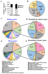
NGS gene analysis of mouse and human origin differentially expressed in si-hVDAC1-2A-TTs. RNA, isolated from tumors treated with si-hVDAC1-2A or si-NT (75 nM), was subjected to NGS with data subjected to bioinformatics analysis. (A) % of uniquely mapped reads for mouse and human. (B) No. of significantly up- and downregulated genes of human and mouse origin with a p-value < 0.05, and a linear fold change >1.5 or <−1.5 in si-hVDAC1-2A-TTs. (CF) Classification of the human (C,D) and mouse (E,F) differentially expressed genes to biological processes, with the number of genes related to each process indicated inside the chart. Further breakdown of the metabolic processes to sub-classes is presented in (E,F) for human and mice, respectively. The analysis was carried out using the Panther gene list analysis, applying functional classification against GO-Slim Biological Process.
Figure 3
Figure 3
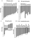
Differentially expressed ECM structure-related genes of murine origin. ECM structure-related genes found to be enriched using DAVID Gene Ontology analysis are presented as the fold change of the expression in si-hVDAC1-2A-TTs relative to si-NT-TTs. (A) Collagen genes. (B) Glycoprotein genes. (C) Integrin genes. (D) ECM organization genes. In all cases, * p ≤ 0.05, ** p ≤ 0.01. Two selected genes, periostin and tenascin C, whose expression was further analyzed at the protein level (Figure 3), are indicated by the dashed frames.
Figure 4
Figure 4
IF analysis of periostin and tenascin C expression in si-NT-TTs and si-hVDAC1-2A-TTs. (A,B) Representative IF staining using specific antibodies against periostin and tenascin C derived from A549 xenografts in si-NT-TT and si-hVDAC1-2A-TT sections. In (C), an enlargement of the area (a,b) in (A,B), respectively, is shown. (D) Quantitative analysis of fluorescence intensity in (A,B). The results are the means ± SD, ** p ≤ 0.01, *** p ≤ 0.001. Furthermore, according to the TCGA database, lung cancer patients with elevated serum levels of periostin have a significantly lower overall survival rate than patients with low serum periostin levels (Figure S2A). The same correlation was also found with serum tenascin C, where high levels in the serum were linked to poor overall survival probability (Figure S2B).
Figure 5
Figure 5
Figure 5. si-hVDAC1-2A reduces the expression of mouse genes involved in ECM deposition and degradation of TME factors. Differentially expressed ECM deposition and degradation genes of murine origin found to be enriched using DAVID Gene Ontology analysis are presented as the fold change in si-VDAC1-2A-TTs relative to si-NT-TTs. (A) Metalloproteinase genes. (B) ADAM and ADAMTS genes. (C) Lysyl oxidase and heparan sulphate genes. (D) Cell–matrix interactions- and stromal factor-related genes. In all cases, * p ≤ 0.05, ** p ≤ 0.01.
Figure 6
Figure 6
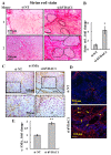
si-hVDAC1-2A-TTs show altered ECM organization and encapsulate cancer cells in the tumor. (A) Representative sections from two mice of si-NT-TT and si-hVDAC1-2A-TT sections derived from A549 xenografts stained with Sirius red. The dashed lines surround the encapsulated cancer cells within the residual tumor. (B) Quantitative analysis of Sirius red intensity. (C,D) Representative IHC (C) and IF (D) staining of si-NT-TT and si-hVDAC1-2A-TT sections with anti-α-SMA antibodies. Arrows point to the spherical organization of fibroblasts. (E) Quantitative analysis of α-SMA presented in (D). The results are the means ± SD, * p ≤ 0.05, ** p ≤ 0.01.
Figure 7
Figure 7
si-hVDAC1-2A tumor treatment alters the expression of angiogenesis-related mouse genes. Differentially expressed angiogenesis-related genes of murine origin found to be enriched using DAVID Gene Ontology analysis are presented as the fold change in si-VDAC1-2A-TTs relative to si-NT-TTs. (A) Pro-angiogenesis genes. (B) Anti-angiogenesis genes, as well as genes encoding for anti- or pro-angiogenesis factors, depending on the conditions. In all cases, * p ≤ 0.05, ** p ≤ 0.01. Representative IF stained sections from si-NT-TTs and si-hVDAC1-2A-TTs derived from A549 xenografts stained with anti-CD-31 antibodies (C). Quantitative analysis of CD-31 staining intensity, presented as means ± SEM, **** p < 0.0001 (n = 3) (D).
Figure 8
Figure 8
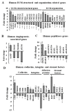
Altered expression of TME-related human genes in si-hVDAC1-2A-TTs. Differentially expressed genes of human origin found to be enriched using DAVID Gene Ontology analysis are presented as the fold change in si-VDAC1-2A-TTs relative to si-NT-TTs. (A) ECM structure- and organization-related genes. (B) Angiogenesis-associated genes. (C) Peptidase genes. (D) Cadherin, integrin, and stromal growth factor genes. In all cases, * p ≤ 0.05, ** p ≤ 0.01, *** p ≤ 0.001, **** p ≤ 0.0001.
Figure 9
Figure 9
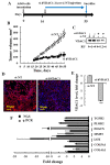
qRT-PCR validation of altered expression of selected TME-related genes. (A) Schematic presentation of the course of the experiment and peptide treatment initiation. (B) A549 cell-derived xenografts were established, and when tumor volume reached ~60 mm3, the mice were split into two groups and injected every 3 days with si-NT or si-hVDAC1-2A (200 nM), and tumor volume was measured. (B) Tumors in the si-NT-treated group reached an average of 490 mm3 at day 55 post-cell inoculation, while tumors in the si-hVDAC1-2A-treated group measured 140 mm3. Mice were sacrificed, and the tumors were excised and frozen in liquid nitrogen until used. (C) Tissue samples from excised tumors analyzed for VDAC1 levels by immunoblotting using anti-VDAC1 antibodies with the relative expression levels (RU) are presented in the blot (Figure S3 for uncropped Western Blot). (D,E) Representative staining of si-NT-TT and si-hVDAC1-2A-TT sections with anti-VDAC1 antibodies with confocal images (D) and quantitative analysis of the fluorescence intensity (E) are shown. The results are the means ± SD, ** p ≤ 0.01. (F) The fold of change in the expression level of selected proteins was analyzed by q-PCR (gray bars), and data obtained from the NGS analysis (black bars) are presented. RNA isolation and qPCR of key TME genes using mouse-specific primers (Table S2) were performed as described in Section 2.
Figure 10
Figure 10
A schematic presentation of mitochondrial VDAC1 depletion and metabolic reprogramming leading to TME remodeling in A549-derived tumors. The overexpressed VDAC1 in mitochondria affects the homeostatic energy and metabolic states of cancer cells. Silencing hVDAC1 expression leads to a reprogramming of metabolism and to TME remodeling. This is reflected in alterations in the expression of genes related to: 1. stromal factors, 2. stroma activity, 3. cell–ECM interactions, 4. ECM structure and organization, 5. ECM degradation and deposition, 6. angiogenesis, and 7. immune system suppression. In each group, the major genes with enhanced expression (blue) or reduced expression (red) are presented. As tumor progression requires a cooperative interplay between host and cancer cells, and the ECM is intensively remodeled during cancer progression, VDAC1-induced alteration in the whole spectrum of TME-related genes affects both the tumor and reciprocal feedback between ECM molecules, host cells, and cancer cells. Tumors can influence the microenvironment by releasing extracellular signals, promoting tumor angiogenesis and inducing peripheral immune tolerance, while the tumor surrounding the microenvironment as immune cells can affect the growth and evolution of cancerous cells.

References (V体育2025版)

    1. Hanahan D., Weinberg R.A. Hallmarks of cancer: The next generation. Cell. 2011;144:646–674. doi: 10.1016/j.cell.2011.02.013. - DOI - PubMed
    1. Koppenol W.H., Bounds P.L., Dang C.V. Otto Warburg’s contributions to current concepts of cancer metabolism. Nat. Rev. Cancer. 2011;11:325–337. doi: 10.1038/nrc3038. - DOI - PubMed
    1. Gogvadze V., Zhivotovsky B., Orrenius S. The Warburg effect and mitochondrial stability in cancer cells. Mol. Asp. Med. 2010;31:60–74. doi: 10.1016/j.mam.2009.12.004. - DOI - PubMed
    1. Cantor J.R., Sabatini D.M. Cancer cell metabolism: One hallmark, many faces. Cancer Discov. 2012;2:881–898. doi: 10.1158/2159-8290.CD (V体育2025版)-12-0345. - DOI - PMC - PubMed
    1. Nakano I. Therapeutic potential of targeting glucose metabolism in glioma stem cells. Expert Opin. Ther. Targets. 2014;18:1233–1236. doi: 10.1517/14728222.2014.944899. - DOI - PubMed

"VSports最新版本" LinkOut - more resources