Skip to main page content
U.S. flag

An official website of the United States government

Dot gov

The . gov means it’s official VSports app下载. Federal government websites often end in . gov or . mil. Before sharing sensitive information, make sure you’re on a federal government site. .

Https

The site is secure. The https:// ensures that you are connecting to the official website and that any information you provide is encrypted and transmitted securely V体育官网. .

. 2020 Nov 24;117(47):29609-29617.
doi: 10.1073/pnas.2016270117. Epub 2020 Nov 9.

Reversing the direction of drug transport mediated by the human multidrug transporter P-glycoprotein

Affiliations

Reversing the direction of drug transport mediated by the human multidrug transporter P-glycoprotein

Andaleeb Sajid et al. Proc Natl Acad Sci U S A. .

Abstract

P-glycoprotein (P-gp), also known as ABCB1, is a cell membrane transporter that mediates the efflux of chemically dissimilar amphipathic drugs and confers resistance to chemotherapy in most cancers. Homologous transmembrane helices (TMHs) 6 and 12 of human P-gp connect the transmembrane domains with its nucleotide-binding domains, and several residues in these TMHs contribute to the drug-binding pocket. To investigate the role of these helices in the transport function of P-gp, we substituted a group of 14 conserved residues (seven in both TMHs 6 and 12) with alanine and generated a mutant termed 14A. Although the 14A mutant lost the ability to pump most of the substrates tested out of cancer cells, surprisingly, it acquired a new function. It was able to import four substrates, including rhodamine 123 (Rh123) and the taxol derivative flutax-1. Similar to the efflux function of wild-type P-gp, we found that uptake by the 14A mutant is ATP hydrolysis-, substrate concentration-, and time-dependent. Consistent with the uptake function, the mutant P-gp also hypersensitizes HeLa cells to Rh123 by 2- to 2. 5-fold VSports手机版. Further mutagenesis identified residues from both TMHs 6 and 12 that synergistically form a switch in the central region of the two helices that governs whether a given substrate is pumped out of or into the cell. Transforming P-gp or an ABC drug exporter from an efflux transporter into a drug uptake pump would constitute a paradigm shift in efforts to overcome cancer drug resistance. .

Keywords: ABC transporter; P-glycoprotein; drug transport; mechanism; multidrug resistance. V体育安卓版.


Conflict of interest statement

The authors declare no competing interest.

Figures
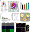
Fig. 1.
Fig. 1.
Transport direction is reversed by mutations in TMHs 6 and 12 of P-gp. (A) Inward open human P-gp structure (PDB ID: 6QEX) highlighting the location of residues mutated in 14A. The residues selected for mutation in TMH 6 and TMH 12 are shown as red and magenta spheres, respectively. (B) Relative surface expression of 14A detected by human P-gp–specific antibodies MRK-16, 4E3, and UIC2, with binding to WT P-gp taken as 100%. (C) Relative efflux of various substrates by 14A, with efflux by WT P-gp taken as 100%. Although 25 substrates (list given in ref. 10) were tested, results for only the top 10 are shown (mean ± SD, n ≥ 3). (D) A typical histogram showing uptake of Rh123 by 14A. Untransduced cells were used as a control for equilibration (referred to as control cells here and in subsequent figures), and Rh123 efflux from WT P-gp–expressing cells is shown for comparison. (E) Cartoon representation of substrate uptake by 14A as compared to efflux by WT protein. (F) Accumulation of Rh123 was determined by fluorescence microscopy. In the top row are phase contrast images, in the middle row are nuclei stained with Hoechst dye, and the bottom row of images show Rh123 fluorescence signal. (G) Confocal microscopy of HeLa cells expressing 14A after uptake of Rh123 (green). MitoTracker (pink), and Hoechst (blue) are markers for mitochondria and nuclei, respectively. The merged image shows colocalized sections (white), and a single cell is enlarged in the inset. GFP, green fluorescent protein; TMR-CI, tetramethylrosamine chloride; TMRE, tetramethylrhodamine, ethyl ester.
Fig. 2.
Fig. 2.
Characterization of Rh123 uptake by the 14A mutant. (A) Efflux of Rh123 from ATP-depleted WT P-gp–expressing cells after 20 min equilibration at different concentrations of Rh123. Efflux was calculated as the difference in mean fluorescence intensity (MFI) of cells at 0 and 3 min. (B) Uptake was determined by measuring the MFI of 14A-expressing cells when incubated with various Rh123 concentrations for 3 min. (C) Cells (control or 14A-expressing) were incubated with Rh123 (1.3 µM) for the indicated times, and MFI was measured. MFI of control cells (after 45 min) was taken as 100%, and relative fluorescence was calculated. (D) Same as in C, except with ATP-depleted cells. (E) Accumulation of Rh123, Rhod-2 AM, and Flutax-1 by 14A (solid bars) and ATP hydrolysis-deficient 14A-EQ (E556Q/E1201Q, diagonal bars). (F) ATP-depleted cells (untransduced control and expressing 14A or 14A-EQ) were equilibrated with Rh123 (1.3 µM). After washing, cells were exposed to [3H]-Rh123 (0.25 µM) for 20 min, and intracellular radioactivity was measured. The significance was calculated using one-way ANOVA (**P < 0.005). (G) Schematic of cell hypersensitization to anticancer drug substrates. Efflux activity of WT P-gp confers resistance to drugs compared with cells that do not express P-gp (sensitive). However, uptake mediated by the 14A mutant increases the intracellular concentration of drugs, resulting in hypersensitization. (H) The 14A mutant P-gp hypersensitizes HeLa cells to Rh123. Cytotoxicity assay with HeLa cells (untransduced or expressing either WT, TMH 1,7, or 14A). After 48 h incubation with 0–50 µM Rh123, survival was estimated by calculating IC50 (mean ± SD of at least three replicates) values given in the box.
Fig. 3.
Fig. 3.
Identification of the residues of TMH 6 and TMH 12 that govern the import of substrates by the 14A mutant. (A) Schematic showing residues mutated in different P-gp variants in TMH 6 (red ovals) and TMH 12 (magenta-filled ovals). (B) Heatmap showing the accumulation of Rh123, DHR123, Rhod-2 AM, and Flutax-1 by 14A and other mutants. The heat map shows the relative levels of accumulation of each substrate, with the darkest being the highest level of uptake of a given substrate (white panels indicate no accumulation).
Fig. 4.
Fig. 4.
Conformational changes in the 14A mutant are very similar to those occurring in WT P-gp. (A) WT and 14A show comparable binding to UIC2 Fab in different steps of the ATP hydrolysis cycle. The histogram shows the quantification of bands corresponding to WT or 14A P-gp: UIC2-Fab complex (native-gels presented in SI Appendix, Fig. S8B). (B) WT and 14A proteins show similar thermal stability in the presence or absence of ATP. The disappearance of monomeric WT or 14A P-gp bands as a function of incubation temperature (37–80 °C) was determined as described in Materials and Methods. Data presented as mean ± SD, n = 3. P-gp NDs, P-gp nanodiscs.
Fig. 5.
Fig. 5.
MD simulations of TMH 6 and 12 of the 14A mutant and WT P-gp. Cartoon representations of Apo-WT (A) and Apo-14A (B) using the paclitaxel-bound cryo-EM structure (PDB ID: 6QEX) of P-gp after 1,000-ns MD simulation: different views (side, top, and bottom) relative to the lipid bilayer. TMD1 helices are presented in green, and TMD2 are in cyan. Mutated residues from TMH 6 and TMH 12 are colored red and magenta, respectively. The gray surface depicts the binding cavity/empty space inside the transmembrane region. WT and 14A views from the extracellular region (top) and from the cytosolic end (bottom) highlight the depth and size of the binding cavity.
Fig. 6.
Fig. 6.
Identification of a switch region that controls the direction of transport mediated by P-gp and the proposed mechanism of substrate uptake by the 14A mutant. (A) Proposed switch in the central region of TMH 6 and 12. The transport profile of several mutants with mutations ranging from a total of 6 to 16 residues in TMH 6 and 12 (Figs. 2 and 3 and SI Appendix, Figs. S5–S7) suggests the presence of a switch that governs whether the substrate is transported out of or into the cell. Residues substituted with Ala, resulting in loss of efflux and gain of uptake function, are shown in spheres with different shades, with darker colors representing a greater contribution to the switch. The relative position of paclitaxel (yellow spheres) is based on the PDB ID: 6QEX structure. (B) Schematic of proposed Rh123 uptake mechanism. Rh123 has access to the binding site either directly from the extracellular region or through the membrane. ATP binding induces the dimerization of the NBDs. Subsequent conformational change leads to intracellular translocation of Rh123 with concomitant ATP hydrolysis. (How the efflux of Rh123 by the 14A mutant is blocked remains to be elucidated.) ADP release and separation of NBDs resets the protein to the inward-open conformation.

References

    1. Ambudkar S. V., Kimchi-Sarfaty C., Sauna Z. E., Gottesman M. M., P-glycoprotein: From genomics to mechanism. Oncogene 22, 7468–7485 (2003). - PubMed
    1. Eckford P. D., Sharom F. J., ABC efflux pump-based resistance to chemotherapy drugs. Chem. Rev. 109, 2989–3011 (2009). - PubMed (VSports)
    1. Gottesman M. M., Fojo T., Bates S. E., Multidrug resistance in cancer: Role of ATP-dependent transporters. Nat. Rev. Cancer 2, 48–58 (2002). - PubMed
    1. Juliano R. L., Ling V., A surface glycoprotein modulating drug permeability in Chinese hamster ovary cell mutants. Biochim. Biophys. Acta 455, 152–162 (1976). - PubMed
    1. Alam A., Kowal J., Broude E., Roninson I., Locher K. P., Structural insight into substrate and inhibitor discrimination by human P-glycoprotein. Science 363, 753–756 (2019). - PMC - PubMed

Publication types

"VSports最新版本" MeSH terms

Substances (VSports)