<noscript lang="hoFViFVo"></noscript><ins id="m0bGG"></ins> "VSports手机版" Skip to main page content
U.S. flag

An official website of the United States government

Dot gov

The . gov means it’s official. Federal government websites often end in . gov or . mil VSports app下载. Before sharing sensitive information, make sure you’re on a federal government site. .

Https

The site is secure V体育官网. The https:// ensures that you are connecting to the official website and that any information you provide is encrypted and transmitted securely. .

. 2020 Aug;25(8):e1221-e1232.
doi: 10.1634/theoncologist.2020-0085. Epub 2020 Jun 18.

"V体育官网" Defining Treatment-Related Adverse Effects in Patients with Glioma: Distinctive Features of Pseudoprogression and Treatment-Induced Necrosis

Affiliations

Defining Treatment-Related Adverse Effects in Patients with Glioma: Distinctive Features of Pseudoprogression and Treatment-Induced Necrosis

Sebastian F Winter et al. Oncologist. 2020 Aug.

"VSports在线直播" Abstract

Background: Pseudoprogression (PP) and treatment-induced brain tissue necrosis (TN) are challenging cancer treatment-related effects. Both phenomena remain insufficiently defined; differentiation from recurrent disease frequently necessitates tissue biopsy. We here characterize distinctive features of PP and TN to facilitate noninvasive diagnosis and clinical management. VSports手机版.

Materials and methods: Patients with glioma and confirmed PP (defined as appearance <5 months after radiotherapy [RT] completion) or TN (>5 months after RT) were retrospectively compared using clinical, radiographic, and histopathological data V体育安卓版. Each imaging event/lesion (region of interest [ROI]) diagnosed as PP or TN was longitudinally evaluated by serial imaging. .

Results: We identified 64 cases of mostly (80%) biopsy-confirmed PP (n = 27) and TN (n = 37), comprising 137 ROIs in total V体育ios版. Median time of onset for PP and TN was 1 and 11 months after RT, respectively. Clinically, PP occurred more frequently during active antineoplastic treatment, necessitated more steroid-based interventions, and was associated with glioblastoma (81 vs. 40%), fewer IDH1 mutations, and shorter median overall survival. Radiographically, TN lesions often initially manifested periventricularly (n = 22/37; 60%), were more numerous (median, 2 vs. 1 ROIs), and contained fewer malignant elements upon biopsy. By contrast, PP predominantly developed around the tumor resection cavity as a non-nodular, ring-like enhancing structure. Both PP and TN lesions almost exclusively developed in the main prior radiation field. Presence of either condition appeared to be associated with above-average overall survival. .

Conclusion: PP and TN occur in clinically distinct patient populations and exhibit differences in spatial radiographic pattern. Increased familiarity with both conditions and their unique features will improve patient management and may avoid unnecessary surgical procedures. VSports最新版本.

Implications for practice: Pseudoprogression (PP) and treatment-induced brain tissue necrosis (TN) are challenging treatment-related effects mimicking tumor progression in patients with brain cancer. Affected patients frequently require surgery to guide management V体育平台登录. PP and TN remain arbitrarily defined and insufficiently characterized. Lack of clear diagnostic criteria compromises treatment and may adversely affect outcome interpretation in clinical trials. The present findings in a cohort of patients with glioma with PP/TN suggest that both phenomena exhibit unique clinical and imaging characteristics, manifest in different patient populations, and should be classified as distinct clinical conditions. Increased familiarity with PP and TN key features may guide clinicians toward timely noninvasive diagnosis, circumvent potentially unnecessary surgical procedures, and improve response assessment in neuro-oncology. .

Keywords: Malignant glioma; Neurotoxicity; Pseudoprogression; Tissue necrosis; Treatment-related effects. VSports注册入口.


VSports在线直播 - Conflict of interest statement

Disclosures of potential conflicts of interest may be found at the end of this article.

Figures

Figure 1
Figure 1
Kaplan‐Meier survival analysis depicting PP and TN groups. PP group (red line): 16 progression events; 11 censored. Median overall survival was 3.25 years (95% confidence interval, 2.16–4.9). TN group (blue line): 10 progression events; 27 censored. Median overall survival was not reached. Last observation was censored at 24.5 years, with an overall survival estimate of 62%. For comparison, p < .0001. Abbreviations: PP, pseudoprogression; TN, treatment‐induced brain tissue necrosis.
Figure 2
Figure 2
Spatiotemporal radiographic pattern of PP and TN lesions. (A): Temporal distribution of first region of interest (ROI) manifestation on magnetic resonance imaging (MRI) after RT completion. (B): Temporal distribution of overall ROI manifestation on MRI after RT completion. (C): Spatial distribution of ROIs relative to the tumor resection cavity (RC), illustrating shortest ROI‐to‐RC distances for each ROI. (D): Cumulative frequency of TN group ROI onset latency from RT completion. Abbreviations: PP, pseudoprogression; RT, radiotherapy; TN, treatment‐induced brain tissue necrosis.
Figure 3
Figure 3
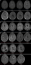
Radiographic evolution of TN and PP over time. T1+ contrast axial magnetic resonance imaging scans from representative patients with TN (A–C) and PP (D–F) depicting radiographic evolution of treatment‐related changes over time. (A): Woman aged 37 years with anaplastic oligoastrocytoma status post (s/p) chemoradiation. Onset of biopsy‐confirmed TN at 13 months after radiotherapy (RT), presenting as multiple contrast‐enhancing lesions (seven total) associated with new neurological symptoms. Gradual regression of lesions under bevacizumab treatment. (B): Man aged 64 years with anaplastic astrocytoma s/p RT. At 28 months after RT, onset of multiple periventricularly located, contrast‐enhancing lesions (four total) was noted. Follow‐up by imaging surveillance showed near total radiographic resolution of all lesions within 1 year of onset without treatment. The dominant left periventricular enhancing lesion is highlighted in the serial scans. (C): Woman aged 43 years with anaplastic astrocytoma s/p chemoradiation. Onset of biopsy‐confirmed TN at 11 months after chemoradiation (chemo‐RT), presenting as multiple contrast‐enhancing lesions (nine total) associated with new neurological symptoms, managed with steroids. Eight of nine lesions radiographically resolved within 6 to 26 months of onset. The dominant right periventricular lesion is shown. (D): Man aged 39 years with glioblastoma multiforme (GBM) s/p chemoradiation. Increased contrast enhancement around the resection cavity (RC) noted at 3 months after chemo‐RT during active antineoplastic treatment. The lesion was associated with new neurological symptoms and was managed with steroids and surgical debulking at 7 months after onset, revealing extensive tissue necrosis. (E): Woman aged 65 years with GBM s/p chemoradiation. Increased contrast enhancement around the RC noted at 1 month after chemo‐RT during active antineoplastic treatment. The lesion was associated with new neurological symptoms, managed with steroids, partially debulked (4 months after onset), and resolved at 9 months after onset. Histopathology revealed predominant tissue necrosis with few and scattered residual tumor cells. (F): Man aged 66 years with GBM s/p chemoradiation. Increased contrast enhancement around the RC noted at 1 month after chemo‐RT during active antineoplastic treatment. The lesion was associated with new neurological symptoms, managed with steroids, and fully resected at 4 months after onset, revealing extensive tissue necrosis. Abbreviations: PP, pseudoprogression; TN, treatment‐induced brain tissue necrosis.
Figure 4
Figure 4
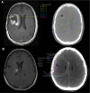
Typical observed radiographic features of treatment‐induced brain tissue necrosis (TN) and pseudoprogression (PP). (A): Axial T1+ contrast magnetic resonance imaging (MRI) (left) showing a non‐nodular focus of enhancement around the tumor resection cavity (RC) in the right frontal lobe, first manifesting at 3 months after radiotherapy (RT), consistent with PP. Corresponding radiotherapy dose distribution overlay on axial computed tomography (right) demonstrates the main radiation field encompassing the RC and surrounding brain parenchyma (60 Gy; green line). (B): T1+ contrast MRI (left) showing multiple small nodular foci of enhancement located medially to the RC in the right frontal region with involvement of the periventricular white matter, manifesting at 11 months after RT, consistent with TN. RT dose distribution overlay on axial computed tomography (right) demonstrates prior exposure of these regions of interest to the main radiation field (59.4 Gy; green line).

References

    1. Winter SF, Loebel F, Loeffler J et al. Treatment‐induced brain tissue necrosis: A clinical challenge in neuro‐oncology. Neuro Oncol 2019. [Epub ahead of print]. - PMC - PubMed
    1. Dietrich J, Winter S, Klein J. Neuroimaging of brain tumors: Pseudoprogression, pseudores‐ponse, and delayed effects of chemotherapy and radiation. Semin Neurol 2017;37:589–596. - PubMed
    1. Karschnia P, Parsons MW, Dietrich J. Pharmacologic management of cognitive impairment induced by cancer therapy. Lancet Oncol 2019;20:e92–e102. - PubMed
    1. Brandsma D, Stalpers L, Taal W et al. Clinical features, mechanisms, and management of pseudoprogression in malignant gliomas. Lancet Oncol 2008;9:453–461. - PubMed
    1. Eisele SC, Dietrich J. Cerebral radiation necrosis: Diagnostic challenge and clinical management. Rev Neurol 2015;61:225–232. - PubMed