Skip to main page content (VSports)
U.S. flag

An official website of the United States government

Dot gov

The . gov means it’s official. Federal government websites often end in . gov or . mil VSports app下载. Before sharing sensitive information, make sure you’re on a federal government site. .

Https

The site is secure. The https:// ensures that you are connecting to the official website and that any information you provide is encrypted and transmitted securely. V体育官网.

. 2020 Feb 21;15(2):469-484.
doi: 10.1021/acschembio.9b00939. Epub 2020 Jan 14.

Radiation-Induced Lipid Peroxidation Triggers Ferroptosis and Synergizes with Ferroptosis Inducers

Affiliations

Radiation-Induced Lipid Peroxidation Triggers Ferroptosis and Synergizes with Ferroptosis Inducers

Ling F Ye et al. ACS Chem Biol. .

Abstract

Although radiation is widely used to treat cancers, resistance mechanisms often develop and involve activation of DNA repair and inhibition of apoptosis. Therefore, compounds that sensitize cancer cells to radiation via alternative cell death pathways are valuable. We report here that ferroptosis, a form of nonapoptotic cell death driven by lipid peroxidation, is partly responsible for radiation-induced cancer cell death. Moreover, we found that small molecules activating ferroptosis through system xc- inhibition or GPX4 inhibition synergize with radiation to induce ferroptosis in several cancer types by enhancing cytoplasmic lipid peroxidation but not increasing DNA damage or caspase activation. Ferroptosis inducers synergized with cytoplasmic irradiation, but not nuclear irradiation. Finally, administration of ferroptosis inducers enhanced the antitumor effect of radiation in a murine xenograft model and in human patient-derived models of lung adenocarcinoma and glioma. These results suggest that ferroptosis inducers may be effective radiosensitizers that can expand the efficacy and range of indications for radiation therapy VSports手机版. .


Conflict of interest statement

CONFLICT OF INTERESTS

B V体育安卓版. R. S. holds equity in and serves as a consultant to Inzen Therapeutics.

Figures
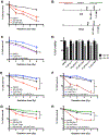
Figure 1.
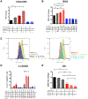
Figure 1.. IKE and RSL3 increase radiation sensitivity in cancer cell lines through lipid peroxidation.
A) Dose response of HT-1080 cells treated with DMSO, IKE, or RSL3 to radiation measured by clonogenic assays. **p<0.01. B) Coefficients of interaction between IKE (top) or RSL3 (bottom) and radiation observed for 5 tested cancer cell lines measured by clonogenic assays. C) Dose response of HT-1080 cells treated with DMSO, ferrostatin-1, or Z-VAD-FMK to radiation measured by clonogenic assays. *p<0.05, n.s.: p>0.05. D) Cell viability of HT-1080 cells treated with DMSO, ferrostatin-1, deferoxamine, Z-VAD-FMK, necrostatin-1S, or 3-methyladenine and co-treated with 0 or 4 Gy radiation for 24 hours. Data normalized to 0 Gy unirradiated controls for each treatment group. **p<0.01, *p<0.05, n.s.: p>0.05. E) Dose response of HT-1080 cells treated with DMSO, IKE, ferrostatin-1, or IKE and ferrostatin-1 measured by clonogenic assays. **p<0.01. Significance is calculated between the group treated with ferroptosis inducer and the group co-treated with ferroptosis inducer and ferroptosis inhibitor in E), F), G) and H). F) Dose response of HT-1080 cells treated with DMSO, RSL3, ferrostatin-1, or RSL3 and ferrostatin-1 measured by clonogenic assays. ***p<0.001. G) Dose response of HT-1080 cells treated with DMSO, IKE, Trolox, or IKE and Trolox measured by clonogenic assays. **p<0.01. H) Dose response of HT-1080 cells treated with DMSO, RSL3, Trolox, or RSL3 and Trolox measured by clonogenic assays. ***p<0.001. Data are plotted as mean ± SEM; n = 3 side-by-side experiments for A), C), D), E), F), G), H). Three biologically independent experiments were performed with similar results.
Figure 2.
Figure 2.. Markers of ferroptosis are elevated in HT-1080 cells treated with radiation.
A) PTGS2 mRNA fold change measured by RT-qPCR in HT-1080 cells treated with DMSO, RSL3, or ferrostatin-1 and co-treated with 0 or 6 Gy radiation for 24 hours. ****p<0.0001. B) MDA levels measured using the TBARS assay in HT-1080 cells treated with DMSO, IKE, or ferrostatin-1 and co-treated with 0 or 6 Gy radiation for 24 hours. ***p<0.001, **p<0.01, *p<0.05. C) Representative histograms of HT-1080 cells treated with DMSO, IKE, or IKE + ferrostatin-1 and co-treated with 0 or 6 Gy radiation for 24 hours and stained with C-11 BODIPY measured by flow cytometry. Horizontal bars indicate C-11 BODIPY-positive cell populations. D) C11-BODIPY staining of HT-1080 cells treated with ferroptosis modulators and co-treated with 0 or 6 Gy radiation measured by flow cytometry. **p<0.01. E) Glutathione (GSH) level is detected in HT-1080 cells treated with DMSO or IKE and co-treated with radiation for 24 hours using a fluorometric assay. ****p<0.0001, **p<0.01, *p<0.05, n.s.: p>0.05. Data are plotted as mean ± SEM; n = 3 technical replicates for A), B), D) and E). Three biologically independent experiments were performed with similar results.
Figure 3.
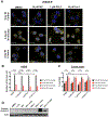
Figure 3.. IKE and RSL3 enhance radiation-induced cell death in HT-1080 cells through mechanisms independent of DNA damage or apoptosis.
A) Representative images of γH2AX immunofluorescence staining in HT-1080 cells treated with DMSO, IKE, RSL3, or ferrostatin-1 and co-treated with 0 or 6 Gy radiation for 30 minutes or 6 hours. Blue: DAPI; Yellow: γH2AX-FITC. Scale bar, 10 μm. B) Quantification of γH2AX immunofluorescence staining in HT-1080 cells treated with DMSO, IKE, RSL3, or ferrostatin-1 and co-treated with 0 or 6 Gy radiation for 30 minutes or 6 hours. ****p<0.0001, n.s.: p>0.05. C) Quantification of percent tail DNA in HT-1080 cells using the comet assay. Cells were treated with DMSO, IKE, RSL3 or ferrostatin-1 and co-treated with 0 or 6 Gy radiation for 30 minutes or 4 hours. ***p<0.001, **p<0.01, *p<0.05, n.s.: p>0.05. D) Western blot of cleaved caspase-3 in HT-1080 cells treated with DMSO, 1 μM IKE, 50 nM RSL3 and co-treated with 0 or 6 Gy radiation for 24 hours. Cells treated with 500 nM staurosporine and 500 nM staurosporine + 100 μM Z-VAD-FMK were included as positive and negative controls. Data are plotted as mean ± SEM. Three biologically independent experiments were performed with similar results for B), C) and D).
Figure 4.
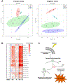
Figure 4.. Untargeted lipidomic study reveals enhanced ferroptosis lipid signatures in cells co-treated with IKE and radiation in HT-1080 cells.
A) Principal component analysis of the extracted lipid features in samples treated with DMSO or 5 μM IKE for 12 hours, with or without 6 Gy radiation for 24 hours, in both positive and negative electrospray ionization modes. B) Fold change heatmap of significantly changed lipid features from both IKE treatment and radiation treatment determined by two-way ANOVA (FDR corrected p value < 0.05) combined from both positive and negative ionization modes. Blue indicates decreased abundance compared to DMSO-treated controls (fold changes between 0.3 and 0.8); white indicates no change (fold changes between 0.8 and 1.2); red indicates increased abundance (fold changes between 1.2 and 10). n = 3 biologically independent samples. Abbreviations: FA, fatty acid; LysoPC, lysophosphatidylcholine; LysoPE, lysophosphatidylethanolamine; LysoPI, lysophosphatidylinositol; DAG, diacylglycerol. C) Proposed model of how oxidation of membrane polyunsaturated fatty acids by IKE and radiation cause elevated lysophospholipids and cell death. Abbreviations: PUFA, polyunsaturated fatty acid; PLA2, phospholipase A2; Lyso-PL, lysophospholipid.
Figure 5.
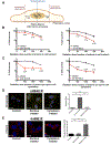
Figure 5.. Ferroptosis inducers synergize with cytoplasmic irradiation but not nuclear irradiation in HT-1080 cells.
A) Diagram of microbeam setup showing locations of beam spots targeting either the nucleus or cytoplasm. B) Clonogenic cell survival of HT-1080 cells treated with nuclear radiation and IKE or RSL3. CDI values are indicated. C) Clonogenic cell survival of HT-1080 cells treated with cytoplasmic radiation and IKE or RSL3. CDI values are indicated. D) Immunofluorescence staining of γH2AX in untreated cells, and cells treated with 100 protons to the nucleus or 2000 protons to the cytoplasm for 30 minutes. Blue: DAPI; Yellow: γH2AX-FITC. ****p<0.0001, n.s.: p>0.05. Scale bar, 10 μm. E) Immunofluorescence staining of 4-HNE in untreated cells, and cells treated with 100 protons to the nucleus or 2000 protons to the cytoplasm for 2 hours. Blue: DAPI; Red: 4-HNE-Rhodamine Red. ****p<0.0001, n.s.: p>0.05. Scale bar, 10 μm. Data are plotted as mean ± SEM; n = 3 side-by-side experiments. Three biologically independent experiments were performed with similar results.
Figure 6.
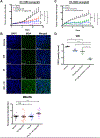
Figure 6.. IKE and sorafenib, combined with stereotactic radiation therapy, suppress tumor growth in a mouse xenograft model of sarcoma and a patient-derived xenograft model of lung adenocarcinoma.
A) Tumor volume ratio change in HT-1080 xenograft tumors treated with vehicle or IKE for 14 days and co-treated with 0 or 6 Gy radiation on days 2 and 4. n = 7 or 8 mice per group. **p<0.01, *p<0.05, n.s.: p>0.05. B) Immunofluorescence staining and quantification of MDA on paraffin-embedded tumor tissue sections measured by confocal microscopy. Blue: DAPI; Green: MDA-FITC. Scale bar, 50 μm. ***p<0.01, n.s.: p>0.05. n = 20 images with sections cut from four randomly chosen mice from each group, and five images captured from each section. C) Tumor volume ratio change in HT-1080 xenograft tumors treated with vehicle or sorafenib for 14 days and co-treated with 0 or 6 Gy radiation on days 1 and 3. n = 4 or 5 mice per group. **p<0.01, *p<0.05, n.s.: p>0.05. D) Glutathione (GSH) level is detected in HT-1080 xenograft tumors treated with vehicle or sorafenib for 14 days and co-treated with 0 or 6 Gy radiation on days 1 and 3, using a fluorometric assay. ****p<0.0001, **p<0.01, *p<0.05. n = 3 tumor samples from different animals per group.
Figure 7.
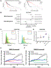
Figure 7.. SLC7A11 is a target for radiosensitization in human models of glioma and lung adenocarcinoma.
A) Kaplan-Meier survival analysis of overall survival of TCGA glioma patients in quartile 1 (low) and quartile 4 (high) of SLC7A11 RNA expression (left) or DNA methylation (right). B) Hazard ratios for disease-free survival between patients in quartile 1 (low) and quartile 4 (high) of SLC7A11 RNA expression or DNA methylation either in the case of radiation treatment, no radiation treatment, or all cases. C) Representative histograms of a human diffuse astrocytoma slice culture sample treated with DMSO, 10 μM IKE, or 10 μM IKE + 10 μM ferrostatin-1, co-treated with 0 or 2 Gy radiation for 24 hours, dissociated, stained with H2DCFDA, and measured by flow cytometry. Horizontal bars indicate H2DCFDA-positive cell populations. D) H2DCFDA staining of three human glioma slice culture samples treated with DMSO, 10 μM IKE, or 10 μM IKE + 10 μM ferrostatin-1, co-treated with 0 or 2 Gy radiation for 24 hours, dissociated, stained with H2DCFDA, and measured by flow cytometry. *p<0.05. n = 3 samples from different human glioma patients. E) Tumor volume ratio change in TM00219 patient-derived xenograft tumors treated with vehicle or IKE for 14 days and co-treated with 0 or 6 Gy radiation on day 1. n = 5 or 6 mice per group. ****p<0.0001, **p<0.01. F) Tumor volume ratio change in TM00219 patient-derived xenograft tumors treated with vehicle or sorafenib for 14 days and co-treated with 0 or 6 Gy radiation on day 1. n = 5 or 6 mice per group. ***p<0.001, **p<0.01. Data are plotted as mean ± SEM.

References

    1. Delaney G, Jacob S, Featherstone C, and Barton M (2005) The role of radiotherapy in cancer treatment: estimating optimal utilization from a review of evidence-based clinical guidelines, Cancer 104, 1129–1137. - PubMed
    1. Morgan MA, and Lawrence TS (2015) Molecular Pathways: Overcoming Radiation Resistance by Targeting DNA Damage Response Pathways, Clin Cancer Res 21, 2898–2904. - PMC - PubMed
    1. Souhami RL, and Tobias JS (2003) Cancer and its management, 4th ed., Blackwell Science, Malden, Mass.
    1. Tang Z, Zeng Q, Li Y, Zhang X, Ma J, Suto MJ, Xu B, and Yi N (2017) Development of a radiosensitivity gene signature for patients with soft tissue sarcoma, Oncotarget 8, 27428–27439. - PMC - PubMed
    1. Gerszten PC, Mendel E, and Yamada Y (2009) Radiotherapy and radiosurgery for metastatic spine disease: what are the options, indications, and outcomes?, Spine (Phila Pa 1976) 34, S78–92. - PubMed

Publication types

"V体育2025版" MeSH terms

LinkOut - more resources (VSports在线直播)