<u date-time="2gqXAqIi"></u><sub date-time="elsdqHf"></sub><sub date-time="4hSuVR"><small dropzone="BAxrqe"></small></sub> Skip to main page content
U.S. flag

An official website of the United States government

Dot gov

The . gov means it’s official. Federal government websites often end in . gov or . mil. Before sharing sensitive information, make sure you’re on a federal government site VSports app下载. .

Https

The site is secure. The https:// ensures that you are connecting to the official website and that any information you provide is encrypted and transmitted securely V体育官网. .

. 2019 Dec;13(12):2554-2573.
doi: 10.1002/1878-0261.12555. Epub 2019 Sep 8.

Down-regulation of long noncoding RNA PVT1 inhibits esophageal carcinoma cell migration and invasion and promotes cell apoptosis via microRNA-145-mediated inhibition of FSCN1 (V体育官网)

Affiliations

Down-regulation of long noncoding RNA PVT1 inhibits esophageal carcinoma cell migration and invasion and promotes cell apoptosis via microRNA-145-mediated inhibition of FSCN1

Si-Ning Shen (VSports) et al. Mol Oncol. 2019 Dec.

Abstract (VSports)

Accumulating evidence has established that long noncoding RNA (lncRNA) plasmacytoma variant translocation 1 (PVT1) is a tumor regulator in many cancers. Here, we aimed to investigate the possible function of lncRNA PVT1 in esophageal carcinoma (EC) via targeting of microRNA-145 (miR-145). Initially, microarray-based gene expression profiling of EC was employed to identify differentially expressed genes. Moreover, the expression of lncRNA PVT1 was examined and the cell line presenting with the highest level of lncRNA PVT1 expression was selected for subsequent experiments. We then proceeded to examine interaction among lncRNA PVT1, FSCN1, and miR-145. The effect of lncRNA PVT1 on viability, migration, invasion, apoptosis, and tumorigenesis of transfected cells was examined with gain-of-function and loss-of-function experiments. We observed that lncRNA PVT1 was robustly induced in EC. lncRNA PVT1 could bind to miR-145 and regulate its expression, and FSCN1 is a target gene of miR-145 VSports手机版. Overexpression of miR-145 or silencing of lncRNA PVT1 was revealed to suppress cell viability, migration, and invasion abilities, while also stimulating cell apoptosis. Furthermore, our in vivo results showed that overexpression of miR-145 or silencing of lncRNA PVT1 resulted in decreased tumor growth in nude mice. In conclusion, our research reveals that down-regulation of lncRNA PVT1 could potentially promote expression of miR-145 to repress cell migration and invasion, and promote cell apoptosis through the inhibition of FSCN1. This highlights the potential of lncRNA PVT1 as a therapeutic target for EC treatment. .

Keywords: FSCN1; apoptosis; esophageal carcinoma; long noncoding RNA; microRNA-145; plasmacytoma variant translocation 1 V体育安卓版. .


Conflict of interest statement

The authors declare no conflict of interest.

Figures

Figure 1
Figure 1
lncRNA PVT1 is highly expressed in EC tissues and cells. A, Wayne chart of top 100 differential genes of datasets http://www.ncbi.nlm.nih.gov/geo/query/acc.cgi?acc=GSE23400, http://www.ncbi.nlm.nih.gov/geo/query/acc.cgi?acc=GSE38129, and http://www.ncbi.nlm.nih.gov/geo/query/acc.cgi?acc=GSE38129; B, expression level of FSCN1 on http://www.ncbi.nlm.nih.gov/geo/query/acc.cgi?acc=GSE20347; C, expression level of FSCN1 on TCGA database; D, target of FSCN1 on four bioinformatics; E, expression level of miR‐145 on TCGA database; F, thermal map of dataset http://www.ncbi.nlm.nih.gov/geo/query/acc.cgi?acc=GSE45168; G, expression‐level scatter diagram of lncRNA PVT1 on TCGA database; H, expression of lncRNA PVT1 in cancer tissues and adjacent normal tissues detected by RTqPCR. Statistical values were measurement data and expressed as mean ± standard deviation. t‐Test was used to conduct data analysis, n = 50; I, expression of lncRNA PVT1 in KYSE‐30, KYSE‐70, KYSE‐109, Eca109, TE‐1, and HHEC cell lines detected by RTqPCR; J, the correlation between lncRNA PVT1 expression and DFS and OS analyzed by the Kaplan–Meier method (n = 50). The results were measurement data, expressed as mean ± standard deviation, and analyzed using one‐way ANOVA. The experiment was repeated three times independently; # vs. adjacent normal tissues, < 0.05; * vs. HEEC cell lines, < 0.05.
Figure 2
Figure 2
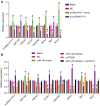
Silencing of lncRNA PVT1 and overexpressed miR‐145 down‐regulate mRNA expression of FSCN1, Bcl‐2, CD147, VEGFR2, and MTA1 yet up‐regulate Bax expression. A, mRNA expression of FSCN1, Bcl‐2, CD147, VEGFR2, MTA1, and Bax upon lncRNA PVT1 interference treatment determined by RTqPCR; B, mRNA expression of FSCN1, Bcl‐2, CD147, VEGFR2, MTA1, and Bax upon miR‐145 interference treatment determined by RTqPCR. The results were measurement data, expressed as mean ± standard deviation, and analyzed using one‐way ANOVA. The experiment was repeated three times independently; * vs. the blank group, < 0.05; # vs. the NC group, < 0.05.
Figure 3
Figure 3
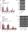
Silencing of lncRNA PVT1 and overexpressed miR‐145 decrease protein expression of FSCN1, Bcl‐2, CD147, VEGFR2, and MTA1 yet increase Bax expression. A and B, western blot analysis of FSCN1, Bcl‐2, CD147, VEGFR2, MTA1, and Bax proteins upon lncRNA PVT1 interference treatment; C and D, western blot analysis of FSCN1, Bcl‐2, CD147, VEGFR2, MTA1, and Bax proteins upon miR‐145 interference treatment. The results were measurement data, expressed as mean ± standard deviation, and analyzed using one‐way ANOVA. The experiment was repeated three times independently; * vs. the blank group, < 0.05; # vs. the NC group, < 0.05.
Figure 4
Figure 4
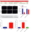
miR‐145 is a target of lncRNA PVT1. A, Subcellular location of lncRNA PVT1 measured by FISH; B, predicted binding site of miR‐145 in 3′ UTR of lncRNA PVT1 determined by FISH (×400, scale bar = 25 μm); C, specific binding regions between lncRNA PVT1 sequence and miR‐145 sequence analyzed by online analysis software; D, the binding of lncRNA PVT1 to miR‐145 verified by dual‐luciferase reporter gene assay; E, the binding of lncRNA PVT1 with Ago2 detected by RIP assay; F, the enrichment of lncRNA PVT1 by miR‐145 detected by RNA pull‐down assay. The values of luciferase activity were measurement data, expressed as mean ± standard deviation, and analyzed using unpaired t‐test. The experiment was repeated three times independently; * vs. the NC group, < 0.05.
Figure 5
Figure 5
FSCN1 is a target gene of miR‐145. A, Specific binding regions between FSCN1 sequence and miR‐145 sequence detected by the online analysis software microRNA; B, the binding of miR‐145 to FSCN1 verified by dual‐luciferase reporter gene assay; the values of luciferase activity were measurement data, expressed as mean ± standard deviation, and analyzed using unpaired t‐test. The experiment was repeated three times independently; * vs. the NC group, < 0.05.
Figure 6
Figure 6
Silencing of lncRNA PVT1 and overexpressed miR‐145 inhibit EC cell viability. A and B, Cell viability determined by MTT assay. Results of viability capacity in each group were measurement data, expressed as mean ± standard deviation, and analyzed using repeated measurement of one‐way ANOVA. The experiment was repeated three times independently; * vs. the blank group, < 0.05; # vs. the NC group, < 0.05.
Figure 7
Figure 7
Silencing of lncRNA PVT1 and overexpressed miR‐145 hinder EC cell migration. A and B, Migration of EC cells after lncRNA PVT1 interference treatment determined by scratch test (×40, scale bar = 250 μm); C and D, migration of EC cells after miR‐145 interference treatment determined by scratch test (×40, scale bar = 250 μm). The results were measurement data, expressed as mean ± standard deviation, and analyzed using one‐way ANOVA. The experiment was repeated three times independently; * vs. the blank group, < 0.05; # vs. the NC group, < 0.05.
Figure 8
Figure 8
Silencing of lncRNA PVT1 and overexpressed miR‐145 decrease EC cell invasion. A and B, Cell invasion in response to lncRNA PVT1 interference treatment determined by Transwell assay (×100, scale bar = 100 μm); C and D, cell invasion in response to miR‐145 interference treatment determined by Transwell assay (×100, scale bar = 100 μm). Statistical results were measurement data, expressed as mean ± standard deviation, and analyzed using one‐way ANOVA. The experiment was repeated three times independently; * vs. the blank group, < 0.05; # vs. the NC group, < 0.05.
Figure 9
Figure 9
Silencing of lncRNA PVT1 stimulates EC cell apoptosis. A and B, Cell cycle distribution in response to lncRNA PVT1 interference treatment measured using PI single staining; C and D, Cell apoptosis in response to lncRNA PVT1 interference treatment measured using Annexin V/PI double staining. Statistical results of panel B were measurement data, expressed as mean ± standard deviation, and analyzed using repeated measurement of one‐way ANOVA. Statistical results of panel D were measurement data, expressed as mean ± standard deviation, and analyzed using one‐way ANOVA. The experiment was repeated three times independently; * vs. the blank group, < 0.05; # vs. the NC group, < 0.05.
Figure 10
Figure 10
Overexpressed miR‐145 promotes cell apoptosis. A and B, Cell cycle in response to miR‐145 interference treatment determined by PI staining; C and D, cell apoptosis in response to miR‐145 interference treatment determined by Annexin V/PI double staining. Statistical results of panel B were measurement data, expressed as mean ± standard deviation, and analyzed using repeated measurement of one‐way ANOVA. Statistical results of panel D were measurement data, expressed as mean ± standard deviation, and analyzed using one‐way ANOVA. The experiment was repeated three times independently * vs. the blank group, < 0.05; # vs. the NC group, < 0.05.
Figure 11
Figure 11
Silencing of lncRNA PVT1 and overexpressed miR‐145 disrupt tumor growth. (A–C) Xenograft tumors and quantitative analysis of tumor growth after lncRNA PVT1 interference treatment; D‐F, xenograft tumors and quantitative analysis of tumor growth after miR‐145 interference treatment. The above results were measurement data, expressed as mean ± standard deviation, and analyzed using one‐way ANOVA. The experiment was repeated three times independently; * vs. the blank group, < 0.05; # vs. the NC group, < 0.05.
Figure 12
Figure 12
The molecular mechanism involved in the contribution of lncRNA PVT1 down‐regulation to alleviated EC progression by regulating miR‐145 and FSCN1. lncRNA PVT1 can specifically compete with miR‐145, and silencing of lncRNA PVT1 can up‐regulate the expression of miR‐145 and down‐regulate FSCN1 expression, thus inhibiting invasion, migration, survival, and viability and promoting apoptosis of EC cells.

References

    1. Akanuma N, Hoshino I, Akutsu Y, Murakami K, Isozaki Y, Maruyama T, Yusup G, Qin W, Toyozumi T, Takahashi M et al (2014) MicroRNA‐133a regulates the mRNAs of two invadopodia‐related proteins, FSCN1 and MMP14, in esophageal cancer. Br J Cancer 110, 189–198. - PMC - PubMed
    1. Chai J, Guo D, Ma W, Han D, Dong W, Guo H and Zhang Y (2018) A feedback loop consisting of RUNX2/LncRNA‐PVT1/miR‐455 is involved in the progression of colorectal cancer. Am J Cancer Res 8, 538–550. - PMC - PubMed
    1. Chang L, Yuan Z, Shi H, Bian Y and Guo R (2017) miR‐145 targets the SOX11 3′UTR to suppress endometrial cancer growth. Am J Cancer Res 7, 2305–2317. - "VSports最新版本" PMC - PubMed
    1. Chen X, Gong J, Zeng H, Chen N, Huang R, Huang Y, Nie L, Xu M, Xia J, Zhao F et al (2010) MicroRNA145 targets BNIP3 and suppresses prostate cancer progression. Cancer Res 70, 2728–2738. - PubMed
    1. Cui XB, Li S, Li TT, Peng H, Jin TT, Zhang SM, Liu CX, Yang L, Shen YY, Li SG et al (2016) Targeting oncogenic PLCE1 by miR‐145 impairs tumor proliferation and metastasis of esophageal squamous cell carcinoma. Oncotarget 7, 1777–1795. - PMC - PubMed

Publication types

"VSports在线直播" MeSH terms

"VSports app下载" LinkOut - more resources