Skip to main page content
U.S. flag

An official website of the United States government

Dot gov

The . gov means it’s official. Federal government websites often end in . gov or . mil. Before sharing sensitive information, make sure you’re on a federal government site. VSports app下载.

Https

The site is secure. The https:// ensures that you are connecting to the official website and that any information you provide is encrypted and transmitted securely V体育官网. .

. 2019 Apr 8;11(4):502.
doi: 10.3390/cancers11040502.

VSports手机版 - A Novel ALDH1A1 Inhibitor Targets Cells with Stem Cell Characteristics in Ovarian Cancer

Affiliations

A Novel ALDH1A1 Inhibitor Targets Cells with Stem Cell Characteristics in Ovarian Cancer

Nkechiyere G Nwani et al. Cancers (Basel). .

Abstract

A small of population of slow cycling and chemo-resistant cells referred to as cancer stem cells (CSC) have been implicated in cancer recurrence. There is emerging interest in developing targeted therapeutics to eradicate CSCs. Aldehyde-dehydrogenase (ALDH) activity was shown to be a functional marker of CSCs in ovarian cancer (OC). ALDH activity is increased in cells grown as spheres versus monolayer cultures under differentiating conditions and in OC cells after treatment with platinum. Here, we describe the activity of CM37, a newly identified small molecule with inhibitory activity against ALDH1A1, in OC models enriched in CSCs. Treatment with CM37 reduced OC cells' proliferation as spheroids under low attachment growth conditions and the expression of stemness-associated markers (OCT4 and SOX2) in ALDH+ cells fluorescence-activated cell sorting (FACS)-sorted from cell lines and malignant OC ascites. Likewise, siRNA-mediated ALDH1A1 knockdown reduced OC cells' proliferation as spheres, expression of stemness markers, and delayed tumor initiation capacity in vivo. Treatment with CM37 promoted DNA damage in OC cells, as evidenced by induction of γH2AX. This corresponded to increased expression of genes involved in DNA damage response, such as NEIL3, as measured in ALDH+ cells treated with CM37 or in cells where ALDH1A1 was knocked down. By inhibiting ALDH1A1, CM37 augmented intracellular ROS accumulation, which in turn led to increased DNA damage and reduced OC cell viability. Cumulatively, our findings demonstrate that a novel ALDH1A1 small molecule inhibitor is active in OC models enriched in CSCs. Further optimization of this new class of small molecules could provide a novel strategy for targeting treatment-resistant OC. VSports手机版.

Keywords: ALDH1A1; CM37; cancer stem cells; ovarian cancer V体育安卓版. .


"V体育2025版" Conflict of interest statement

The authors declare no conflict of interest.

Figures

Figure 1
Figure 1
Effects of CM37 on ovarian cancer (OC) sphere formation and stemness markers. (A) The chemical structure of CM37; (B) percent inhibition of aldehyde-dehydrogenase (ALDH) enzymatic activity by 20 µM CM37 measured in vitro for the different orthologues; (C) spheres derived from primary OC cells isolated from ascites fluid and treated with control or increasing doses of CM37 were photographed with an inverted microscope at 100× magnification. (D) Numbers of live cells growing as spheres were assessed by CCK-8 colorimetric assay in patient-derived OC cells. (E) Percentage of ALDH+ cells in untreated/or CM37-treated (500 nM–5 µM) patient-derived OC cells. (F) Percentage of ALDH+ cells in untreated/or CM37-treated (2.5–10 µM) OVCAR5 cells. (G) OVCAR5 cells were plated under low attachment conditions for six days; numbers of live cells were assessed by using the CCK8 colorimetric assay. (H) Relative expression of stem cell markers KLF4, Nanog, Oct4, Sox2 as measured by qRT-PCR in ALDH+ FACS-sorted OVCAR5 cells treated with CM37 (1 µM) for 24 h. Bars represent averages of triplicate measurements; **** corresponds to p < 0.0001; *** corresponds to p < 0.001.
Figure 2
Figure 2
Effects of CM37 on OC sphere formation: CM37 disrupts ALDH1A1-mediated sphere formation and growth under low attachment conditions. (A,B) OVCAR8 cells were treated with DMSO or 1–20 µM CM37 for six days, and numbers of live cells were assessed by quantifying ATP production via Cell-Titer Glo assay. Spheres were photographed with an inverted microscope at 100× magnification. (C,D) OVCAR3 cells were treated with control or 1–20 µM CM37 for six days, and numbers of live cells were assessed by quantifying ATP production by using the Cell-Titer Glo assay. Spheres were photographed with an inverted microscope at 100× magnification. (EH) Relative expression of ALDH1A isoforms in OVCAR3, SKOV3, OVCAR5, and COV362 cells grown as spheres as measured by qRT-PCR. Bars represent averages of triplicate measurements; ** corresponds to p < 0.01; **** corresponds to p < 0.0001.
Figure 3
Figure 3
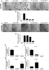
Effects of ALDH1A1 knock down on stemness phenotype. (A) OVCAR3 cells were transfected with nontargeting shRNA (Sh-Control) or shRNA targeting ALDH1A1 (sh-ALDH1A1), and ALDH1A1 knockdown was verified by qRT-PCR. (B,D) Sphere formation in OVCAR3 cells stably transfected with sh-Control and sh-ALDH1A1 and plated under low attachment conditions for six days. Spheres were photographed (100× magnification, B); numbers of live cells were assessed by using the Cell-Titer Glo assay (C), and the numbers of sphere per well were counted (D). (E) OVCAR5 cells were transfected with sh-Control or sh-ALDH1A1 and assessed for ALDH1A1 knockdown by qRT-PCR analysis. (FH) OVCAR5 cells were plated under low attachment conditions for six days; spheres were photographed (100× magnification, F); numbers of live cells were assessed by using the Cell-Titer Glo assay (G), and the numbers of sphere per well were counted (H). Bars represent averages of triplicate measurements; *** corresponds to p < 0.001; ** corresponds to p < 0.01; * corresponds to p < 0.05. (I) Percentage of ALDH+ cells in sh-Control or sh-ALDH1A1 transfected OVCAR3 cells. (J) OVCAR3 sh-Control and sh-ALDH1A1 were subcutaneously injected into the flanks of nude mice and tumor initiation was assessed; data captures the total number of tumors detectable at week #3 and at week #4. (K) Time to tumor initiation measured in days after subcutaneous injection of sh-Control and sh-ALDH1A1 transfected cells in the flanks of nude mice (n = 6 per group).
Figure 4
Figure 4
Effects of ALDH1A1 depletion on sphere formation. (A) COV362 cells were transfected with 50 nM scrambled siRNA (si-Control) or siRNA targeting ALDH1A1 (si-ALDH1A1 sequences #6 and #9); ALDH1A1 knockdown was assessed by qRT-PCR. (B) COV362 were plated under low attachment conditions for six days; spheres were photographed under 100× magnification. (C) The numbers of spheres per well were counted (five fields per well); graph depicts fold-change. (D) OVCAR5 cells were transfected with 50 nM si-Control or si-ALDH1A1 siRNA; cells were assessed for ALDH1A1 knockdown by qRT-PCR analysis. (E) OVCAR5 cells were plated under low attachment conditions for six days; spheres were photographed under 100× magnification. (F) The number of spheres per well were counted (five fields per well); graph depicts fold-change between cells transfected with control and ALDH-targeting siRNA. Bars represent averages of triplicate measurements; *** corresponds to p < 0.001; ** corresponds to p < 0.01; * corresponds to p < 0.05.
Figure 5
Figure 5
CM37-induced DNA damage response in ovarian cancer cell lines. (A) Immunofluorescent staining illustrates increased γH2AX abundance in OVCAR5 and SKOV3 cells cultured as spheres and treated with 100 nM CM37 for 72 h; 60× magnification. (B,C) Western blot demonstrating increased γH2AX protein levels in OC cell lines cultured as spheres and treated with CM37 for 45 min. (D,E) OVCAR3 ALDH+ cells were treated with 1 µM CM37 for 6 h prior to measurement of genes implicated in DNA damage response. (F,G) qRT-PCR measured expression of NEIL3, RAD21, RAD23 mRNA expression levels in ALDH+ OVCAR3 and OVCAR5 cells treated with 1 µM CM37. (H) qRT-PCR measured expression of RAD9A, RAD9B, RAD51 mRNA expression levels in OVCAR5 cells treated with 10 µM CM37. (I,J) QRT-PCR measured mRNA expression levels of NEIL3, RAD21, and RAD23 in OVCAR3 and OVCAR5 cells stably transduced with sh-Control or sh-ALDH1A1 lentiviral particles. Bars represent averages of triplicate measurements; *** corresponds to p < 0.001; ** corresponds to p < 0.01; * corresponds to p < 0.05.
Figure 6
Figure 6
CM37 increases reactive oxygen species (ROS) levels in OC cells. (A) Representative flow cytometry for total intracellular ROS levels in OVCAR5 cells treated with DMSO or 1 µM CM37. (BD) Fold change in ROS levels measured by flow cytometry in SKOV3, OVCAR3, and OVCAR5 treated with DSMO or 1 µM CM37 for 1 h. (E,F) Sphere formation in OVCAR5 cancer cells treated with 100 nM CM37 +/− 50 µM trolox for three days. Spheres were photographed under an inverted microscope (E) and counted (five fields per well; (F). Bars represent averages of triplicate measurements; * corresponds to p < 0.05. (G,H) Western blot measures γH2AX, H2AX, and β-actin protein levels in OVCAR5 and OVCAR3 cells cultured as spheres for six days and treated with 5 µM CM37 for 45 min, after 1 h or not of pre-treatment with Trolox (20 µM).

References

    1. Jemal A., Siegel R., Ward E., Hao Y., Xu J., Thun M.J. Cancer statistics, 2009. CA Cancer J. Clin. 2009;59:225–249. doi: 10.3322/caac.20006. - V体育2025版 - DOI - PubMed
    1. Berek J.S., Bertelsen K., du Bois A., Brady M.F., Carmichael J., Eisenhauer E.A., Gore M., Grenman S., Hamilton T.C., Hansen S.W., et al. Advanced epithelial ovarian cancer: 1998 consensus statements. Ann. Oncol. 1999;10:87–92. doi: 10.1023/A:1008323922057. - DOI - PubMed
    1. Lengyel E., Burdette J.E., Kenny H.A., Matei D., Pilrose J., Haluska P., Nephew K.P., Hales D.B., Stack M.S. Epithelial ovarian cancer experimental models. Oncogene. 2013:33. doi: 10.1038/onc.2013.321. - DOI - PMC - PubMed
    1. Silva I.A., Bai S., McLean K., Yang K., Griffith K., Thomas D., Ginestier C., Johnston C., Kueck A., Reynolds R.K., et al. Aldehyde dehydrogenase in combination with CD133 defines angiogenic ovarian cancer stem cells that portend poor patient survival. Cancer Res. 2011;71:3991–4001. doi: 10.1158/0008-5472.CAN-10-3175. - DOI - PMC - PubMed
    1. Zhang S., Balch C., Chan M.W., Lai H.C., Matei D., Schilder J.M., Yan P.S., Huang T.H., Nephew K.P. Identification and characterization of ovarian cancer-initiating cells from primary human tumors. Cancer Res. 2008;68:4311–4320. doi: 10.1158/0008-5472.CAN-08-0364. - "VSports" DOI - PMC - PubMed

LinkOut - more resources