Skip to main page content
U.S. flag

An official website of the United States government

Dot gov

The . gov means it’s official VSports app下载. Federal government websites often end in . gov or . mil. Before sharing sensitive information, make sure you’re on a federal government site. .

Https

The site is secure. The https:// ensures that you are connecting to the official website and that any information you provide is encrypted and transmitted securely V体育官网. .

. 2018 Dec;298(6):1181-1194.
doi: 10.1007/s00404-018-4912-4. Epub 2018 Sep 28.

"VSports在线直播" Morin decreases galectin-3 expression and sensitizes ovarian cancer cells to cisplatin

Affiliations

Morin decreases galectin-3 expression and sensitizes ovarian cancer cells to cisplatin

Dominik Bieg et al. Arch Gynecol Obstet. 2018 Dec.

V体育2025版 - Abstract

Purpose: This study aimed at evaluating whether morin (a natural flavonoid and a known inhibitor of NF-κB) can sensitize ovarian cancer cells to cisplatin by decreasing the expression of galectin-3, which is an anti-apoptotic protein regulated by NF-κB transcription factor VSports手机版. .

Methods: To assess the possibility of augmentation the activity of cisplatin by morin, we studied the separate and the combined effect of morin and cisplatin on viability, proliferation, and apoptosis of TOV-21G (cisplatin-sensitive) and SK-OV-3 (cisplatin-resistant) ovarian cancer cells V体育安卓版. We also analysed the effect of morin and cisplatin on galectin-3 expression at the mRNA and protein levels. .

Results: We demonstrated that morin possess antitumor activity against TOV-21G and SK-OV-3 ovarian cancer cells by reducing cell viability and proliferation as well as increasing the induction of apoptosis. Co-treatment of the cells with selected concentrations of morin and cisplatin, accordingly to specific treatment approaches, reveals a synergism, which leads to sensitization of the cells to cisplatin. During this sensitization, morin significantly reduces the expression of galectin-3 at the mRNA and protein level, regardless of the presence of cisplatin. V体育ios版.

Conclusions: Morin sensitizes TOV-21G and SK-OV-3 ovarian cancer cells to cisplatin, what is associated with a decrease of the expression of galectin-3 VSports最新版本. .

Keywords: Cisplatin; Drug resistance; Galectin-3; Morin; Ovarian cancer. V体育平台登录.


Conflict of interest statement

Conflict of interest

The authors declare that they have no conflict of interest.

Ethical approval

This article does not contain any studies with human participants or animals performed by any of the authors.

Figures
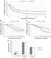
Fig. 1
Fig. 1
Effects of morin and cisplatin on viability of TOV-21G (cisplatin-sensitive) and SK-OV-3 (cisplatin-resistant) human ovarian cancer cells. The cells were treated with indicated concentrations of the drugs in indicated periods of time: a TOV-21G and SK-OV-3 cells treated with cisplatin (3.125–200 µM) for 24 h, b TOV-21G cells treated with morin (50–500 µM) for 24 h and 48 h, c SK-OV-3 cells treated with morin (50–500 µM) for 24 h and 48 h. Cell viability was analysed by XTT assay. The data are shown as mean ± SD of triplicate experiments. Asterisk: ANOVA p < 0.05 between the cells treated with the different concentrations of drug and the untreated control group in the same period of time (dose-dependence). Double asterisk: ANOVA p < 0.05 between the cells treated with the same concentration of drug in the different period of time (time-dependence). The cytotoxicity of morin was dose- and time-dependent, and cisplatin was dose-dependent. d Comparison of IC50 values obtained for TOV-21G and SK-OV-3 cells treated with morin for 24 h and 48 h or cisplatin for 24 h. Triple asterisk: T test p < 0.05 between IC50 values obtained for the same drug in the same period of time in different cell lines (TOV-21G or SK-OV-3)
Fig. 2
Fig. 2
Cytotoxic effect of morin-cisplatin combination on a TOV-21G (cisplatin-sensitive) and b SK-OV-3 (cisplatin-resistant) human ovarian cancer cells. The cells were treated with different combinations of morin–cisplatin according to three approaches: APP:1—simultaneous treatment with both drugs for 24 h; APP:2—pre-treatment with morin for 24 h, followed by treatment with cisplatin alone for the another 24 h; APP:3—pre-treatment with morin for 24 h, followed by co-treatment with morin and cisplatin for another 24 h. After the treatments the cell viability was determined (XTT assay), followed by the calculation of combination index (CI). The CI values are shown as heat maps, where the effect of a drug combination is synergistic if CI < 0.9 (green colour); additive if 0.9 ≤ CI ≤ 1.1 (yellow colour); antagonistic if CI > 1.1 (red colour)
Fig. 3
Fig. 3
Effects of morin and cisplatin on proliferation of TOV-21G (cisplatin-sensitive) and SK-OV-3 (cisplatin-resistant) human ovarian cancer cells. The cells were treated with indicated concentrations of the drugs in indicated periods of time: a TOV-21G and SK-OV-3 cells treated with cisplatin (3.125–50 µM) for 24 h, b TOV-21G cells treated with morin (100–250 µM) for 24 h, c SK-OV-3 cells treated with morin (100–250 µM) for 24 h and 48 h. Cell proliferation was analysed by EdU incorporation assay. The data are shown as mean ± SD of triplicate experiments. Asterisk: ANOVA p < 0.05 between the cells treated with the different concentrations of drug and the untreated control group in the same period of time (dose-dependence). Double asterisk: ANOVA p < 0.05 between the cells treated with the same concentration of drug in the different period of time (time-dependence). The anti-proliferative effect of morin was dose- (in both cell lines) and time-dependent (in SK-OV-3 cells), and cisplatin was dose-dependent (in both cell lines). d Comparison of GI50 values obtained for TOV-21G and SK-OV-3 cells treated with morin for 24 h and/or 48 h or cisplatin for 24 h. Triple asterisk: T test p < 0.05 between GI50 values obtained for the same drug in the same period of time in different cell lines (TOV-21G or SK-OV-3)
Fig. 4
Fig. 4
Anti-proliferative effect of morin–cisplatin combination on a TOV-21G (cisplatin-sensitive) and b SK-OV-3 (cisplatin-resistant) human ovarian cancer cells. The cells were treated with different combinations of morin–cisplatin according to selected approaches: APP:1—simultaneous treatment with both drugs for 24 h; APP:3—pre-treatment with morin for 24 h, followed by co-treatment with morin and cisplatin for another 24 h. After the treatments the cell proliferation was determined (EdU incorporation assay), followed by the calculation of combination index (CI). The CI values are shown as heat maps, where the effect of a drug combination is synergistic if CI < 0.9 (green colour); additive if 0.9 ≤ CI ≤ 1.1 (yellow colour); antagonistic if CI > 1.1 (red colour)
Fig. 5
Fig. 5
Effects of morin and cisplatin on apoptosis of TOV-21G (cisplatin-sensitive) and SK-OV-3 (cisplatin-resistant) human ovarian cancer cells. The cells were treated with indicated concentrations of the drugs in indicated periods of time: a TOV-21G and SK-OV-3 cells treated with cisplatin (6.25–100 µM) for 24 h, b TOV-21G cells treated with morin (100–400 µM) for 24 h, c SK-OV-3 cells treated with morin (100–400 µM) for 48 h. Cell apoptosis was analysed by FITC annexin V and PI staining. The data are shown as mean ± SD of triplicate experiments. Asterisk: ANOVA p < 0.05 between the cells treated with the different concentrations of drug and the untreated control group (dose-dependence). The pro-apoptotic effect of morin and cisplatin was dose-dependent in both cell lines
Fig. 6
Fig. 6
Pro-apoptotic effect of morin–cisplatin combination on a TOV-21G (cisplatin-sensitive) and b SK-OV-3 (cisplatin-resistant) human ovarian cancer cells. The cells were treated with different combinations of morin–cisplatin according to selected approaches: APP:1—simultaneous treatment with both drugs for 24 h; APP:3—pre-treatment with morin for 24 h, followed by co-treatment with morin and cisplatin for another 24 h. After the treatments the cell apoptosis was determined (FITC annexin V and PI staining), followed by the calculation of combination index (CI). The CI values are shown as heat maps, where the effect of a drug combination is synergistic if CI < 0.9 (green colour); additive if 0.9 ≤ CI ≤ 1.1 (yellow colour); antagonistic if CI > 1.1 (red colour)
Fig. 7
Fig. 7
Effects of morin and cisplatin on the expression of galectin-3 at the mRNA and protein level in TOV-21G (cisplatin-sensitive) and SK-OV-3 (cisplatin-resistant) human ovarian cancer cells. The cells were treated with indicated concentrations of the drugs in indicated periods of time: a, b TOV-21G cells treated with morin (100–250 µM) and with or without cisplatin (6.25 µM) for 24 h (APP:1), c, d SK-OV-3 cells pre-treated with morin (100–250 µM) for 24 h and then treated with morin (100–250 µM) and with or without cisplatin (12.5 µM) for another 24 h (APP:3). At the mRNA level, the expression was analysed by Real Time™ RT-PCR technique. At the protein level, the expression was assessed by ELISA assay. The data are shown as mean ± SD of triplicate experiments. Asterisk: ANOVA p < 0.05 between the cells treated with the different concentrations of morin and the untreated control group (dose-dependence). Double asterisk: ANOVA p < 0.05 between the cells treated with the same concentration of morin in the presence or absence of cisplatin. Morin significantly reduced the expression of galectin-3 at mRNA and protein level in a dose-dependent manner in both cell lines. The presence of cisplatin significantly increased the level of galectin-3 at mRNA and protein level in both cell lines, without significantly affecting the ability of morin to reduce the expression of galectin-3

References

    1. Arzuman L, Beale P, Chan C, et al. Synergism from combinations of tris (benzimidazole) monochloroplatinum(II) chloride with capsaicin, quercetin, curcumin and cisplatin in human ovarian cancer cell lines. Anticancer Res. 2014;34:5453–5464. - PubMed
    1. Long Q, Xie Y, Huang Y, et al. Induction of apoptosis and inhibition of angiogenesis by PEGylated liposomal quercetin in both cisplatin-sensitive and cisplatin-resistant ovarian cancers. J Biomed Nanotechnol. 2013;9:965–975. doi: 10.1166/jbn.2013.1596. - DOI - PubMed
    1. Yang Z, Liu Y, Liao J, et al. Quercetin induces endoplasmic reticulum stress to enhance cDDP cytotoxicity in ovarian cancer: involvement of STAT3 signaling. FEBS J. 2015;282:1111–1125. doi: 10.1111/febs.13206. - DOI - PubMed
    1. Zhu X, Ji M, Han Y, et al. PGRMC1-dependent autophagy by hyperoside induces apoptosis and sensitizes ovarian cancer cells to cisplatin treatment. Int J Oncol. 2017;50:835–846. doi: 10.3892/ijo.2017.3873. - DOI - PubMed
    1. Kang HG, Kim S, Cho Y, et al. Galectin-3 supports stemness in ovarian cancer stem cells by activation of the Notch1 intracellular domain. Oncotarget. 2016;7:68229–68241. - PMC - PubMed

Publication types

MeSH terms