<kbd date-time="zQnfFcAn"></kbd><del id="vvWYo"></del> <b draggable="HfcUeJA6"><bdo draggable="Zqhvft"></bdo></b><area dropzone="fljDg25N"></area> Skip to main page content
U.S. flag

An official website of the United States government

Dot gov

The . gov means it’s official. Federal government websites often end in . gov or . mil. Before sharing sensitive information, make sure you’re on a federal government site. VSports app下载.

Https

The site is secure. The https:// ensures that you are connecting to the official website and that any information you provide is encrypted and transmitted securely V体育官网. .

. 2018 Jul 15;24(14):3334-3347.
doi: 10.1158/1078-0432.CCR-17-2452. Epub 2018 Apr 10.

Resistance Mechanisms to Targeted Therapies in ROS1+ and ALK+ Non-small Cell Lung Cancer

Affiliations

Resistance Mechanisms to Targeted Therapies in VSports - ROS1+ and ALK+ Non-small Cell Lung Cancer

Caroline E McCoach et al. Clin Cancer Res. .

Abstract

Purpose: Despite initial benefit from tyrosine kinase inhibitors (TKIs), patients with advanced non-small cell lung cancer (NSCLC) harboring ALK (ALK+) and ROS1 (ROS1+) gene fusions ultimately progress. Here, we report on the potential resistance mechanisms in a series of patients with ALK+ and ROS1+ NSCLC progressing on different types and/or lines of ROS1/ALK-targeted therapy. Experimental Design: We used a combination of next-generation sequencing (NGS), multiplex mutation assay, direct DNA sequencing, RT-PCR, and FISH to identify fusion variants/partners and copy-number gain (CNG), kinase domain mutations (KDM), and copy-number variations (CNVs) in other cancer-related genes. We performed testing on 12 ROS1+ and 43 ALK+ patients. Results: One of 12 ROS1+ (8%) and 15 of 43 (35%) ALK + patients harbored KDM. In the ROS1+ cohort, we identified KIT and β-catenin mutations and HER2-mediated bypass signaling as non-ROS1-dominant resistance mechanisms. In the ALK+ cohort, we identified a novel NRG1 gene fusion, a RET fusion, 2 EGFR, and 3 KRAS mutations, as well as mutations in IDH1, RIT1, NOTCH, and NF1 In addition, we identified CNV in multiple proto-oncogenes genes including PDGFRA, KIT, KDR, GNAS, K/HRAS, RET, NTRK1, MAP2K1, and others. Conclusions: We identified a putative TKI resistance mechanism in six of 12 (50%) ROS1 + patients and 37 of 43 (86%) ALK+ patients. Our data suggest that a focus on KDMs will miss most resistance mechanisms; broader gene testing strategies and functional validation is warranted to devise new therapeutic strategies for drug resistance VSports手机版. Clin Cancer Res; 24(14); 3334-47. ©2018 AACR. .


Conflict of interest statement

Conflicts of Interest.

CEM- honoraria for Takeda, Guardant Health, DRC- honoraria from Takeda; RD – Pfizer speaker’s bureau and advisory board, Novartis, Roche, Ignyta – advisory board; RCD – AstraZeneca – advisory board, Guardant Health – honoraria, Pfizer – honoraria, Ignyta – advisory board and sponsored research grant, Takeda – advisory board, Spectrum Pharmaceuticals – advisory board, Abbott Molecular – remuneration for patent license, Rain Therapeutics – stock owner; PAB – consultant for Genentech and Pfizer; ATL – Abbott Molecular – remuneration for patent license; DTM, ATL, KG, KJ, LS, AD, AEB, KDD, MV-G, WTP, DLA - declare no potential conflicts of interest;

Figures

Figure 1
Figure 1
ROS1 and ALK fusion partner and variant frequencies. A, ROS1 fusion partner frequencies in the 10 patients whose tumor samples underwent NGS. Three patient’s tumor samples were positive by FISH but negative by NGS. B, ALK fusion variant frequencies in the 28 patients whose tumor samples underwent NGS. ○ All NGS negative and NGS not done patients responded to crizotinib ❖ Though fusion partner not identified, one patient found to have two ROS1 kinase domain mutations, illustrated in figure 2 * One patient whose fusion partner was not identifiable by NGS underwent additional testing with Archer DX and was found to have an E2;A20 fusion variant. **One patient’s NGS and FISH was negative for ALK fusion however the patient’s pre-treatment tumor sample was positive for ALK fusion indicating loss of ALK during treatment.
Figure 2
Figure 2
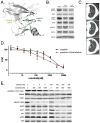
ROS1 resistance in human-derived cell lines. A, Crystal structure of ROS1 bound to crizotinib (PDB 3ZBF) highlighting the two mutations identified in this tumor sample showing the L2026M gatekeeper mutation (yellow) and the L1951R mutation (green). VAF is shown in parentheses for each mutation. B, Western blot using protein extract from cell line derived from patient’s crizotinib-resistant tumor sample (CUTO16) demonstrating only partial inhibition of pROS1, pSHP2 and pERK1/2 with 1 mm crizotinib or ceritinib following 2 hour drug treatment. C, CT scan of the ROS1+ patient after development of resistance to crizotinib (top) and the first interval scan at 1 month (middle) after starting ceritinib and the same nodule after 3 months on ceritinib with progression (bottom). D, Cell viability of CUTO23 treated with increasing doses of crizotinib alone (black) or in the presence of 100 nM afatinib (red) for 72 hours. The IC50 for CUTO23 cells in crizotinib alone was 1959 ± 31nM and for crizotinib with 100nM afatinib 438 ± 79nM. Error bars, mean ± SEM for three triplicate experiments (n = 9). E, Western blot analysis of CUTO23 cells treated with the indicated doses of crizotinib in the absence (left) or presence (right) of 100 nM afatinib for 2 hours demonstrating inhibition of pERBB2 with afatinib and concomitant rescue of inhibition of downstream signaling by AKT and ERK1/2 with the addition of afatinib to crizotinib.
Figure 3
Figure 3
Kinase Domain Mutation Distribution in ALK+ Resistance samples. A, Distribution of kinase domain mutations across ALK+ samples. One sample with G1202* (VAF 19%) in combination with G1202R (VAF 25%). 3 patients demonstrated compound mutation after multiple ALK-targeted TKIs. B, Distribution of kinase domain mutations in those patient who had a post progression biopsy after crizotinib, firstline therapy for all patients. C, Distribution of kinase domain mutations in patients who had post progression biopsy after >1 TKI (D) Distribution of kinase domain mutations in patients who had post progression biopsy after ceritinib. E, Distribution of kinase domain mutations in patients who had post progression biopsy after brigatinib. F, Distribution of kinase domain mutations in patients who had post progression biopsy after alectinib. G, One patient had a post progression biopsy after treatment with lorlatinib. Prior treatments for each patient listed in subscript. Patients with serial biopsies on different treatments indicated with Asterisk. If multiple biopsies KDM listed for biopsy on which it appeared. Abbreviations: C- crizotinib, B- brigatinib, A-Alectinib, TKI – tyrosine kinase inhibitor, X- stop codon.
Figure 4
Figure 4
RALGAPA1-NRG1 fusion induces drug resistance in ALK+ cancer. A, Cellular proliferation of the EML4-ALK cell lines, H3122 (blue)or its derivative H3122-NRG1 (red) which harbors a CRISPR induced RALGAPA1-NRG1 fusion using increasing doses of crizotinib demonstrating marked resistance crizotinib resistance. Addition of afatinib (100nM) resensitized the H3122-NRG1 (black) to crizotinib. IC50 for crizotinib was H3122 cells was 187 ± 3.7nM, for H3122-NRG1 was 1182 ± 5.3nM and for H3122-NRG1 with afatinib was 200 ± 10.5nM. Error bars, mean ± SEM for three triplicate experiments (n = 9). B, Western blot analysis of H3122 or H3122-NRG1 cells treated with crizotinib 500 nM and/or afatinib 100nM as indicated for 2 hours. H3122-NRG1 cells demonstrated increased levels of phosphorylated and total ERBB3, pAKT and pERK1/2 compared to H3122 cell lines, but lack of pSHP2. Crizotinib inhibits SHP2, AKT and ERK1/2 in H3122 cells. AKT and ERK are not inhibited by crizotinib alone in H3122-NRG1, but are inhibited by afatinib as is ERBB3 indicating oncogene switch from ALK to HER3, the receptor for neuregulin. C, Proximity ligation assay (PLA) of ALK and GRB2 from ALK-22_2 FFPE tumor sample showing functional ALK signaling. D, PLA of ERBB2 and GRB2 from ALK-22_2 sample demonstrating functional ERBB2 signaling.
Figure 5
Figure 5
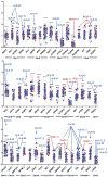
Copy Number Variation Distribution. Median read depths across the 48 gene NGS panel illustrating outliers in our cohort. Genes are arranged by chromosome. Patients who have a gene with a read depth > 2.5 SD above the median are annotated. ROS1 samples (red), ALK samples (blue), and other NSCLC samples (open circles).
Figure 6
Figure 6
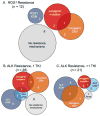
Resistance Mechanisms after TKI in ROS1+ ALK+ patients. Venn Diagrams demonstrating the distribution of resistance mechanisms identified in each cohort. A, ROS1 cohort (n = 12). B, ALK 1 TKI cohort (n = 28). C, ALK >1 TKI cohort (n = 21).

References

    1. Rikova K, Guo A, Zeng Q, Possemato A, Yu J, Haack H, et al. Global survey of phosphotyrosine signaling identifies oncogenic kinases in lung cancer. Cell. 2007;131(6):1190–203. doi: 10.1016/j.cell.2007.11.025. - "VSports注册入口" DOI - PubMed
    1. Bergethon K, Shaw AT, Ou SH, Katayama R, Lovly CM, McDonald NT, et al. ROS1 rearrangements define a unique molecular class of lung cancers. J Clin Oncol. 2012;30(8):863–70. doi: 10.1200/JCO.2011.35.6345. - DOI - PMC - PubMed
    1. Shaw AT, Ou SH, Bang YJ, Camidge DR, Solomon BJ, Salgia R, et al. Crizotinib in ROS1-rearranged non-small-cell lung cancer. The New England journal of medicine. 2014;371(21):1963–71. doi: 10.1056/NEJMoa1406766. - DOI - PMC - PubMed
    1. Soda M, Choi YL, Enomoto M, Takada S, Yamashita Y, Ishikawa S, et al. Identification of the transforming EML4-ALK fusion gene in non-small-cell lung cancer. Nature. 2007;448(7153):561–6. doi: 10.1038/nature05945. - DOI (V体育2025版) - PubMed
    1. Birchmeier C, Sharma S, Wigler M. Expression and rearrangement of the ROS1 gene in human glioblastoma cells. Proceedings of the National Academy of Sciences of the United States of America. 1987;84(24):9270–4. - V体育官网 - PMC - PubMed

V体育官网 - Publication types

MeSH terms