Skip to main page content (VSports最新版本)
U.S. flag

An official website of the United States government

Dot gov

The . gov means it’s official. Federal government websites often end in . gov or . mil. Before sharing sensitive information, make sure you’re on a federal government site VSports app下载. .

Https

The site is secure. The https:// ensures that you are connecting to the official website and that any information you provide is encrypted and transmitted securely. V体育官网.

. 2018 Feb 20;57(7):1262-1273.
doi: 10.1021/acs.biochem.7b01176. Epub 2018 Jan 30.

A Small-Molecule Inhibitor of Human DNA Polymerase η Potentiates the Effects of Cisplatin in Tumor Cells

Affiliations

A Small-Molecule Inhibitor of Human DNA Polymerase η Potentiates the Effects of Cisplatin in Tumor Cells

Maroof K Zafar et al. Biochemistry. .

Abstract

Translesion DNA synthesis (TLS) performed by human DNA polymerase eta (hpol η) allows tolerance of damage from cis-diamminedichloroplatinum(II) (CDDP or cisplatin). We have developed hpol η inhibitors derived from N-aryl-substituted indole barbituric acid (IBA), indole thiobarbituric acid (ITBA), and indole quinuclidine scaffolds and identified 5-((5-chloro-1-(naphthalen-2-ylmethyl)-1H-indol-3-yl)methylene)-2-thioxodihydropyrimidine-4,6(1H,5H)-dione (PNR-7-02), an ITBA derivative that inhibited hpol η activity with an IC50 value of 8 μM and exhibited 5-10-fold specificity for hpol η over replicative pols. We conclude from kinetic analyses, chemical footprinting assays, and molecular docking that PNR-7-02 binds to a site on the little finger domain and interferes with the proper orientation of template DNA to inhibit hpol η. A synergistic increase in CDDP toxicity was observed in hpol η-proficient cells co-treated with PNR-7-02 (combination index values = 0 VSports手机版. 4-0. 6). Increased γH2AX formation accompanied treatment of hpol η-proficient cells with CDDP and PNR-7-02. Importantly, PNR-7-02 did not impact the effect of CDDP on cell viability or γH2AX in hpol η-deficient cells. In summary, we observed hpol η-dependent effects on DNA damage/replication stress and sensitivity to CDDP in cells treated with PNR-7-02. The ability to employ a small-molecule inhibitor of hpol η to improve the cytotoxic effect of CDDP may aid in the development of more effective chemotherapeutic strategies. .


"VSports手机版" Conflict of interest statement

The authors declare no competing financial interest.

"V体育平台登录" Figures
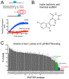
Figure 1.
Figure 1.
Structure–function studies identified PNR-7-02 as an inhibitor of hpol η with improved potency relative to other indole-derived compounds. (A) Schematic illustration of the pol activity assay. (B) General chemical structure of the parent indole barbituric acid (IBA) scaffold that incorporates all analogues tested for inhibition of TLS pol action. The availability of multiple sites of modification (R1–R3, A, and X positions are noted on the parent scaffold) allowed a useful structure–activity study to be performed. (C) Catalytic activity of hpol η (2.5 nM) was measured in the presence of 85 IBA and indole thiobarbituric acid (ITBA) derivatives (20 μM) to identify structure–activity relationships important for inhibition of this TLS pol. The results represent the mean (±SD) for experiments performed in triplicate. See Supporting Information Table S1 for the chemical structure and details regarding pol activity for each compound.
Figure 2.
Figure 2.
PNR-7-02 inhibits hpol η activity. (A) Chemical structure of PNR-7-02. (B) Dose–response curve for PNR-7-02 inhibition of hpol η. The IC50 value reported represents the best fit of the data (95% confidence interval). PNR-7-02 inhibits dNTP binding to hpol η through a partial competitive mechanism of action. (C) Kd,DNA for hpol η binding to primer–template DNA plotted as a function of PNR-7-02 concentration. (D) Michaelis constant (KM,dTTP) for hpol η activity exhibits a nonlinear increase as a function of PNR-7-02 concentration. (E) Turnover number (kcat) does not change at concentrations of PNR-7-02 at or below the IC50 for inhibition of hpol η activity. The results in panels B–E represent the mean (±SD) for three independent experiments.
Figure 3.
Figure 3.
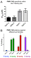
PNR-7-02 inhibits hpol η, hRev1, and hpol λ with similar potency. (A) IC50 value for PNR-7-02-mediated inhibition of the four human Y-family pols measured using gel-based analysis of pol activity. The IC50 values were 8 (7–9) μM for hpol η, 8 (6–11) μM for hRev1, 21 (19–23) μM for hpol β, and 22 (18–26) μM for hpol κ, where the values reported represent the best-fit value (95% confidence interval) derived from the results of three independent experiments. (B) Pol activity measured in the presence of PNR-7-02 (20 μM) using the fluorescence-based assay. Pol activity was 4.4 (±3.8)% for hpol η, 86 (±1)% for hpol θ, 93 (±2)% for TbPol I, 32 (±5)% for hpol ε, 118 (±15)% for Dpo1, 103 (±8)% for hpol β, and 5.9 (±2.1)% for hpol λ. The values reported represent the mean (±SD) activity relative to the DMSO control for three independent experiments. A two-tailed Student’s t test was used to compare hpol η activity in the presence of PNR-7-02 to that observed for each of the other pols (** = P < 0.001, *** = P < 0.0001).
Figure 4.
Figure 4.
Protein footprinting identifies the PNR-7-02 binding site on the hpol η little finger. (A) Selected ion chromatogram for the HPG-modified peptide containing Arg351 in the presence of HPG alone (blue), HPG + 20 μM PNR-7-02 (red), and 100 μM PNR-7-02 (dark red). (B) Relative abundance of HPG-modified arginine was quantified from MS data. The fraction of HPG-modified arginine observed in the presence of PNR-7-02 was normalized to that observed in the absence of the compound. The modified arginine residues in the finger, palm, thumb, and little finger domains of hpol η are shown in blue, red, green, and purple, respectively. (C) Cartoon representation of hpol η (PDB ID 3MR2) shown with regions of the protein containing HPG-modified residues in cyan and regions that were not modified by HPG in gray. The addition of PNR-7-02 caused changes in HPG reactivity for multiple arginines. Peptides with arginine residues that displayed a greater than 2-fold reduction in HPG reactivity (i.e., protection) are shown in red, whereas regions that exhibited a greater than 2-fold increase in HPG reactivity (i.e., exposure) are shown in blue.
Figure 5.
Figure 5.
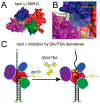
Molecular docking analysis of IBA/ITBA binding and model for inhibition of hpol η. (A) PNR-7-01 and PNR-7-02, the two most potent inhibitors of hpol η, were docked onto the target (PDB code 3MR2) using SwissDock. The finger, palm, thumb, and little finger domains of hpol η are shown in blue, red, green, and purple, respectively, as is the top binding pose for PNR-7-01 (green carbons) and the two top binding poses for PNR-7-02 (yellow carbons). (B) More detailed view of PNR-7-01 and PNR-7-02 (pose 2) binding to the cleft between the finger and little finger domains of hpol η. The location of Arg351 is noted as a yellow patch, and the primer–template DNA, which was removed for docking, is shown superimposed on the target in semitransparent cartoon form. (C) Model depicting the mechanism of hpol η inhibition by IBA/ITBA compounds based on the kinetic analysis, chemical footprinting, and molecular docking results.
Figure 6.
Figure 6.
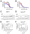
PNR-7-02 potentiates the cytotoxic effects of CDDP in a manner largely dependent on hpol η expression. Cell viability was measured for parental HAP-1 cells expressing hpol η (panel A) and hpol η-deficient cells (panel B) treated with CDDP alone (black circles), CDDP + 0.1 μM PNR-7-02 (red triangles), or CDDP + 1 μM PNR-7-02 (blue diamonds). The values reported represent the mean (±SEM) of n =12 for panel A and 6 for panel B. The EC50 values derived from the dose–response curves for HAP-1 cells expressing hpol η (panel A) were 0.31 (0.27–0.37) for CDDP alone, 0.14 (0.08–0.23) for CDDP + 0.1 μM PNR-7-02, and 0.12 (0.08–0.17) for CDDP + 1 μM PNR-7-02. The EC50 values derived from the dose–response curves for hpol η-deficient cells (panel B) were 0.10 (0.07–0.14) for CDDP alone, 0.10 (0.08–0.15) for CDDP + 0.1 μM PNR-7-02, and 0.07 (0.06–0.09) for CDDP + 1 μM PNR-7-02. The reported EC50 values represent the mean (95% confidence interval). hpol η-proficient HAP-1 (C) and hpol η-deficient cells (D) were treated with either DMSO or CDDP (0.5 μM) in combination with PNR-7-02 (0.1 and 1 μM) at the indicated concentrations. Immunoblotting was performed to assess changes in γH2AX levels. Quantification of γH2AX expression (normalized against β-actin and relative to DMSO-treated cells) is shown for hpol η-proficient HAP-1 (E) and hpol η-deficient knockout cells (F). The mean (±SD) is shown for three biological replicates. A Student’s t test was used to calculate the P values.

References

    1. Friedberg EC, Aguilera A, Gellert M, Hanawalt PC, Hays JB, Lehmann AR, Lindahl T, Lowndes N, Sarasin A, and Wood RD (2006) DNA repair: from molecular mechanism to human disease. DNA Repair 5, 986–996. - PubMed
    1. Chang DJ, and Cimprich KA (2009) DNA damage tolerance: when it’s OK to make mistakes. Nat. Chem. Biol 5, 82–90. - PMC (V体育官网入口) - PubMed
    1. Xie K, Doles J, Hemann MT, and Walker GC (2010) Error-prone translesion synthesis mediates acquired chemoresistance. Proc. Natl. Acad. Sci U. S. A 107, 20792–20797. - V体育安卓版 - PMC - PubMed
    1. Albertella MR, Green CM, Lehmann AR, and O’Connor MJ (2005) A role for polymerase eta in the cellular tolerance to cisplatin-induced damage. Cancer Res. 65, 9799–9806. - PubMed
    1. Albertella MR, Lau A, and O’Connor MJ (2005) The overexpression of specialized DNA polymerases in cancer. DNA Repair 4, 583–593. - PubMed

Publication types

MeSH terms

LinkOut - more resources (VSports手机版)