Skip to main page content
U.S. flag

An official website of the United States government

Dot gov

The . gov means it’s official VSports app下载. Federal government websites often end in . gov or . mil. Before sharing sensitive information, make sure you’re on a federal government site. .

Https

The site is secure. The https:// ensures that you are connecting to the official website and that any information you provide is encrypted and transmitted securely V体育官网. .

. 2017 Nov 1;12(11):e0187213.
doi: 10.1371/journal.pone.0187213. eCollection 2017.

Essential roles of aspartate aminotransferase 1 and vesicular glutamate transporters in β-cell glutamate signaling for incretin-induced insulin secretion

Affiliations

VSports - Essential roles of aspartate aminotransferase 1 and vesicular glutamate transporters in β-cell glutamate signaling for incretin-induced insulin secretion

Naoya Murao et al. PLoS One. .

VSports最新版本 - Abstract

Incretins (GLP-1 and GIP) potentiate insulin secretion through cAMP signaling in pancreatic β-cells in a glucose-dependent manner. We recently proposed a mechanistic model of incretin-induced insulin secretion (IIIS) that requires two critical processes: 1) generation of cytosolic glutamate through the malate-aspartate (MA) shuttle in glucose metabolism and 2) glutamate transport into insulin granules by cAMP signaling to promote insulin granule exocytosis. To directly prove the model, we have established and characterized CRISPR/Cas9-engineered clonal mouse β-cell lines deficient for the genes critical in these two processes: aspartate aminotransferase 1 (AST1, gene symbol Got1), a key enzyme in the MA shuttle, which generates cytosolic glutamate, and the vesicular glutamate transporters (VGLUT1, VGLUT2, and VGLUT3, gene symbol Slc17a7, Slc17a6, and Slc17a8, respectively), which participate in glutamate transport into secretory vesicles VSports手机版. Got1 knockout (KO) β-cell lines were defective in cytosolic glutamate production from glucose and showed impaired IIIS. Unexpectedly, different from the previous finding that global Slc17a7 KO mice exhibited impaired IIIS from pancreatic islets, β-cell specific Slc17a7 KO mice showed no significant impairment in IIIS, as assessed by pancreas perfusion experiment. Single Slc17a7 KO β-cell lines also retained IIIS, probably due to compensatory upregulation of Slc17a6. Interestingly, triple KO of Slc17a7, Slc17a6, and Slc17a8 diminished IIIS, which was rescued by exogenously introduced wild-type Slc17a7 or Slc17a6 genes. The present study provides direct evidence for the essential roles of AST1 and VGLUTs in β-cell glutamate signaling for IIIS and also shows the usefulness of the CRISPR/Cas9 system for studying β-cells by simultaneous disruption of multiple genes. .


Conflict of interest statement

Competing Interests: The authors have declared that no competing interests exist.

VSports手机版 - Figures

Fig 1
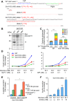
Fig 1. Establishment and characterization of Got1 KO β-cell lines.
(A) Mutations in Got1 exon 1 in two Got1 KO cell lines induced by the CRISPR/Cas9 nickase system. WT sequence is shown with target sites of sgRNAs. Protospacer adjacent motif (PAM) and mutations are shown in red. (B) Absence of AST1 protein in Got1 KO cell lines revealed by western blotting. (C) Cytosolic glutamate content in Got1 KO cell line. WT MIN6-K8 or Got1 KO (A64) cell lines were stimulated with [U-13C]-glucose and 13C-enriched glutamate isotopomers M+2 to M+5 (two to five substitutions of 12C by 13C) were quantified by mass spectrometry (n = 3). (D) Insulin secretory response in Got1 KO cell lines. The cell lines were stimulated with glucose and incretin (GLP-1 or GIP) (n = 4). Insulin secretion was normalized by cellular insulin content and presented as fold-change relative to the amount of insulin secretion at 16.7 mM glucose. (E) Rescue of the AST1 activity by introducing WT Got1 into Got1 KO cell line. The Got1 KO (A60) cell line was transfected with INS1 along with Got1 or empty construct and stimulated with glucose and GLP-1 (n = 4). C-peptide secretion was normalized by cellular C-peptide content and the data are presented as fold-change relative to the amount of C-peptide secretion at 16.7 mM glucose. (F) The effect of dimethyl glutamate (dmGlu) on insulin secretion. The Got1 KO (A60) cell line was stimulated with glucose and dmGlu (n = 4). Insulin secretion was normalized by cellular insulin content. The data are expressed as means ± SEM. Representative results are shown (C, D, E, and F). Similar results were found in 3 independent experiments. Welch’s t-test was used for evaluation of statistical significance vs. 2.8 mM glucose in (C) and vs. control in (E). Dunnett's method was used for evaluation of statistical significance vs. WT in (D) and vs. 16.7 mM glucose in (F). *p < 0.05; ***p < 0.001; n.s., not significant.
Fig 2
Fig 2. Characterization of β-cell specific Slc17a7 KO mice.
(A) Immunohistochemical analysis of the pancreas of βSlc17a7−/− mice. Green, Insulin; red, VGLUT1; blue, 4',6-diamidino-2-phenylindole (DAPI). Scale bars, 100 μm. (B) Pancreas perfusion experiment of Slc17a7flox/flox and βSlc17a7−/− mice at 17–18 weeks of age (n = 4). (C) mRNA expression levels of Slc17a7, Slc17a6, and Slc17a8 in βSlc17a7−/− mice and Slc17a7flox/flox mice (control) at 33 weeks of age (n = 3–4 for each). Welch’s t-test was used for comparisons between the two groups. **p < 0.01; n.s., not significant.
Fig 3
Fig 3. Establishment and characterization of Slc17a7 single KO β-cell lines.
(A) Mutations in Slc17a7 exon 2 in Slc17a7 single KO cell lines induced by the CRISPR/Cas9 nickase system. WT sequence is shown with target sites of sgRNAs. Protospacer adjacent motif (PAM) and mutations are shown in red. Allele 2 of both KO cell lines were not detected by PCR probably due to large deletions. (B) mRNA expression levels of Slc17a6 in Slc17a7 KO cell lines. The mRNA expression levels of KO cell lines are presented as fold increase relative to those of WT (n = 4). (C) Insulin secretory response in Slc17a7 single KO cell lines. Cells were stimulated with glucose and GLP-1 (n = 4). Insulin secretion was normalized by cellular insulin content and the data are presented as fold-change relative to the amount of insulin secretion at 16.7 mM glucose. The data are expressed as means ± SEM. Representative results are shown (B and C). Similar results were found in 3 independent experiments. Welch’s t-test with Bonferroni correction was used for evaluation of statistical significance vs. WT in (B). Dunnett's method was used for evaluation of statistical significance vs. WT in (C). **p < 0.01; ***p < 0.001.
Fig 4
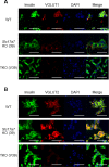
Fig 4. Immunofluorescence staining of Slc17a7 single KO and Slc17a7, Slc17a6, and Slc17a8 triple KO β-cell lines.
Green, Insulin; red, VGLUT1 (A) or VGLUT2 (B); blue, 4',6-diamidino-2-phenylindole (DAPI). Scale bars, 50 μm.
Fig 5
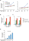
Fig 5. Characterization of Slc17a7, Slc17a6, and Slc17a8 triple KO β-cell lines.
(A) Insulin secretory response in Slc17a7, Slc17a6, and Slc17a8 triple KO (TKO) cell lines. WT and TKO cell lines were stimulated with glucose and incretin (GLP-1 or GIP) (n = 4). Insulin secretion was normalized by cellular insulin content and the data are presented as fold-change relative to the amount of insulin secretion at 16.7 mM glucose. (B, C) Rescue of the VGLUT1 (B) or VGLUT2 (C) activity by introducing WT Slc17a7 or Slc17a6 into triple KO cell line, respectively. The cell line V39 was transfected with INS1 (control) or INS1 and rescue construct and stimulated with glucose and GLP-1 (n = 4). C-peptide secretion was normalized by cellular C-peptide content and the data are presented as fold-change relative to the amount of C-peptide secretion at 16.7 mM glucose. (D) The effect of dimethyl glutamate (dmGlu) on insulin secretion. The cell line V39 was stimulated with glucose and dmGlu (n = 4). Insulin secretion was normalized by cellular insulin content. The data are expressed as means ± SEM. Representative results are shown. Similar results were found in 3 independent experiments. Dunnett's method was used for evaluation of statistical significance vs. WT in (A) and vs. 16.7 mM glucose in (D). Welch’s t-test was used for evaluation of statistical significance vs. control in (B) and (C). **p < 0.01; ***p < 0.001.

References

    1. Henquin JC. Regulation of insulin secretion: a matter of phase control and amplitude modulation. Diabetologia. 2009;52: 739–751. doi: 10.1007/s00125-009-1314-y - DOI - PubMed
    1. Yokoi N, Gheni G, Takahashi H, Seino S. β-cell glutamate signaling: Its role in incretin-induced insulin secretion. J Diabetes Investig. 2016;7: 38–43. doi: 10.1111/jdi.12468 - DOI - PMC - PubMed
    1. Drucker DJ. The biology of incretin hormones. Cell Metab. 2006;3: 153–165. doi: 10.1016/j.cmet.2006.01.004 - DOI - PubMed
    1. Holst JJ. The physiology of glucagon-like peptide 1. Physiol Rev. 2007;87: 1409–1439. doi: VSports注册入口 - 10.1152/physrev.00034.2006 - DOI - PubMed
    1. Siegel EG, Creutzfeldt W. Stimulation of insulin release in isolated rat islets by GIP in physiological concentrations and its relation to islet cyclic AMP content. Diabetologia. 1985;28: 857–861. - PubMed

MeSH terms (VSports在线直播)

"VSports手机版" LinkOut - more resources