Skip to main page content
U.S. flag

An official website of the United States government

Dot gov

The . gov means it’s official. Federal government websites often end in . gov or . mil. Before sharing sensitive information, make sure you’re on a federal government site. VSports app下载.

Https

The site is secure. The https:// ensures that you are connecting to the official website and that any information you provide is encrypted and transmitted securely. V体育官网.

Clinical Trial
. 2017 Sep;7(9):984-998.
doi: 10.1158/2159-8290.CD-17-0419. Epub 2017 Jun 6.

Secondary Somatic Mutations Restoring RAD51C and RAD51D Associated with Acquired Resistance to the PARP Inhibitor Rucaparib in High-Grade Ovarian Carcinoma

Affiliations
Clinical Trial

Secondary Somatic Mutations Restoring V体育ios版 - RAD51C and RAD51D Associated with Acquired Resistance to the PARP Inhibitor Rucaparib in High-Grade Ovarian Carcinoma

Olga Kondrashova et al. Cancer Discov. 2017 Sep.

Abstract

High-grade epithelial ovarian carcinomas containing mutated BRCA1 or BRCA2 (BRCA1/2) homologous recombination (HR) genes are sensitive to platinum-based chemotherapy and PARP inhibitors (PARPi), while restoration of HR function due to secondary mutations in BRCA1/2 has been recognized as an important resistance mechanism. We sequenced core HR pathway genes in 12 pairs of pretreatment and postprogression tumor biopsy samples collected from patients in ARIEL2 Part 1, a phase II study of the PARPi rucaparib as treatment for platinum-sensitive, relapsed ovarian carcinoma. In 6 of 12 pretreatment biopsies, a truncation mutation in BRCA1, RAD51C, or RAD51D was identified. In five of six paired postprogression biopsies, one or more secondary mutations restored the open reading frame. Four distinct secondary mutations and spatial heterogeneity were observed for RAD51CIn vitro complementation assays and a patient-derived xenograft, as well as predictive molecular modeling, confirmed that resistance to rucaparib was associated with secondary mutations. Significance: Analyses of primary and secondary mutations in RAD51C and RAD51D provide evidence for these primary mutations in conferring PARPi sensitivity and secondary mutations as a mechanism of acquired PARPi resistance. PARPi resistance due to secondary mutations underpins the need for early delivery of PARPi therapy and for combination strategies. Cancer Discov; 7(9); 984-98. ©2017 AACR. See related commentary by Domchek, p. 937See related article by Quigley et al. , p. 999See related article by Goodall et al. , p. 1006This article is highlighted in the In This Issue feature, p. 920 VSports手机版. .


Conflict of interest statement

Disclosure of Potential Conficts of Interest: A. V. Tinker reports receiving commercial research support from AstraZeneca. M. Friedlander is a consultant/advisory board member for AstraZeneca. D. M. O'Malley is a consultant/advisory board member for Clovis, AstraZeneca, Tesaro, Novocure, Genentech/Roche, Janssen, and Eisai, and is on the steering committee for Amgen. T. C. Harding has ownership interest (including patents) in Clovis Oncology. I. A. McNeish is a consultant/advisory board member for Clovis Oncology. C. L. Scott has received speakers bureau honoraria from Prime Oncology and is a consultant/advisory board member for Clovis Oncology and AstraZeneca. No potential conficts of interest were disclosed by the other authors V体育ios版.

Figures (VSports)
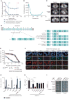
Figure 1
Figure 1
Identification and functional assessment of RAD51C secondary mutations identified in the postprogression biopsy in case 5. A, RECIST measurements of three metastatic sites, which were monitored in the patient identified to have a germline RAD51C mutation (c.577C>T). None of these lesions progressed during rucaparib treatment. After 11 months of rucaparib treatment, the patient developed a new enlarged groin lymph node, which was biopsied. B, Serum CA-125 levels monitored during the ARIEL2 Part 1 trial in the patient with the germline RAD51C mutation (c.577C>T). C, CT scans prior to (Baseline), during (Cycles 7 and 9), and following (Cycle 11) treatment of the patient with the germline RAD51C mutation (c.577C>T). D, Diagram of the predicted RAD51C protein sequence changes caused by the primary (c.577C>T) and the secondary mutations (c.577_579delinsTGG, c.577C>A, c.574_577delinsGGCG, and c.577_578delinsTT) detected in the progressing groin lymph node biopsy from the patient with the germline RAD51C mutation (c.577C>T). Examination of the parental OVCAR8, OVCAR8 RAD51C KO clone 2-130, and OVCAR8 RAD51C KO clone 2-130 transduced with wild-type (WT), primary mutant, or secondary mutant RAD51C transcripts using (E) cell viability assay after treatment with rucaparib for 6 days and (F) γH2AX and RAD51 foci formation 48 hours after rucaparib (10 μmol/L) exposure: γH2AX foci are observed at the sites of DNA damage, and RAD51 foci are observed at the sites of HR pathway repair. G, Quantification of RAD51 foci formation in geminin-positive cells (mean ± SEM). OVCAR8 RAD51C KO cells were transfected with plasmids expressing the WT, primary mutant, or secondary mutant RAD51C transcripts. The response of these cells to 10 μmol/L rucaparib was compared after 48 hours with the parental OVCAR8 cell line or OVCAR8 RAD51C KO clone. n = 8 fields of view (4 fields of view from 2 independent experiments) for each cell type and treatment. ***, P < 0.001. H, RAD51C secondary mutants restore HR as well as WT RAD51C in RAD51C-mutant cells. RAD51C−/− MCF10A cells containing the DR-GFP reporter were infected with an I-SceI expressing lentivirus and cultured for 48 hours. GFP+ cells were quantified by flow cytometry. n > 4 independent experiments. ***, P < 0.001. I, RAD51C secondary mutants restore RAD51C-R193* Y2H interactions with RAD51C binding partners RAD51B and XRCC3. RAD51C and the corresponding mutants were cloned into the Y2H plasmids expressing the GAL4 activating domain (AD), whereas RAD51B and XRCC3 were cloned into GAL4 binding domain (BD) expressing plasmids. Empty AD and BD vectors were used as negative controls. A Y2H interaction was observed as growth on medium lacking histidine, leucine, and tryptophan (interaction), whereas equal cell loading was observed on medium lacking leucine and tryptophan (control). ns, not significant; PD, progressive disease; PR, partial response.
Figure 2
Figure 2
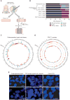
Tumor heterogeneity analysis of the postprogression biopsy with secondary RAD51C mutations. A, Model of the postprogression groin lymph node biopsy cores collected for analysis. Two postprogression core biopsies of the enlarging lymph node were obtained. Core 1 was used for genomic DNA analysis. Two ends of core 2, annotated as sections 1 and 2, were used for separate DNA extractions and subsequent analysis. The middle section of core 2 was used to generate the PDX, where 6 small pieces were subcutaneously transplanted into recipient mice. The leftover section of core 2 adjacent to section 1 was frozen in OCT and sectioned for direct PCR library preparation. B, Variant allele frequencies detected by sequencing in the pretreatment and postprogression biopsies, and in the generated PDX samples. Deep amplicon sequencing of RAD51C exon 4 (minimum coverage of 10,000×) was performed on these cores and on three recipient first passage (T1) mice. Although the first core analyzed contained all four RAD51C secondary mutations, only two of these were detected in the second core, which was used to generate the PDX. Spatial heterogeneity was even observed within the second core biopsy, with the c.577C>A mutation evident on one side of the core biopsy and at decreasing frequency toward the center of the core. The other side of the core biopsy predominantly contained the c.574_577delinsGGCG mutation, as did PDX tumors expanded in three T1 mice implanted with tissue from the same core. PDX T2 was treated with 450 mg/kg rucaparib for 2 weeks. C, Circos plot of the copy-number alterations detected by WGS in the postprogression biopsy (core 2 section 2) showing high levels of genomic instability. Losses are depicted in red, and gains in blue. D, Circos plot of copy-number alterations detected by WGS in the two analyzed PDX tumor samples obtained from the postprogression biopsy (core 2 section 2). The outer and inner copy-number tracks show the two analyzed tumor samples, and the middle track shows differences between them. E, RAD51C FISH assay of the postprogression biopsy (core 2 OCT block for serial sectioning) and the PDX sample treated with 450 mg/kg rucaparib for 2 weeks. Arrows point to the cells with three distinct signals visible for the postprogression biopsy and the PDX sample. H&E, hematoxylin and eosin.
Figure 3
Figure 3
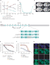
Identification and functional assessment of RAD51D secondary mutation identified in the postprogression biopsy in case 6. A, RECIST measurements of three tumor deposits in the patient with a germline RAD51D mutation (c.770_776del). The metastasis in the left lobe of the liver was biopsied prior to treatment. Surgery was performed following progression on rucaparib in order to remove the enlarging splenic lesion. The tumor deposit in the liver, which was still responding to rucaparib treatment, was also excised. B, Serum CA-125 levels monitored during the ARIEL2 Part 1 trial in the patient with the germline RAD51D mutation (c.770_776del). C, CT images obtained prior to (Baseline), during, and following (Cycle 7) treatment of the patient with the germline RAD51D mutation (c.770_776del). D, Diagram of the predicted RAD51D protein sequence changes caused by the primary mutation (c.770_776del) and the secondary mutation (c.770_776delinsA) detected in the patient with the germline RAD51D mutation. E, In vitro response to rucaparib in parental CHO cell line, CHO RAD51D KO clone, and CHO RAD51D KO clone transduced with WT, primary mutant, or secondary mutant RAD51D transcripts after treatment for 6 days. F, In vitro response to rucaparib in parental PEO4 cell line, PEO4 cells with the homozygous frameshift RAD51D mutation (c.762_763del), and PEO4 cells with the homozygous secondary RAD51D mutation (c.770_776delinsA) after treatment for 7 days. G, RAD51 foci formation 48 hours after rucaparib (10 μmol/L) exposure in geminin-positive cells in parental PEO4, PEO4 cells with the homozygous frameshift RAD51D mutation (c.762_763del), and PEO4 cells with the homozygous secondary RAD51D mutation (c.770_776delinsA). SD, stable disease.

Comment in

References

    1. Scott CL, Swisher EM, Kaufmann SH. Poly (ADP-Ribose) polymerase inhibitors: recent advances and future development. J Clin Oncol. 2015;33:1397–406. - PMC - PubMed
    1. Curtin NJ. DNA repair dysregulation from cancer driver to therapeutic target. Nat Rev Cancer. 2012;12:801–17. - PubMed (V体育官网入口)
    1. Konstantinopoulos PA, Ceccaldi R, Shapiro GI, D'Andrea AD. Homologous recombination deficiency: exploiting the fundamental vulnerability of ovarian cancer. Cancer Discov. 2015;5:1137–54. - PMC - PubMed
    1. Pommier Y, O'Connor MJ, De Bono J. Laying a trap to kill cancer cells: PARP inhibitors and their mechanisms of action. Sci Transl Med. 2016;8:362ps17. - "VSports" PubMed
    1. Lord CJ, Ashworth A. PARP inhibitors: synthetic lethality in the clinic. Science. 2017;355:1152–8. - PMC (V体育平台登录) - PubMed

"VSports手机版" Publication types