Skip to main page content
U.S. flag

An official website of the United States government

Dot gov

The VSports app下载. gov means it’s official. Federal government websites often end in . gov or . mil. Before sharing sensitive information, make sure you’re on a federal government site. .

Https

The site is secure. The https:// ensures that you are connecting to the official website and that any information you provide is encrypted and transmitted securely. V体育官网.

. 2016 Nov;2(2):10.16966/2472-6990.111.
doi: 10.16966/2472-6990.111. Epub 2016 May 30.

Temporary, Systemic Inhibition of the WNT/β-Catenin Pathway promotes Regenerative Cardiac Repair following Myocardial Infarct (VSports注册入口)

Affiliations

Temporary, Systemic Inhibition of the WNT/β-Catenin Pathway promotes Regenerative Cardiac Repair following Myocardial Infarct

Dikshya Bastakoty et al. Cell Stem Cells Regen Med. 2016 Nov.

Abstract

Aims: The WNT/β-catenin pathway is temporarily activated in the heart following myocardial infarction (MI). Despite data from genetic models indicating both positive and negative roles for the WNT pathway depending on the model used, the effect of therapeutic inhibition of WNT pathway on post-injury outcome and the cellular mediators involved are not completely understood VSports手机版. Using a newly available, small molecule, GNF-6231, which averts WNT pathway activation by blocking secretion of all WNT ligands, we sought to investigate whether therapeutic inhibition of the WNT pathway temporarily after infarct can mitigate post injury cardiac dysfunction and fibrosis and the cellular mechanisms responsible for the effects. .

Methods and results: Pharmacologic inhibition of the WNT pathway by post-MI intravenous injection of GNF-6231 in C57Bl/6 mice significantly reduced the decline in cardiac function (Fractional Shortening at day 30: 38. 71 ± 4. 13% in GNF-6231 treated vs. 34. 89 ± 4. 86% in vehicle-treated), prevented adverse cardiac remodeling, and reduced infarct size (9. 07 ± 3 V体育安卓版. 98% vs. 17. 18 ± 4. 97%). WNT inhibition augmented proliferation of interstitial cells, particularly in the distal myocardium, inhibited apoptosis of cardiomyocytes, and reduced myofibroblast proliferation in the peri-infarct region. In vitro studies showed that WNT inhibition increased proliferation of Sca1+ cardiac progenitors, improved survival of cardiomyocytes, and inhibited collagen I synthesis by cardiac myofibroblasts. .

Conclusion: Systemic, temporary pharmacologic inhibition of the WNT pathway using an orally bioavailable drug immediately following MI resulted in improved function, reduced adverse remodeling and reduced infarct size in mice. Therapeutic WNT inhibition affected multiple aspects of infarct repair: it promoted proliferation of cardiac progenitors and other interstitial cells, inhibited myofibroblast proliferation, improved cardiomyocyte survival, and reduced collagen I gene expression by myofibroblasts. Our data point to a promising role for WNT inhibitory therapeutics as a new class of drugs to drive post-MI repair and prevent heart failure V体育ios版. .

Keywords: Myocardial infarct; Regenerative cardiac repair; WNT/β-Catenin Pathway VSports最新版本. .


Conflict of interest statement

Conflicts of Interests JL and JLH are employees of the Novartis Research Foundation. PPY is listed as inventor for a WNT inhibitory topical therapeutic V体育平台登录.

Figures (VSports app下载)

Figure 1
Figure 1. GNF-6231 inhibits canonical WNT pathway activity in vitro
(A) Schematic of the WNT pathway and point of action of WNT inhibitors, GNF-6231 and C-113. (B) Fold change in Axin2 gene expression in WNT3a overexpressing cardiac cells showing GNF-6231 treatment reduced WNT target gene expression (N=3 replicates from independent experiments; ***p ≤ 0.0001; Repeated measures ANOVA with Bonferroni correction for multiple comparisons). (C) IV free plasma level of GNF-6231 after a single intravenous injection of 5 mg/kg. The plasma half-life of the drug was approximately 2.3 hours; GNF-6231 free plasma concentrations were above the in vitro Porcupine IC50 for >12 h. (D) qRT-PCR showed inhibition of Axin2 gene expression in liver at different time points following a single 5 mg/kg intravenous treatment with GNF-6231 (N=2 mice per timepoint). Bars represent Mean ± SD.
Figure 2
Figure 2. Porcupine inhibitor treatment inhibits WNT pathway activity in the infarcted heart
(A) Schematic summarizing animal study timelines. Mice were treated with daily intravenous injection of 5 mg/kg drug or vehicle following MI and continued through day 6. For cardiac recovery studies, mice underwent echocardiography at days 7 and 30. For histology, a separate cohort of mice was sacrificed on days 3, 7 and 15. (B) β-catenin immunostaining of peri- infarct region of ventricles at day 7 showed reduction in β-catenin levels with GNF-6231 treatment. (B′) High magnification image of vehicle-treated tissue showed nuclear localization of β-catenin signifying WNT pathway activation. (C) β-galactosidase immunostaining in ventricle sections from WNT reporter, TOPGAL mice demonstrated inhibition in WNT activity at day 7 post-infarct with GNF-6231 treatment. Scale bars equal 50 μm. Images are representative of sections from N ≥ 3 mice; at least 4 areas were imaged from each mouse.
Figure 3
Figure 3. Porcupine inhibition improves cardiac function and reduces adverse remodeling after MI
Left ventricular remodeling was measured as % change in (A) LVIDd and (B) LVIDs. LV function was measured as % change in (C) FS and (D) EF. Data showed no increase in left ventricular diameter (A and B), and improved cardiac function (C and D) with GNF-6231 treatment compared to vehicle. (E) Masson’s trichrome stained representative sections of the left ventricle at day 30 depicted more collagen stained (blue) area in vehicle-treated LV compared to GNF-6231-treated. (F) Quantification of infarct size. Each data point on graphs represents individual mouse; *p ≤ 0.05, **p ≤ 0.01 or ***p ≤ 0.005; unpaired t-test.
Figure 4
Figure 4. WNT inhibition promotes proliferation of interstitial αSMA negative cells in the infarcted heart
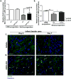
(A) H&E stained cross-section of the heart demarcating peri-infarct and distal regions of the left ventricle as defined in the study. Representative pHisH3 stained sections of the ventricles at (B) day 3 and (C) day 7 showing more proliferative cells in the distal myocardium of GNF-6231 treated hearts. (D) Quantification of percent pHisH3+ cells. Bars represent mean ± SD; N ≥ 4 images of sections from N ≥ 3 mice per group were imaged; *p ≤ 0.05, ***p ≤ 0.005; One-Way ANOVA with Bonferroni Correction for multiple comparisons. Representative sections of the distal myocardium at day 3 post-MI (E) co-stained with αSarcomeric Actin and pHisH3, and (F) high magnification confocal microscopy image of ventricle co-stained with cTnI and Ki67, demonstrating that the majority of proliferative cells in the GNF-6231-treated tissue localized to the interstitium of myofibers. (G) αSarcomeric Actin/pHisH3 co-stained LV depicting the rare proliferating cardiomyocytes (white arrows). (H) Proliferating myofibroblasts were identified by αSMA/Ki67 co-staining as depicted in the representative section from the peri-infarct region at day 7. (I) Quantification of αSMA/Ki67 co-stained cells revealed that the percentage of proliferating myofibroblasts (grey shaded portion of the bars) was significantly lower in GNF-6231-treated peri-infarct tissue than control at day 3 (**p=0.0013) and lower (#p=0.0587) at day 7. In contrast, the percentage of proliferating non-myofibroblasts (αSMA cells; lower white portion of the graphs) was significantly higher (*p=0.0135) in GNF-6231 treated ventricles compared to control at day 7. Bars represent mean ± SD. N ≥ 12: at least 3 separate sections from at least 3 mice per group were imaged. P-values for individual comparisons between each two groups of data were calculated using Mann-Whitney test. Scale bars equal 50 μm.
Figure 5
Figure 5. WNT inhibition increases proliferation of progenitor cells that may contribute to myogenesis
(A and B) Relative BrdU incorporation by Sca1+ progenitor cells stably expressing LZRS (empty vector) or Wnt3a-LZRS revealed that proliferation was reduced by Wnt3a overexpression and this effect was reversed by (A) GNF-6231 treatment and (B) C-113 treatment. Data are presented as Mean ± SD. (A) N=5 and (B) N=3 replicates from independent experiments; *p ≤ 0.05 and **p ≤ 0.01; Kruskal-Wallis test with Dunns correction for multiple comparisons. (C and D) Representative GATA4 immunostained sections of infarct border zone at day 3 (left panels) and day 7 (right panels) post-MI of (C) vehicle-treated hearts and (D) GNF-6231 treated hearts. White arrows point to GATA4 stained nuclei. Scale bars equal 50 μm; the images are representative of at least 4 sections each from N ≥ 3 mice per group.
Figure 6
Figure 6. WNT inhibition reduces cardiomyocyte cell death
(A and B) Representative sections co-stained with cTnI and TUNEL of infarct border zone of (A) vehicle-treated hearts, and (B) GNF-6231 treated hearts. (C) Quantification of percent TUNEL positive cardiomyocytes revealed significantly fewer apoptotic cardiomyocytes in GNF-6231 treated hearts. N=5 mice per group, at least 4 sections imaged per mouse, *p=0.022, unpaired t-test. (D) Percent cell survival of HL-1 rat cardiomyocytes as measured by metabolic uptake (MTT) assay showed significant reduction in survival with recombinant WNT3A treatment, which was rescued by addition of C-113. Bars represent mean ± SD; N=3 replicates from independent experiments; *P ≤ 0.05; Kruskal- Wallis test with Dunns correction for multiple comparisons. (E) Representative images of human iPSC-derived iCell cardiomyocytes treated with recombinant WNT3A or WNT3A and C-113 in presence of 250 μM H2O2 showing that under stress, treatment with recombinant WNT3A increased cell death, which was rescued by WNT inhibition with C-113. (F) Quantification of %TUNEL positive iCell cardiomyocytes per 20× field. N ≥ 12 per group, at least 4 areas imaged in each replicate from 3 independently run experiments; **p ≤ 0.01 and ***p ≤ 0.001; One-Way ANOVA with Bonferroni correction for multiple comparisons. Scale bars in A-B and E represent 50 μm.
Figure 7
Figure 7. WNT inhibition reduces collagen synthesis activity in vitro
(A) Primary myofibroblasts in culture confirmed by αSMA (green) and Periostin (red) staining; scale bar equals 50 μm. (B) Relative Col1α1 gene expression in primary cardiac myofibroblasts with and without WNT inhibitor treatement revealed significant reduction in Col1α1 gene expression in response to WNT inhibitor treatment. Bars represent mean ± SD. N=4 replicates from independent experiments; *p=0.0265; Mann-Whitney test.

References

    1. Mozaffarian D, Benjamin EJ, Go AS, Arnett DK, Blaha MJ, et al. Heart disease and stroke statistics–2015 update: a report from the American Heart Association. Circulation. 2015;131:e29–e322. - PubMed
    1. Mill JG, Stefanon I, dos Santos L, Baldo MP. Remodeling in the ischemic heart: the stepwise progression for heart failure. Braz J Med Biol Res. 2011;44:890–898. - PubMed (V体育ios版)
    1. Sutton MG, Sharpe N. Left ventricular remodeling after myocardial infarction: pathophysiology and therapy. Circ. 2000;101:2981–2988. - PubMed
    1. Oerlemans MI, Goumans MJ, van Middelaar B, Clevers H, Doevendans PA, et al. Active WNT signaling in response to cardiac injury. Basic Res Cardiol. 2010;105:631–641. - PMC - PubMed
    1. Aisagbonhi O, Rai M, Ryzhov S, Atria N, Feoktistov I, et al. Experimental myocardial infarction triggers canonical WNT signaling and endothelial-to-mesenchymal transition. Dis Model Mech. 2011;4:469–483. - PMC - PubMed

LinkOut - more resources