Skip to main page content
U.S. flag

An official website of the United States government

Dot gov

The . gov means it’s official. Federal government websites often end in VSports app下载. gov or . mil. Before sharing sensitive information, make sure you’re on a federal government site. .

Https

The site is secure. The https:// ensures that you are connecting to the official website and that any information you provide is encrypted and transmitted securely V体育官网. .

. 2017 Apr 20;45(7):4021-4035.
doi: 10.1093/nar/gkw1201.

Identification of senescence-associated circular RNAs (SAC-RNAs) reveals senescence suppressor CircPVT1

Affiliations

Identification of senescence-associated circular RNAs (SAC-RNAs) reveals senescence suppressor CircPVT1

Amaresh C Panda et al. Nucleic Acids Res. .

Abstract (V体育安卓版)

Using RNA sequencing (RNA-Seq), we compared the expression patterns of circular RNAs in proliferating (early-passage) and senescent (late-passage) human diploid WI-38 fibroblasts. Among the differentially expressed senescence-associated circRNAs (which we termed 'SAC-RNAs'), we identified CircPVT1, generated by circularization of an exon of the PVT1 gene, as a circular RNA showing markedly reduced levels in senescent fibroblasts. Reducing CircPVT1 levels in proliferating fibroblasts triggered senescence, as determined by a rise in senescence-associated β-galactosidase activity, higher abundance of CDKN1A/P21 and TP53, and reduced cell proliferation. Although several microRNAs were predicted to bind CircPVT1, only let-7 was found enriched after pulldown of endogenous CircPVT1, suggesting that CircPVT1 might selectively modulate let-7 activity and hence expression of let-7-regulated mRNAs VSports手机版. Reporter analysis revealed that CircPVT1 decreased the cellular pool of available let-7, and antagonizing endogenous let-7 triggered cell proliferation. Importantly, silencing CircPVT1 promoted cell senescence and reversed the proliferative phenotype observed after let-7 function was impaired. Consequently, the levels of several proliferative proteins that prevent senescence, such as IGF2BP1, KRAS and HMGA2, encoded by let-7 target mRNAs, were reduced by silencing CircPVT1. Our findings indicate that the SAC-RNA CircPVT1, elevated in dividing cells and reduced in senescent cells, sequesters let-7 to enable a proliferative phenotype. .


Figures (VSports注册入口)

Figure 1.
Figure 1.
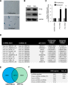
Identification and annotation of senescence-associated circRNAs in WI-38 fibroblasts. (A) Micrographs to visualize SA-β-gal activity in proliferating, early-passage, and senescent, late-passage WI-38 cells. (B) Western blot analysis of the levels of P53, P21, and loading control HSP90 in proliferating and senescent cells. (C) RT-qPCR analysis of the levels of P16 mRNA, P21 mRNA, and loading control GAPDH mRNA in proliferating and senescent cells. (D) Table of highly expressed circRNAs in proliferating WI-38 cells. (E) Venn diagram depicting the overlap between the two different circRNA prediction algorithms. (F) Number of circRNAs identified by the CIRCexplorer and find_circ algorithms. Data in (C) represent the means ± S.E.M. from three independent experiments. **P <0.01 (Student's t-test).
Figure 2.
Figure 2.
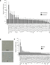
Characterization of senescence-associated circRNAs in WI-38 fibroblasts. (A) RT-qPCR analysis of changes in circRNA expression in proliferating and senescent WI-38 cells. (B) Representative images of SA-βgal staining in mock-treated and IR-treated (senescent) WI-38 cells. (C) RT-qPCR analysis of circRNA levels in mock-treated and IR-treated (senescent) WI-38 cells. Data in A and C are the means ± S.E.M. from three independent experiments.
Figure 3.
Figure 3.
CircPVT1 inhibits WI-38 cell senescence. (A) RT-qPCR results showing the abundance of circRNAs and linear RNAs in WI-38 cells treated with RNase R. The levels of CircPVT1 and PVT1 lncRNA were normalized to the values measured after mock treatments. (B) The RT-qPCR product of CircPVT1 (± RT, with or without reverse transcription) was visualized by electrophoresis in ethidium bromide-stained 2.5% agarose gels. (C) qPCR products were purified and sequenced to confirm CircPVT1 junction sequences. (D) Absolute quantification for CircPVT1 and PVT1 lncRNA in proliferating WI-38 cells. (E) RT-qPCR analysis of the levels of CircPVT1 and PVT1 lncRNA in proliferating WI-38 cells 4 days after transfection of Ctrl siRNA or CircPVT1 siRNA. (F, G) Western blot analysis of the senescence marker TP53 (F) and SA-βgal staining (G) in WI-38 cells 4 days after transfection with Ctrl siRNA or CircPVT1 siRNA. Data in A, D–F are the means ± S.E.M. from three independent experiments. *P < 0.05 (Student's t-test).
Figure 4.
Figure 4.
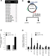
let-7 associates with CircPVT1 in WI-38 cells. (A) List of microRNAs predicted to target CircPVT1. (B) Schematic of CircPVT1 pulldown for specific detection of microRNAs associated with CircPVT1 in WI-38 cells. (C) RT-qPCR analysis of the enrichment of CircPVT1 in CircPVT1 pulldown compared with control. (D) Enrichment of microRNAs predicted to target CircPVT1 in CircPVT1 pulldown analyzed by RT-qPCR. Data in (C, D) are the means ± S.E.M. from at least three independent experiments.
Figure 5.
Figure 5.
CircPVT1 regulates cellular senescence by inhibiting let-7 function. (A) Proliferating WI-38 cells were transfected with CircPVT1 siRNA or Ctrl siRNA, in the presence or absence of anti-let-7. The graph shows RT-qPCR measurements of the senescence marker CDKN1A (P21) mRNA in WI-38 cells 4 days after transfection with the two siRNAs above. N.S., not significant (P = 0.06); *P < 0.05; **P < 0.01 (Student's t-test). (B) Western blot analysis of senescence marker P21, TP53, and loading control GAPDH in WI-38 cells 4 days after transfection with two siRNAs indicated. (C) SA-βgal staining in WI-38 cells 4 days after transfection with the siRNAs shown. (D) RT-qPCR analysis of let-7 levels in proliferating and senescent WI-38 cells. (E, F) Four days after transfection of WI-38 cells with the siRNAs indicated, cell numbers were counted (E) and measurements were taken for [3H]-thymidine incorporation (F). Data in A, D–F are the means ± S.E.M. from three or four independent experiments. N.S., not significant; *P < 0.05; **P < 0.01 (Student's t-test).
Figure 6.
Figure 6.
CircPVT1 promotes translation of let-7 targets encoding proliferative proteins. (A) Top, schematic of the dual luciferase reporter plasmids derived from the parent vector psiCHECK2 (psi), which expresses renilla luciferase (RL) and the internal control firefly luciferase (FL), and psiCHECK2-derived plasmids bearing the target sequence of let-7. Bottom, 48 h after transfection of HeLa cells with CircPVT1 siRNA, anti-let-7 or let-7 mimic, reporter plasmid was transfected, and 16 h later the ratio of RL activity to FL activity was measured. The changes in RL/FL ratios after the various small RNA transfections relative to RL/FL ratios of Ctrl siRNA-transfected cells are indicated. (B) RT-qPCR analysis of the levels of CircPVT1 in proliferating WI-38 cells 4 days after transfection with Ctrl siRNA or CircPVT1 siRNA. (C) Western blot analysis of the let-7 targets HMGA2, IGF2BP1, and KRAS, as well as loading control GAPDH in WI-38 cells 4 days after transfection with Ctrl siRNA or CircPVT1 siRNA. (D) RT-qPCR analysis of the levels of CircPVT1 and lncRNA PVT1 in three sets of cancerous and noncancerous lines from lung [BEAS-2B versus A549 (left) and IMR-90 versus H1299 (center)] and breast [MCF10A vs MCF7 (right)]. ACTB mRNA was used as a normalization control. Data in A–D are the means ± S.E.M. from three independent experiments. *P < 0.05; **P < 0.01 (Student's t-test).
Figure 7.
Figure 7.
CircPVT1 action model. Proposed model whereby CircPVT1 acts as a competing endogenous RNA, sponging let-7; CircPVT1 promotes the expression of target genes required for cell cycle progression by preventing let-7 from acting on such target mRNAs. The decreased expression levels of CircPVT1 in senescent cells allows higher levels of functional let-7, which in turn suppresses let-7 target expression leading to growth inhibition and cellular senescence.

References

    1. Campisi J. Aging, cellular senescence, and cancer. Annu. Rev. Physiol. 2013; 75:685–705. - PMC - PubMed
    1. Hayflick L. The Limited in Vitro Lifetime of Human Diploid Cell Strains. Exp. Cell Res. 1965; 37:614–636. - PubMed
    1. Toussaint O., Remacle J., Dierick J.F., Pascal T., Frippiat C., Royer V., Magalhacs J.P., Zdanov S., Chainiaux F.. Stress-induced premature senescence: from biomarkers to likeliness of in vivo occurrence. Biogerontology. 2002; 3:13–17. - PubMed
    1. Busuttil R.A., Dolle M., Campisi J., Vijg J.. Genomic instability, aging, and cellular senescence. Ann. N. Y. Acad. Sci. 2004; 1019:245–255. - "V体育官网" PubMed
    1. Marcotte R., Wang E.. Replicative senescence revisited. J. Gerontol. A Biol. Sci. Med. Sci. 2002; 57:B257–B269. - PubMed

Substances