Skip to main page content
U.S. flag

An official website of the United States government

Dot gov

The . gov means it’s official. Federal government websites often end in . gov or . mil. Before sharing sensitive information, make sure you’re on a federal government site VSports app下载. .

Https

The site is secure. The https:// ensures that you are connecting to the official website and that any information you provide is encrypted and transmitted securely. V体育官网.

. 2016 Nov;143(2):379-388.
doi: 10.1016/j.ygyno.2016.08.328. Epub 2016 Sep 8.

In vivo anti-tumor activity of the PARP inhibitor niraparib in homologous recombination deficient and proficient ovarian carcinoma

Affiliations

"V体育ios版" In vivo anti-tumor activity of the PARP inhibitor niraparib in homologous recombination deficient and proficient ovarian carcinoma

Mariam M AlHilli et al. Gynecol Oncol. 2016 Nov.

Abstract (VSports手机版)

Objective: Poly(ADP-ribose) polymerase (PARP) inhibitors have yielded encouraging responses in high-grade serous ovarian carcinomas (HGSOCs), but the optimal treatment setting remains unknown. We assessed the effect of niraparib on HGSOC patient-derived xenograft (PDX) models as well as the relationship between certain markers of homologous recombination (HR) status, including BRCA1/2 mutations and formation of RAD51 foci after DNA damage, and response of these PDXs to niraparib in vivo. VSports手机版.

Methods: Massively parallel sequencing was performed on HGSOCs to identify mutations contributing to HR deficiency. HR pathway integrity was assessed using fluorescence microscopy-based RAD51 focus formation assays. Effects of niraparib (MK-4827) on treatment-naïve PDX tumor growth as monotherapy, in combination with carboplatin/paclitaxel, and as maintenance therapy were assessed by transabdominal ultrasound. Niraparib responses were correlated with changes in levels of poly(ADP-ribose), PARP1, and repair proteins by western blotting. V体育安卓版.

Results: Five PDX models were evaluated in vivo. Tumor regressions were induced by single-agent niraparib in one of two PDX models with deleterious BRCA2 mutations and in a PDX with RAD51C promoter methylation. Diminished formation of RAD51 foci failed to predict response, but Artemis loss was associated with resistance V体育ios版. Niraparib generally failed to enhance responses to carboplatin/paclitaxel chemotherapy, but maintenance niraparib therapy delayed progression in a BRCA2-deficient PDX. .

Conclusions: Mutations in HR genes are neither necessary nor sufficient to predict response to niraparib. Assessment of repair status through multiple complementary assays is needed to guide PARP inhibitor therapy, design future clinical trials and identify ovarian cancer patients most likely to benefit from PARP inhibition. VSports最新版本.

Keywords: BRCA; DNA repair; Homologous recombination; Niraparib; Ovarian cancer; PARP inhibitors; Xenografts. V体育平台登录.


Conflict of interest statement

Authors' disclosure of potential conflicts of interest: PH was an unpaid consultant for Merck and received research funding from Merck and Tesaro.

Conflict of interest statement

Dr. Becker reports personal fees from Ovarian Cancer Tumorgrafts - Avatar System, outside the submitted work; Dr. Haluska reports non-financial support from Tesaro, during the conduct of the study through the supply of niraparib; and intellectual property (non-patent) involving the use of the PDX models employed in this work, which includes the awarding of royalties. Dr. Kaufmann reports grants from National Cancer Institute and Ovarian Cancer Research Fund during the conduct of the study. In addition, Mayo Clinic has a patent related to methods for assessing responsiveness to PARP inhibitors on which Dr VSports在线直播. Kaufmann is a co-inventor. Dr. Weroha reports grants from Tesaro, during the conduct of the study.

"V体育ios版" Figures
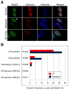
Fig. 1
Fig. 1. RAD51 focus assay on PDX tumor cells
A. Immunofluorescence microscopy of niraparib-treated PDX cells (PH039) and irradiated cells showing RAD51 foci (arrow) within geminin positive cells and lack of RAD51 foci formation in control cells. B. Percentage of geminin positive cells that were RAD51 foci positive after treatment with ionizing irradiation (IR) or niraparib. Similar results were observed using multiple independent tumorgrafts from each model, including 5 separate PH095 xenografts.
Fig. 2
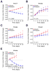
Fig. 2. Response of PDXs to single-agent niraparib
Change in mean tumor area (solid lines) in niraparib treated (blue line) and control (red line) arms. Mean tumor area has been normalized to day 1 mean area as described in the statistical methods. p-Values for differences in growth trajectories in each PDX model are presented. Estimated (predicted) mean tumor areas together with standard error bars from the regression model are included for visual demonstration that the model fits the data well (dashed lines). “P” indicates test for different growth rates (slope), while “Mean P” indicates test for different average area (centered intercept).
Fig. 3
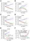
Fig. 3. Response of PDXs to paclitaxel/carboplatin with and without niraparib
AE. Tumor area of carboplatin/paclitaxel-treated models relative to control. Although tumors regressed below baseline for PDXs PH077, PH080, PH087 and PH095, the addition of niraparib (CP + N, red line) did not result in statistically significant differences from carboplatin/paclitaxel without niraparib (CP, green line). Solid lines indicate change in mean tumor area normalized to day 1 mean area as described in the statistical methods, and dashed lines indicate estimated or predicted mean tumor areas together with standard error bars from the regression model. p-Values shown on graphs are for differences in trajectory between PDXs treated with carboplatin/paclitaxel and carboplatin/paclitaxel/niraparib. p-Values for differences in mean area over time also were >0.05. In PH039, the growth trajectories were not statistically significantly different (p = 0.40), but the average change from baseline over time was significantly larger with addition of niraparib (p = 0.003). F. Effect of maintenance niraparib therapy in PH077. Initial treatments (weeks 0 to 3) are denoted as “primary” and consisted of carboplatin/paclitaxel (CP, gray line) or carboplatin/paclitaxel/niraparib (CP + N, black line). Niraparib “maintenance” was initiated following primary carboplatin/paclitaxel/niraparib (Niraparib, green line) and diluent controls included for both carboplatin/paclitaxel CP (Diluent, red line) and CP + N (Diluent, blue line).
Fig. 4
Fig. 4. PARP is inhibited in all PDX models
A. Three in-vivo tumor replicates of treated models and controls were harvested at the end of treatment (day 28), pooled, and subjected immunoblotting of the indicated antigens. Formation of pADPr is inhibited in niraparib treated tumors. Absence of PARP1 protein is seen in model PH039 treated with niraparib. Arrow indicates expected location of 89 kDa caspase-generated PARP1 fragment if it were present. B. Lysates containing 50 μg of protein from one or two separate aliquots of the indicated PDX were subjected to SDS-PAGE and blotted with antibodies to the indicated antigen. PE01 and PE04 cells, which are known to express all of these proteins, were included as positive controls. HSP90β served as a loading control. * indicates nonspecific band. C. Parental PEO1 cells (open circles) or Artemis−/− PEO1 cells derived as described in the Materials and methods (closed circles) were plated (750 cells/plate) and treated beginning 18 h later with diluent (0.1% DMSO) or the indicated concentration of niraparib. Error bars, ±SEM from triplicate aliquots.
Fig. 5
Fig. 5. Gene expression and RAD51C methylation analysis in PDXs
A. Using bisulfite sequencing and methylation specific PCR, methylated (M) and unmethylated (UM) RAD51C and BRCA1 in PDX models is shown with a control (ctrl) for M and UM. Water (H2O) was used as a blank control. B. Relative RAD51C and BRCA1 mRNA levels across the 5 PDX models shows lower expression of RAD51C in niraparib-responsive PDX PH039. C. Immunoblot for RAD51C in xenograft samples.

References

    1. Integrated genomic analyses of ovarian carcinoma. Nature. 2011;474:609–615. - PMC - PubMed
    1. Abkevich V, Timms KM, Hennessy BT, Potter J, Carey MS, Meyer LA, et al. Patterns of genomic loss of heterozygosity predict homologous recombination repair defects in epithelial ovarian cancer. Br J Cancer. 2012;107:1776–1782. - "VSports" PMC - PubMed
    1. Pennington KP, Walsh T, Harrell MI, Lee MK, Pennil CC, Rendi MH, et al. Germline and somatic mutations in homologous recombination genes predict platinum response and survival in ovarian, fallopian tube, and peritoneal carcinomas. Clin Cancer Res. 2014;20:764–775. - PMC - PubMed
    1. Walsh T, Casadei S, Lee MK, Pennil CC, Nord AS, Thornton AM, et al. Mutations in 12 genes for inherited ovarian, fallopian tube, and peritoneal carcinoma identified by massively parallel sequencing. Proc Natl Acad Sci U S A. 2011;108:18032–18037. - PMC - PubMed
    1. Wickramanyake A, Bernier G, Pennil C, Casadei S, Agnew KJ, Stray SM, et al. Loss of function germline mutations in RAD51D in women with ovarian carcinoma. Gynecol Oncol. 2012;127:552–555. - "VSports" PMC - PubMed