Skip to main page content
U.S. flag

An official website of the United States government

Dot gov

The . gov means it’s official. Federal government websites often end in . gov or VSports app下载. mil. Before sharing sensitive information, make sure you’re on a federal government site. .

Https

The site is secure. The https:// ensures that you are connecting to the official website and that any information you provide is encrypted and transmitted securely. V体育官网.

. 2015 Dec 1:5:17605.
doi: 10.1038/srep17605.

Inhibition of Osteoclastogenesis and Bone Resorption in vitro and in vivo by a prenylflavonoid xanthohumol from hops

Affiliations

V体育官网入口 - Inhibition of Osteoclastogenesis and Bone Resorption in vitro and in vivo by a prenylflavonoid xanthohumol from hops

Jing Li et al. Sci Rep. .

Abstract

Excessive RANKL signaling leads to superfluous osteoclast formation and bone resorption, is widespread in the pathologic bone loss and destruction. Therefore, targeting RANKL or its signaling pathway has been a promising and successful strategy for this osteoclast-related diseases. In this study, we examined the effects of xanthohumol (XN), an abundant prenylflavonoid from hops plant, on osteoclastogenesis, osteoclast resorption, and RANKL-induced signaling pathway using both in vitro and in vivo assay systems. In mouse and human, XN inhibited osteoclast differentiation and osteoclast formation at the early stage. Furthermore, XN inhibited osteoclast actin-ring formation and bone resorption in a dose-dependent manner. In ovariectomized-induced bone loss mouse model and RANKL-injection-induced bone resorption model, we found that administration of XN markedly inhibited bone loss and resorption by suppressing osteoclast activity. At the molecular level, XN disrupted the association of RANK and TRAF6, resulted in the inhibition of NF-κB and Ca(2+)/NFATc1 signaling pathway during osteoclastogenesis. As a results, XN suppressed the expression of osteoclastogenesis-related marker genes, including CtsK, Nfatc1, Trap, Ctr VSports手机版. Therefore, our data demonstrated that XN inhibits osteoclastogenesis and bone resorption through RANK/TRAF6 signaling pathways. XN could be a promising drug candidate in the treatment of osteoclast-related diseases such as postmenopausal osteoporosis. .


Conflict of interest statement

The authors declare no competing financial interests.

Figures
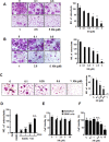
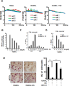
Figure 1
Figure 1. XN suppresses human and mouse osteoclast differentiation.
(A) The effect of xanthohumol (XN) on mouse BMMs differentiation. BMMs were incubated with M-CSF (20 ng/ml) and RANKL (30 ng/ml), followed by addition of different concentrations of XN for 6 days. The cells were stained for TRAP assay and photographed (×40; left). The numbers of TRAP positive multinucleated (>5 nuclei) osteoclasts were counted (right). (B) The effect of XN on RAW264.7 cell differentiation. RAW264.7 cells were treated with RANKL (30 ng/ml) and different doses of XN for 3 days. The cells were stained for TRAP assay and photographed (×40; left) and the numbers of TRAP positive multinucleated (>3 nuclei) osteoclasts were counted (right). (C) The effect of XN on human PBMC differentiation. PBMCs (5 × 105 cells) were stimulated with hRANKL (50 ng/mL) and hCSF1 (20 ng/mL) for 8–9 days. The cells were stained for TRAP assay and photographed (×40; left) and the numbers of TRAP positive multinucleated (>3 nuclei) osteoclasts were counted (right). (D) XN inhibits RANKL-induced osteoclast differentiation at the early stage. Osteoclast precursor BMMs were cultured with M-CSF and RANKL for differentiation into mature osteoclasts in 6 days. XN (5 μM) were added at indicated time (day). The cells were fixed and stained for TRAP activity. (E) The effect of XN on cell viability in BMMs and RAW264.7 cells. BMMs or RAW264.7 cells were treated with different concentrations of XN for 5 days, and the cell viability was measured by SRB assay. (F) The effect of XN on cell viability in human PBMC cells. Human PBMC cells were incubated with hCSF1 (20 ng/mL) and different concentrations of XN for 4 days. The cell viability was measured by SRB assay. Column, means of experiments performed in triplicate; bar, SD. *p < 0.05, **p < 0.01, ***p < 0.001. N.S., no significant.
Figure 2
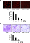
Figure 2. XN inhibits RANKL-induced actin-ring formation and bone resorption.
(A) The effect of XN on actin-ring formation of osteoclast. Mouse BMMs were incubated with RANKL (30 ng/ml) in the presence of M-CSF (20 ng/ml), followed by treatment with indicated doses of XN. Cells were fixed and stained for F-actin (top). Osteoclasts with actin-rings were counted (bottom). (B) The effect of XN on pits formation of osteoclast. Mouse BMMs were cultured with M-CSF (20 ng/ml) and RANKL (30 ng/ml) for 6 days with or without indicated doses of XN. The cells were washed and the resorption pits were stained with Mayer’s hematoxylin and photographed (top, original magnification, ×40). The numbers of pits were analyzed with Image-Pro software (bottom). Column, means of experiments performed in triplicate; bar, SD.
Figure 3
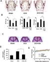
Figure 3. XN inhibits ovariectomy-induced bone loss by inhibiting osteoclast activity in vivo.
Four weeks after ovariectomy or sham-operation, mice were divided into three groups: sham-operated mice (sham), ovariectomized mice treated with vehicle (OVX) and OVX mice treated with XN (OVX + XN) (10 mg/kg, n = 6) for another five weeks. The treated mice were intraperitoneally (i.p.) injected with XN every days. Then, all the mice were euthanized for bone histomorphometry. (A,B) Histomorphometric analysis of lumbar vertebrae from sham, OVX, OVX + XN mice. Bone value/total value (BV/TV), trabecular space (Tb.Sp), and trabecular number (Tb.N) were analyzed as described in Materials and Methods. n = 6. (C,D) TRAP staining of the whole calvaria. The osteoclast area were analyzyed by OsteoMeasure Analysis system as described in Materials and Methods (D). (E) Effect of XN on mouse body weight at the concentrations tested. *p < 0.05, **p < 0.01, ***p < 0.001.
Figure 4
Figure 4. XN prevents RANKL-injection-induced osteoclast activity.
RANKL/vehicle and RANKL/XN were injected into the calvaria of 6-week-old male mice every day, respectively (n = 6). After 15 days, the mice were sacrificed and calvaria were stained by TRAP staining and sectioned. (A) Representative TRAP stained whole calvaria (top) and calvarial section (bottom) from normal mice, RANKL/vehicle injected mice (RANKL) and RANKL/XN injected mice (RANKL + XN). (B) Eroded surface/bone surface (ES/BS); osteoclast surface/bone surface (Oc.S/BS); osteoclast number/bone perimeter (N.Oc/B.Pm) were analyzed by OsteoMeasure Analysis system as described in Materials and Methods.
Figure 5
Figure 5. XN blocks RANKL-induced Ca2+/NFATc1 signaling pathway.
(A) The effect of XN on RANKL-induced Ca2+ oscillation. The M-CSF incubated BMMs were pretreated with or without XN (5 uM) for 24 hours, and then incubated with or without RANKL (30 ng/ml) for another 60 hours. The Ca2+ oscillation was analyzed as described in Materials and Methods. (B) The effect of XN on RANKL-induced activity of NFAT. RAW264.7 cells were cotransfected with NFAT-luciferase reporter gene and Renilla gene. After 36 hours, the cells were treated with RANKL and indicated concentrations of XN for another 24 hours. Cell extracts were collected and luciferase activity was measured as described in Materials and Methods. Results are expressed as fold activity over the activity of the control. (C,D) The effect of XN on NFATc1 binding to Cathepsin K (Ctsk) promoter region by Chromatin immunoprecipitation (ChIP) assays. BMMs incubated with RANKL (30 ng/ml) were treated with or without XN for indicated time (C). Or BMMs incubated with RANKL (30 ng/ml) were treated with indicated concentration of XN for 48 hours (D). The Chromatin DNA were immunoprecipitated with control IgG or the anti-NFATc1 antibody and subjected to quantitative real-time PCR with primers specific for NFATc1-binding sites. (E) NFATc1 prevents the inhibitory effect of XN in RANKL-induced osteoclast differentiation. RAW264.7 cells were transfected with NFATc1 or vector control plasmids, and then incubated with or without XN (5 μM) in the presence of RANKL (30 ng/ml). After 4 days, the cells were fixed and stained for TRAP activity (left). Original magnification, ×40. The numbers of TRAP positive multinucleated (>3 nuclei) osteoclasts were counted (right). Column, means of three experiments conducted in triplicate; bar, SD. ***P < 0.001.
Figure 6
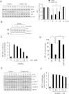
Figure 6. XN suppresses RANKL-induced NF-κB signaling pathway, but has little effect on MAPK/AP-1 signaling.
(A) The effect of XN on RANKL-induced IκBα degradation and p65 phorsphorylation. RAW264.7 cells were pretreated with XN (5 μM) for 3 hours, and then stimulated with RANKL (30 ng/ml) for indicated time. The degradation of IκBα and the phosphorylation of p65 were tested by Western blot analysis (left). The Western blot were performed in triplicate. The IκBα protein level (with β-actin for normalization) were quantified by Quantity One software (right). (B) The effect of XN on RANKL-induced p65 nuclear translocation. RAW264.7 cells were pretreated with different doses of XN for 3 hours, and then stimulated with RANKL for 20 minutes. Cell Nuclear Extracts (NE) and Cytoplasmic Extract (CE) were collected and subjected to Western blot analysis with the indicated antibodies. (C) The effect of XN on RANKL-induced activity of NF-κB. RAW264.7 cells were co-transfected with NF-κB-luciferase reporter gene and Renilla gene. After 48 hours, the cells were treated with RANKL and indicated concentrations of XN for another 24 hours. Cell extracts were collected and luciferase activity was measured. (D) NF-κB (p65) prevents the inhibitory effect of XN in RANKL-induced osteoclast differentiation. RAW264.7 cells were transfected with p65 or control plasmids, and then incubated with or without XN (5 μM) in the presence of RANKL (30 ng/ml). After 4 days, the cells were stained for TRAP activity and the numbers of osteoclasts were counted. (E) XN has little effect on RANKL-induced phosphorylation of MAPKs. BMMs were cultured in the presence of XN (5 μM) for 4 hours, and then RANKL was stimulated at the indicated time points. Cell lysates were extracted for Western blot analysis with indicated antibodies. (F) XN has little effect on the activity of AP-1 induced by RANKL. RAW264.7 cells were co-transfected with AP-1-luciferase reporter gene and Renilla gene. After 48 hours, the cells were treated with RANKL and indicated concentrations of XN for another 24 hours. Cell extracts were collected and luciferase activity was measured. Column, means of experiments conducted in triplicate; bar, SD. *p < 0.05, **p < 0.01, ***p < 0.001.
Figure 7
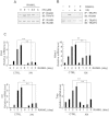
Figure 7. XN represses the association of RANK and TRAF6, and abrogates osteoclastogenesis-related gene expression.
(A,B) XN suppressed the RANKL-induced binding of RANK and TRAF6. The RAW264.7 cells were pretreated with indicated concentration of XN for 4 hours then stimulated with RANKL (30 ng/ml) for another 20 minutes. The cell lysate were harvest and immunoprecipitated with antibody to RANK and then blotted with anti-TRAF6 (A). Or the cell lysates were immunoprecipitated with antibody to TRAF6 and then blotted with anti-RANK (B). (C) XN inhibits the mRNA levels of NFATc1, cathepsin K (CtsK), CTR and TRAP induced by RANKL. Mouse BMMs were incubated with XN (5 μM) and RANKL (30 ng/mg) for indicate day. Total RNA was collected and analyzed by Real time-PCR. Column, means of experiments performed in triplicate; bar, SD. **p < 0.01.

References

    1. Krum S. A., Chang J., Miranda-Carboni G. & Wang C. Y. Novel functions for NFkappaB: inhibition of bone formation. Nat Rev Rheumatol 6, 607–611 (2010). - PMC - PubMed
    1. Lee J. H. et al. Epigallocatechin-3-gallate inhibits osteoclastogenesis by down-regulating c-Fos expression and suppressing the nuclear factor-kappaB signal. Mol Pharmacol 77, 17–25 (2010). - PubMed
    1. Qi B. et al. Ablation of Tak1 in osteoclast progenitor leads to defects in skeletal growth and bone remodeling in mice. Sci Rep 4, 7158, 10.1038/srep07158. (2014). - DOI - PMC - PubMed
    1. Vives V. et al. Pharmacological inhibition of Dock5 prevents osteolysis by affecting osteoclast podosome organization while preserving bone formation. Nat Commun 6, 6218, 10.1038/ncomms7218 (2015). - "V体育2025版" DOI - PubMed
    1. Cummings S. R. et al. Denosumab for prevention of fractures in postmenopausal women with osteoporosis. N Engl J Med 361, 756–765 (2009). - "VSports最新版本" PubMed

"VSports在线直播" Publication types

"VSports注册入口" MeSH terms