Skip to main page content (V体育ios版)
U.S. flag

An official website of the United States government

Dot gov

The . gov means it’s official. Federal government websites often end in . gov or . mil VSports app下载. Before sharing sensitive information, make sure you’re on a federal government site. .

Https

The site is secure. The https:// ensures that you are connecting to the official website and that any information you provide is encrypted and transmitted securely. V体育官网.

. 2016 Jul 7;35(27):3514-23.
doi: 10.1038/onc.2015.412. Epub 2015 Nov 2.

The Arkadia-ESRP2 axis suppresses tumor progression: analyses in clear-cell renal cell carcinoma (V体育2025版)

Affiliations

"V体育官网" The Arkadia-ESRP2 axis suppresses tumor progression: analyses in clear-cell renal cell carcinoma

A Mizutani et al. Oncogene. .

Abstract

Tumor-specific alternative splicing is implicated in the progression of cancer, including clear-cell renal cell carcinoma (ccRCC). Using ccRCC RNA sequencing data from The Cancer Genome Atlas, we found that epithelial splicing regulatory protein 2 (ESRP2), one of the key regulators of alternative splicing in epithelial cells, is expressed in ccRCC. ESRP2 mRNA expression did not correlate with the overall survival rate of ccRCC patients, but the expression of some ESRP-target exons correlated with the good prognosis and with the expression of Arkadia (also known as RNF111) in ccRCC. Arkadia physically interacted with ESRP2, induced polyubiquitination and modulated its splicing function VSports手机版. Arkadia and ESRP2 suppressed ccRCC tumor growth in a coordinated manner. Lower expression of Arkadia correlated with advanced tumor stages and poor outcomes in ccRCC patients. This study thus reveals a novel tumor-suppressive role of the Arkadia-ESRP2 axis in ccRCC. .


Conflict of interest statement

The authors declare no conflict of interest.

Figures
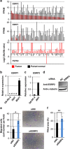
Figure 1
Figure 1
Expression of ESRPs in ccRCC. (a) Expression of ESRP2 (top) and ESRP1 (middle) in 65 tumor tissues and 65 paired normal tissues using the RNA-seq data from TCGA. The log 2 values of FGFR2 IIIc/IIIb ratios are shown in the bottom panel. FPKM: fragments per kilobase of exon per million mapped sequence reads. (b and c) qRT–PCR analysis of ESRP1 (b) and ESRP2 (c) expression in OS-RC-2 and HEK293T cells. MCF7 breast cancer cells were used as a positive control in (b). ND, not detected. (d) Immunoblot analysis to determine ESRP2 protein expression in HEK293T cells treated with ESRP2 siRNA. NC, negative control. (e and f) Transwell cell migration assay (e) and wound healing assay (f) in OS-RC-2 cells treated with siRNA for ESRP2. *P<0.01 and **P<0.05 by two-sided Student's paired t-test. Representative pictures are shown in (e). siNC: negative control siRNA.
Figure 2
Figure 2
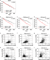
ESRP-splicing function and prognosis in ccRCC. (ad) Kaplan–Meier analysis of overall survival in ccRCC patients enrolled in the TCGA database, with classifications based on the expression of ESRP2 mRNA (a), ENAH exon 11a (b), ITGA6 exon 27 (c) or SLK exon 13 (d) in TCGA ccRCC RNA-seq data. Patients were divided into two equally sized groups based on the expression of ESRP2 mRNA or ESRP-target exons. The half of the patients with higher expression of ESRP2 mRNA or each ESRP-target exon are shown in black, and the half of the patients with lower expression are shown in red; n=466. P-values are based on the log-rank significance tests. (eg) Scatter plots of Arkadia expression and ENAH exon 11a (e), ITGA6 exon 27 (f) or SLK exon 13 (g) expression in TCGA ccRCC RNA-seq data; n=542. (hj) Scatter plots of ESRP2 expression and ENAH exon 11a (h), ITGA6 exon 27 (i) or SLK exon 13 (j) expression in TCGA ccRCC RNA-seq data; n=542. The r-values were determined using Pearson's correlation. FPKM, fragments per kilobase of exon per million mapped sequence reads.
Figure 3
Figure 3
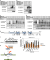
Functional interaction between Arkadia and ESRP2. (a) qRT–PCR analyses of the expression of Arkadia, ITGA6 exon 27, SLK exon 13 and TSC2 exon 27 in HEK293T cells in the presence of negative control siRNA (siNC) or siArkadia. Values were normalized to glyceraldehyde 3-phosphate dehydrogenase (GAPDH), total ITGA6, SLK and TSC2, respectively. (b) qRT–PCR analysis of Arkadia and ENAH exon 11a expression in OS-RC-2 cells in the presence of siNC or siArkadia. Values were normalized to GAPDH and total ENAH, respectively; n=3 in (a) and (b). *P<0.01 and **P<0.05 by two-sided Student's paired t-test. (c and d) Immunoprecipitation assay using HEK293T cells transiently transfected with ESRP1 (c) or ESRP2 (d) and Arkadia. IB, immunoblotting; IP, immunoprecipitation; WT, wild-type. (e) Immunoprecipitation assay using HEK293T cells to determine the interaction between endogenous ESRP2 and Arkadia proteins.
Figure 4
Figure 4
Modulation of the splicing function of ESRP2 by Arkadia through ubiquitination. (a and b) Immunoprecipitation assay using HEK293T cells transiently transfected with ESRP1 (a) or ESRP2 (b), Arkadia wild-type or CA mutant, and ubiquitin. IP, immunoprecipitation; IB, immunoblotting; Ub, ubiquitin; WT, wild-type. (c and d) Immunoprecipitation assay using HEK293T cells transiently transfected with ESRP1 (c) or ESRP2 (d), WT Arkadia, and ubiquitin WT or its mutants. 7KR: all seven lysine residues are substituted by arginine residues; 6, 11, 27, 29, 33, 48 and 63K: one lysine residue is intact, but the others are substituted by arginine residues. KR, lysine-to-arginine mutation. (e) Schematic representation of ESRP2-KR mutants. (f) Immunoblot analysis to examine the protein expression of WT or ESRP2-KR mutants overexpressed in HEK293T cells. (g) qRT–PCR analysis of ENAH exon 11a expression in HEK293T cells transiently transfected with WT or ESRP2-KR mutants upon Arkadia knockdown. Data were normalized to total ENAH. Error bars indicate s.d. siNC, negative control siRNA. Experiments were repeated, and a representative set of data are shown in (g). (h) Schematic representation of the regulation of ESRP2 function by Arkadia through ubiquitination.
Figure 5
Figure 5
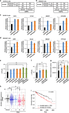
Suppression of tumor growth by Arkadia. (a and b) Analysis using the 2x2 contingency matrix of RNA-seq data upon knockdown of Arkadia or ESRP2 in OS-RC-2 cells (a) and in HEK293T cells (b). We select the isoforms showing the maximum FPKM (fragments per kilobase of exon per million mapped sequence reads) values are z⩾5 among the three samples (siNC-treated, siESRP2-treated and siArkadia-treated) in OS-RC-2 or HEK293T cells. ESRP2-enhanced: siNC isoform/siESRP2 isoform is ⩾2. ESRP2-silenced, siNC isoform/siESRP2 isoform is ⩽0.5. Arkadia-enhanced and -silenced were determined using the same cutoff values as ESRP2-enhanced and -silenced, respectively. The number of appropriate isoforms for each category is described in the 2x2 contingency matrix. P<2.2E−16 by Fisher's exact test in (a). P<1.2E−04 by Fisher's exact test in (b). (c and d) qRT–PCR analyses of the expression of NRP1, GCLM, HBEGF and PPP2CB in OS-RC-2 cells (c) and in HEK293T cells (d) upon knockdown of ESRP2 or Arkadia. Values were normalized to GAPDH. Knockdown efficiency of Arkadia or ESRP2 is shown in Supplementary Figure S5. siNC, negative control siRNA; n=2. (e and f) Cell count assays using HEK293T (e) and OS-RC-2 (f) cells upon knockdown of ESRP2 and/or Arkadia. Cells were transfected with siRNA at the same time as seeding and counted after 48 h. Knockdown efficiency of Arkadia and ESRP2 is shown in Supplementary Figure S6. (g) BrdU assay using OS-RC-2 cells after knocking down ESRP2 and/or Arkadia. (h) Arkadia mRNA expression in 68 normal kidney tissues and 474 ccRCC tumors grouped into stages I–IV by TCGA clinical data. (i) Kaplan–Meier analysis of overall survival in ccRCC patients enrolled in the TCGA database, with classifications based on Arkadia mRNA expression in TCGA ccRCC RNA-seq data. Patients were divided into two equally sized groups based on Arkadia mRNA expression. The half of patients with higher Arkadia expression are shown in black, and the half of patients with lower expression are shown in red. In (eh), *P<0.05 and **P<0.01 by two-sided Student's paired t-test. The P-value in (i) is based on the log-rank significance test.

References

    1. Gamazon ER, Stranger BE. Genomics of alternative splicing: evolution, development and pathophysiology. Hum Genet 2014; 133: 679–687. - PubMed
    1. Kalsotra A, Cooper TA. Functional consequences of developmentally regulated alternative splicing. Nat Rev Genet 2011; 12: 715–729. - "V体育平台登录" PMC - PubMed
    1. Chen J, Weiss WA. Alternative splicing in cancer: implications for biology and therapy. Oncogene 2014; 34: 1–14. - PubMed
    1. Biamonti G, Catillo M, Pignataro D, Montecucco A, Ghigna C. The alternative splicing side of cancer. Semin Cell Dev Biol 2014; 32C: 30–36. - PubMed
    1. Turner N, Grose R. Fibroblast growth factor signalling: from development to cancer. Nat Rev Cancer 2010; 10: 116–129. - PubMed

Publication types (VSports最新版本)

MeSH terms