Skip to main page content
U.S. flag

An official website of the United States government

Dot gov

The . gov means it’s official VSports app下载. Federal government websites often end in . gov or . mil. Before sharing sensitive information, make sure you’re on a federal government site. .

Https

The site is secure V体育官网. The https:// ensures that you are connecting to the official website and that any information you provide is encrypted and transmitted securely. .

. 2016 Jun;27(6):1727-40.
doi: 10.1681/ASN.2015040449. Epub 2015 Oct 9.

Sustained Activation of Wnt/β-Catenin Signaling Drives AKI to CKD Progression

Affiliations

Sustained Activation of Wnt/β-Catenin Signaling Drives AKI to CKD Progression

Liangxiang Xiao et al. J Am Soc Nephrol. 2016 Jun.

Abstract

AKI is increasingly recognized as a major risk factor for progression to CKD. However, the factors governing AKI to CKD progression are poorly understood. In this study, we investigated this issue using moderate (20 minutes) and severe (30 minutes) ischemia/reperfusion injury (IRI) in mice. Moderate IRI led to acute kidney failure and transient Wnt/β-catenin activation, which was followed by the restoration of kidney morphology and function. However, severe IRI resulted in sustained and exaggerated Wnt/β-catenin activation, which was accompanied by development of renal fibrotic lesions characterized by interstitial myofibroblast activation and excessive extracellular matrix deposition. To assess the role of sustained Wnt/β-catenin signaling in mediating AKI to CKD progression, we manipulated this signaling by overexpression of Wnt ligand or pharmacologic inhibition of β-catenin. In vivo, overexpression of Wnt1 at 5 days after IRI induced β-catenin activation and accelerated AKI to CKD progression. Conversely, blockade of Wnt/β-catenin by small molecule inhibitor ICG-001 at this point hindered AKI to CKD progression. In vitro, Wnt ligands induced renal interstitial fibroblast activation and promoted fibronectin expression. However, activated fibroblasts readily reverted to a quiescent phenotype after Wnt ligands were removed, suggesting that fibroblast activation requires persistent Wnt signaling. These results indicate that sustained, but not transient, activation of Wnt/β-catenin signaling has a decisive role in driving AKI to CKD progression. VSports手机版.

Keywords: CKD; acute renal failure; fibroblast; ischemia-reperfusion; renal ischemia V体育安卓版. .


Figures

Figure 1.
Figure 1.
Wnt/β-catenin is activated in both moderate and severe AKI induced by ischemia/reperfusion. (A) Twenty-minute IRI induced moderate AKI followed by rapid restoration of kidney function. Serum creatinine (Scr) levels were assayed at different time points after 20-minute IRI as indicated. Data are expressed as means±SEMs. Ctrl, control; D, day. *P<0.05 versus sham controls (n=5). (B) Thirty-minute IRI caused severe AKI with sustained kidney dysfunction. Scr levels were elevated at different time points after injury. *P<0.05 versus sham controls (n=4). (C) Representative micrographs show the kidney morphology at 10 days after 20- or 30-minute IRI. Kidney sections were subjected to Periodic acid–Schiff staining. Boxed areas are enlarged. Arrows indicate renal tubules. Scale bar, 50 µm. (D) Representative RT-PCR analysis reveals different magnitudes of Wnt induction after 20- and 30-minute IRI. The expression of most Wnt genes was substantially increased in the kidneys at 1 day after 30-minute IRI compared with sham or 20-minute IRI groups. (E and F) Western blot shows that β-catenin protein level was dramatically increased at 3 days after severe AKI induced by 30-minute IRI compared with sham or 20-minute IRI groups. Numbers (1–3) indicate each individual animal in a given group. *P<0.05 versus sham controls; P<0.05 versus 20-minute IRI (n=4).
Figure 2.
Figure 2.
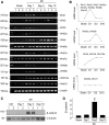
Wnt/β-catenin activation is transient in moderate AKI after 20-minute IRI. (A) Representative RT-PCR analysis of renal Wnt gene expression at different time points after 20-minute IRI. The majority of Wnt genes was transiently induced at 1 day but returned to baselines at 3 and 10 days after injury. Numbers (1–3) indicate each individual animal in a given group. (B) Graphic presentation shows the dynamic pattern of Wnt expression in the kidneys after 20-minute IRI. Different Wnts with similar expression patterns after IRI were grouped. D, day. (C and D) Representative Western blot reveals that the β-catenin protein level was induced with a peak at 3 days after IRI compared with controls. Numbers (1 and 2) indicate each individual animal in a given group. Data are presented as fold induction over sham controls. Ctrl, control. *P<0.05 versus sham controls (n=5).
Figure 3.
Figure 3.
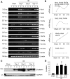
Severe AKI after 30-minute IRI is associated with sustained activation of Wnt/β-catenin. (A) Representative RT-PCR analysis reveals that most Wnt genes were induced at 1 day after severe AKI induced by 30-minute IRI compared with sham controls and that Wnt induction was sustained at 3 and 10 days. Numbers (1–3) indicate each individual animal in a given group. (B) Graphic presentation shows the distinct pattern of Wnt expression in severe AKI after 30-minute IRI. Different Wnts with similar dynamic pattern were grouped. D, day. (C) Representative Western blot and (D) graphic presentation show that β-catenin protein was induced at 1, 3, and 10 days after 30-minute IRI, with a peak expression at 3 days. Data are presented as fold induction over sham controls. *P<0.05 versus sham controls (n=4). GAPDH, glyceraldehyde-3-phosphate dehydrogenase.
Figure 4.
Figure 4.
In vivo expression of Wnt1 accelerates AKI-CKD progression after IRI. (A) Diagram shows the experimental design. Arrows indicate the time point of UIRI and unilateral nephrectomy (UNx). The green arrowhead indicates the time point when pcDNA3 or pHA-Wnt 1 was injected. (B) Representative RT-PCR shows Wnt1 mRNA expression in the kidneys at 6 days after single plasmid injection. Ctrl, control. (C) Representative Western blot shows renal Wnt1 and HA protein expression at 6 days after plasmid injection; kidney lysates were immunoblotted with antibodies against Wnt1 and HA, respectively. (D) Representative micrographs show renal Wnt1 and β-catenin protein expression and localization at 6 days after plasmid injection; kidney sections were stained with specific antibodies against Wnt1 and β-catenin, respectively. Arrows indicate positive staining. Scale bar, 50 μm. (E and F) Graphic presentation shows the levels of serum creatinine (Scr) and BUN in different groups. *P<0.05 versus sham controls; P<0.05 versus pcDNA3 alone (n=5).
Figure 5.
Figure 5.
In vivo expression of exogenous Wnt1 promotes the expression of β-catenin target genes. (A–C) Western blot analyses show the expression of β-catenin and PAI-1 in different groups. Numbers (1–3) indicate each individual animal in a given group. (A) Representative Western blot and quantitative data for (B) β-catenin and (C) PAI-1 are shown. Data are presented as fold induction over sham controls. Ctrl, control. *P<0.05 versus sham controls; P<0.05 versus pcDNA3 alone (n=5). (D and E) qRT-PCR analyses show that expression of Wnt1 in vivo promoted renal expression of (D) MMP-7 and (E) Fsp1 after IRI. *P<0.05 versus sham controls; P<0.05 versus pcDNA3 alone (n=5). (F) Representative micrographs show renal induction of MMP-7 and renin at 6 days after plasmid injection. Arrows indicate positive staining. Open arrow shows renin expression in the juxtaglomerular apparatus. Scale bar, 50 μm.
Figure 6.
Figure 6.
Exogenous Wnt1 in vivo drives CKD progression after AKI. (A–D) qRT-PCR shows the relative mRNA levels of (A) α-SMA, (B) fibronectin, (C) collagen I, and (D) collagen III in different groups. Relative mRNA levels were determined after normalization with β-actin and expressed as fold induction over controls. Ctrl, control. *P<0.05 versus sham controls; P<0.05 versus pcDNA3 (n=5). (E) Western blot analyses of renal expression of fibronectin and α-SMA. Kidney lysates were immunoblotted with specific antibodies against fibronectin (FN), α-SMA, and β-actin. Numbers (1–3) indicate each individual animal in a given group. (F and G) Graphic presentation of renal protein levels of fibronectin and α-SMA. *P<0.05 versus sham controls; P<0.05 versus pcDNA3 (n=5). (H) Immunofluorescence and immunohistochemical staining show fibronectin and α-SMA expression in the kidneys at 6 days after Wnt1 plasmid injection. Kidney sections were stained with specific antifibronectin and α-SMA antibody. Scale bar, 50 μm. (I) Kidney sections were stained with MTS. Representative micrographs from different groups as indicated are shown. Scale bar, 50 μm. (J) Quantitative determination of renal fibrotic lesions in different groups. *P<0.05 versus sham controls; P<0.05 versus pcDNA3 alone (n=5).
Figure 7.
Figure 7.
Blockade of Wnt/β-catenin signaling by small molecule inhibitor ICG-001 hinders AKI-CKD progression. (A) Diagram shows the experimental design. Arrows indicate the time point of UIRI and unilateral nephrectomy (UNx). The green arrowhead indicates the time point when ICG-001 or vehicle was injected. (B and C) Graphic presentation shows that ICG-001 reduced the levels of serum creatinine (Scr) and BUN at 11 days after UIRI compared with vehicle controls. Ctrl, control. *P<0.05 versus sham controls; P<0.05 versus vehicle alone (n=6). (D–G) qRT-PCR shows that ICG-001 reduced the mRNA expression of (D) fibronectin, (E) collagen I, (F) collagen III, and (G) α-SMA in the kidneys after IRI compared with vehicle controls. *P<0.05 versus sham controls; P<0.05 versus vehicle alone (n=6). (H) Kidney sections were stained with MTS. Representative micrographs from different groups as indicated are shown. The arrow indicates positive staining. Scale bar, 50 μm. (I) Quantitative determination of renal fibrotic lesions in different groups. *P<0.05 versus sham controls; P<0.05 versus vehicle alone (n=6).
Figure 8.
Figure 8.
ICG-001 represses Wnt/β-catenin target genes and reduces fibrotic lesions. (A) Western blot analyses show that ICG-001 inhibited renal expression of β-catenin, fibronectin, α-SMA, and PAI-1 after IRI. Numbers (1–3) indicate each individual animal. Ctrl, control. (B–E) Quantitative data for (B) β-catenin, (C) fibronectin, (D) α-SMA, and (E) PAI-1 are shown in different groups. *P<0.05 versus sham controls; P<0.05 versus vehicle alone (n=6). (F) Immunostaining shows that ICG-001 reduced the expression of β-catenin, fibronectin, and α-SMA after IRI; kidney sections from different groups were stained with specific antibodies against β-catenin, fibronectin, and α-SMA, respectively. Scale bar, 50 μm. (G and H) qRT-PCR analyses show that ICG-001 inhibited MMP-7 and Snail1 mRNA expression in vivo. *P<0.05 versus sham controls; P<0.05 versus vehicle (n=6). GAPDH, glyceraldehyde-3-phosphate dehydrogenase.
Figure 9.
Figure 9.
Myofibroblast activation requires sustained Wnt/β-catenin signaling in vitro. (A) Diagram shows the experimental design: a and b show that NRK-49F cells were treated without or with 10% Wnt-CM for 24 hours; c indicates that NRK-49F cells were treated with 10% Wnt-CM for 24 hours, washed, and incubated without Wnt-CM for an additional 24 hours, and d shows that NRK-49F cells were incubated with 40% Wnt-CM continuously for 48 hours. (B–D) Western blot analyses and quantitative data show that myofibroblast activation was dependent on sustained Wnt/β-catenin signaling in vitro. Wnt-CM was collected from HKC-8 cells after transfection with a group of Wnt expression vectors, including Wnt1, Wnt2, Wnt3a, Wnt4, and Wnt16. Some samples were pretreated with ICG-001 (10 µM) for 1 hour. The experimental conditions (a–d) are presented at the bottom of each panel. Fn, fibronectin. *P<0.05 versus controls (n=3); P<0.05 versus Wnt-CM alone (n=3). (E–H) Blockade of Wnt/β-catenin signaling by ICG-001 abolished the Wnt-CM–induced mRNA expression of fibronectin, collagen I, collagen III, and Fsp1 in NRK-49F cells. NRK-49F cells were treated as indicated, and the mRNA expression was assessed by qRT-PCR. *P<0.05 versus controls (n=3); P<0.05 versus Wnt-CM alone (n=3). (I) Immunofluorescence staining shows that Wnt-CM induced fibronectin, collagen III, and vimentin expression in NRK-49F cells and that ICG-001 inhibited their expression; NRK-49F cells cultured on coverslips were stained with specific antibodies against fibronectin, collage III, and vimentin, respectively. Arrows indicate positive staining.

"VSports在线直播" References

    1. Belayev LY, Palevsky PM: The link between acute kidney injury and chronic kidney disease. Curr Opin Nephrol Hypertens 23: 149–154, 2014 - "V体育2025版" PMC - PubMed
    1. Leung KC, Tonelli M, James MT: Chronic kidney disease following acute kidney injury-risk and outcomes. Nat Rev Nephrol 9: 77–85, 2013 - PubMed
    1. Chawla LS, Kimmel PL: Acute kidney injury and chronic kidney disease: An integrated clinical syndrome. Kidney Int 82: 516–524, 2012 - V体育官网 - PubMed
    1. Bonventre JV, Basile D, Liu KD, McKay D, Molitoris BA, Nath KA, Nickolas TL, Okusa MD, Palevsky PM, Schnellmann R, Rys-Sikora K, Kimmel PL, Star RA Kidney Research National Dialogue (KRND) : AKI: A path forward. Clin J Am Soc Nephrol 8: 1606–1608, 2013 - VSports在线直播 - PMC - PubMed
    1. Coca SG, Singanamala S, Parikh CR: Chronic kidney disease after acute kidney injury: A systematic review and meta-analysis. Kidney Int 81: 442–448, 2012 - PMC - PubMed

Publication types

"VSports在线直播" MeSH terms