VSports - Skip to main page content
U.S. flag

An official website of the United States government

Dot gov

The . gov means it’s official. Federal government websites often end in . gov or . mil. Before sharing sensitive information, make sure you’re on a federal government site VSports app下载. .

Https

The site is secure V体育官网. The https:// ensures that you are connecting to the official website and that any information you provide is encrypted and transmitted securely. .

. 2015 Sep 17;59(6):1011-24.
doi: 10.1016/j.molcel.2015.07.029. Epub 2015 Sep 10.

Distinct but Concerted Roles of ATR, DNA-PK, and Chk1 in Countering Replication Stress during S Phase (V体育官网入口)

Affiliations

Distinct but Concerted Roles of ATR, DNA-PK, and Chk1 in Countering Replication Stress during S Phase (VSports)

V体育ios版 - Rémi Buisson et al. Mol Cell. .

Abstract (V体育ios版)

The ATR-Chk1 pathway is critical for DNA damage responses and cell-cycle progression. Chk1 inhibition is more deleterious to cycling cells than ATR inhibition, raising questions about ATR and Chk1 functions in the absence of extrinsic replication stress. Here we show that a key role of ATR in S phase is to coordinate RRM2 accumulation and origin firing VSports手机版. ATR inhibitor (ATRi) induces massive ssDNA accumulation and replication catastrophe in a fraction of early S-phase cells. In other S-phase cells, however, ATRi induces moderate ssDNA and triggers a DNA-PK and Chk1-mediated backup pathway to suppress origin firing. The backup pathway creates a threshold such that ATRi selectively kills cells under high replication stress, whereas Chk1 inhibitor induces cell death at a lower threshold. The levels of ATRi-induced ssDNA correlate with ATRi sensitivity in a panel of cell lines, suggesting that ATRi-induced ssDNA could be predictive of ATRi sensitivity in cancer cells. .


Figures

Fig. 1
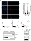
Fig. 1. Acute ATR inhibition exerts two distinct effects on S-phase cells
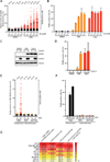
A. U2OS cells were cultured in BrdU for 36 h, treated with DMSO or ATRi (10 μM VE-821), and analyzed for BrdU and γH2AX by immunestaining. B. Quantification of the BrdU intensity of 1,000 U2OS cells treated with DMSO or ATRi. Black lines indicate median BrdU intensities of BrdU-positive cells in various cell populations. C. Quantification of the BrdU and γH2AX intensities of 1,200 U2OS cells treated with DMSO or ATRi. Cell subpopulation 1 displayed less ssDNA at 8 h than at 2h. Cell subpopulation 2 displayed very high levels of ssDNA and became γH2AX-positive at 8h. D. Levels of RPA32, pRPA32, and γH2AX in the soluble and chromatin fractions of ATRi-treated cells were analyzed by Western blot. E. The levels of chromatin-bound RPA32 and pRPA32 were analyzed in cells treated with ATRi#2 (AZ20) and ATRi#3 (EPT-46464). See also Fig. S1.
Fig. 2
Fig. 2. ATR suppresses ssDNA accumulation in early S phase
A-B. Quantification of chromatin-bound RPA, EdU incorporation, and DNA contents of 5,000 U2OS cells treated with DMSO or ATRi (10 μM VE-821). Cells were color-coded according to the intensity of RPA staining as shown in the left panel. C. T98G cells were synchronously released from G0 and analyzed for EdU incorporation at the indicated times. D. Staining intensity of chromatin-bound RPA was analyzed at different stages of the cell cycle after ATRi or DMSO treatment. Red lines indicate mean RPA intensities in various cell populations. **, P<0.01; ***, P<0.001. See also Fig. S2.
Fig. 3
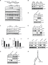
Fig. 3. ATRi suppresses DNA damage by promoting RRM2 accumulation and limiting origin firing
A. T98G cells were synchronously released from G0 in the presence or absence of ATRi (10 μM VE-821). Levels of RRM2, γH2AX, RPA70, RPA32, RPA14 and Cyclin A were analyzed during the time course. B. Asynchronously growing U2OS cells were treated with ATRi (10 μM VE-821) or Chk1i (2 μM MK-8776). Levels of RRM2 and E2F1 were analyzed at the indicated times. C. Levels of RRM2 and E2F1 were analyzed in U2OS cells treated with DMSO or ATRi in the presence of cycloheximide (CHX). Relative levels of RRM2 and E2F1 were quantified from 3 blots (n=3). Error bars: S.D. D. U2OS cells transfected with empty vector or E2F1-expressing plasmids were treated with ATRi for 8 h. Levels of RRM2, E2F1, and γH2AX were analyzed. E. U2OS cells were treated with the indicated inhibitors for 8 h. Levels of RRM2, E2F1, and γH2AX were analyzed. F. U2OS cells were treated with DMSO or ATRi for 8 h in the presence or absence of MG132 or MLN4924. G. U2OS cells infected with HA-RRM2-expressing retrovirus or control virus were treated with ATRi for 8 h. Levels of RRM2 and γH2AX were analyzed at the indicated times. H. A model in which ATR coordinates RRM2 accumulation and origin firing in early S phase. See also Fig. S3.
Fig. 4
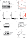
Fig. 4. ATRi-treated cells recover via a Chk1-mediated mechanism
A. U2OS cells were treated with DMSO, ATRi (10 μM VE-821), or Chk1i (2 μM MK-8776). Levels of RPA32, pRPA32, and γH2AX were analyzed at the indicated times. B. U2OS cells were treated with increasing concentrations of ATRi or Chk1i for 24 h and then cultured in inhibitor-free media. Cell survival was analyzed 4 days after treatment. Error bar: S.D. (n=3). C. U2OS cells were treated with DMSO, ATRi, or Chk1i for 8 h. BrdU and γH2AX intensities were quantified in 1,200 cells at the indicated times. D. U2OS cells were treated with ATRi or Chk1i. Levels of chromatin-bound RPA were analyzed at the indicated times. E. U2OS cells were treated with ATRi, and levels of pChk1 and CDC25A were analyzed at the indicated times. F. Levels of CDC25A in U2OS cells treated with ATRi or Chk1i were compared at the indicated times. G. Amodel in which Chk1 promotes the recovery of ATRi-treated cells with moderate ssDNA. See also Fig. S4.
Fig. 5
Fig. 5. Regulation and function of Chk1 during recovery
A. Levels of pDNA-PK, pATM, pChk1 and CDC25A were analyzed in ATRi-treated U2OS cells at the indicated times. B. U2OS cells were treated with ATRi, ATMi, DNA-PKi, or the combinations of these inhibitors. Levels of pDNA-PK, pATM, pChk1 and CDC25A were analyzed 8 h after treatment. C. The percentage of replication tracts containing fired origins was determined in RPE1 cells treated with DMSO or ATRi at the indicated times. Error bars: S.D. (n=3 experiments). **P<0.01; ***P<0.001. D. RPE1 cells were treated with DMSO or various inhibitors as indicated. The inter-origin distance was analyzed using DNA fiber assay at the indicated times. Error bars: S.E.M. (n=25 to 67 as indicated). ****, P<0.0001; n.s., not significant. E-F. RPE1 cells were treated with DMSO or various inhibitors as indicated. The length of continuous replication tracts was determined using DNA fiber assay at the indicated times (>600 forks per condition, n=3 experiments). See also Fig. S5.
Fig. 6
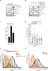
Fig. 6. ATRi selectively kills cells under high replication stress
A. U2OS cells were treated with ATRi for 2 h in the presence of increasing concentrations of HU. The BrdU intensity of 1,000 U2OS cells was quantified. Black lines indicate mean BrdU intensities of BrdU-positive cells in various populations. B. U2OS cells were treated with ATRi or Chk1i for 16 h in the presence of increasing concentrations of HU. Cell death was measured by the TUNEL assay. Error bars: S.D. (n=3). C-D. U2OS cells were induced to overexpress Cyclin E or left uninduced, and treated with ATRi or Chk1i for 16h. Levels of Cyclin E and γH2AX were analyzed by Western (C). Fractions of TUNEL-positive cells were quantified (D). Error bars: S.D. (n=3). E-F. T98G, RPE1, and MCF10A cells were treated with DMSO or ATRi. Levels of ssDNA were analyzed by native BrdU staining 2 h after ATRi treatment (E). Yellow lines indicate mean BrdU intensities of BrdU-positive cells in various populations. Levels of cell death were measured by TUNEL assay at the indicated times (F). Error bars: S.D. (n=3). G. Quantification of ssDNA, γH2AX, and cell survival of 10 colorectal cell lines after ATRi treatment. Levels of ssDNA and γH2AX were analyzed 2 h and 16 h after the indicated treatments, respectively. Fractions of ATRi-treated cells displaying stronger BrdU staining than untreated cells were determined (see Supplemental Methods). Cell survival was analyzed 6 days after the indicated treatments using CellTiter-Glo. See also Fig. S6.
Fig. 7
Fig. 7. Modeling the roles for ATR, DNA-PK, and Chk1 in countering replication stress
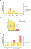
A. A fraction of early S-phase cells are particularly vulnerable to ATR inactivation. B. ATRi selectively kills cells under high replication stress, whereas Chk1i induces cell death even in cells in which replication stress is moderate. C. ATRi-induced ssDNA is an indicator of replication stress that may predict the ATRi sensitivity of cancer cells.

References

    1. Ahmed D, Eide PW, Eilertsen IA, Danielsen SA, Eknaes M, Hektoen M, Lind GE, Lothe RA. Epigenetic and genetic features of 24 colon cancer cell lines. Oncogenesis. 2013;2:e71. - PMC - PubMed
    1. Bartkova J, Horejsi Z, Koed K, Kramer A, Tort F, Zieger K, Guldberg P, Sehested M, Nesland JM, Lukas C, et al. DNA damage response as a candidate anti-cancer barrier in early human tumorigenesis. Nature. 2005;434:864–870. - PubMed
    1. Bartkova J, Rezaei N, Liontos M, Karakaidos P, Kletsas D, Issaeva N, Vassiliou LV, Kolettas E, Niforou K, Zoumpourlis VC, et al. Oncogene-induced senescence is part of the tumorigenesis barrier imposed by DNA damage checkpoints. Nature. 2006;444:633–637. - PubMed
    1. Bastos de Oliveira FM, Kim D, Cussiol JR, Das J, Jeong MC, Doerfler L, Schmidt KH, Yu H, Smolka MB. Phosphoproteomics Reveals Distinct Modes of Mec1/ATR Signaling during DNA Replication. Mol Cell. 2015;57:1124–1132. - PMC (VSports最新版本) - PubMed
    1. Beck H, Nahse-Kumpf V, Larsen MS, O’Hanlon KA, Patzke S, Holmberg C, Mejlvang J, Groth A, Nielsen O, Syljuasen RG, et al. Cyclin-dependent kinase suppression by WEE1 kinase protects the genome through control of replication initiation and nucleotide consumption. Mol Cell Biol. 2012;32:4226–4236. - VSports - PMC - PubMed

Publication types

MeSH terms