Skip to main page content
U.S. flag

An official website of the United States government

Dot gov

The . gov means it’s official. Federal government websites often end in . gov or . mil. Before sharing sensitive information, make sure you’re on a federal government site. VSports app下载.

Https

The site is secure. The https:// ensures that you are connecting to the official website and that any information you provide is encrypted and transmitted securely. V体育官网.

. 2013 Nov 27;32(23):3017-28.
doi: 10.1038/emboj.2013.224. Epub 2013 Oct 18.

VSports - Symbiotic lactobacilli stimulate gut epithelial proliferation via Nox-mediated generation of reactive oxygen species

Affiliations

Symbiotic lactobacilli stimulate gut epithelial proliferation via Nox-mediated generation of reactive oxygen species

Rheinallt M Jones et al. EMBO J. .

Abstract (V体育ios版)

The resident prokaryotic microbiota of the metazoan gut elicits profound effects on the growth and development of the intestine. However, the molecular mechanisms of symbiotic prokaryotic-eukaryotic cross-talk in the gut are largely unknown VSports手机版. It is increasingly recognized that physiologically generated reactive oxygen species (ROS) function as signalling secondary messengers that influence cellular proliferation and differentiation in a variety of biological systems. Here, we report that commensal bacteria, particularly members of the genus Lactobacillus, can stimulate NADPH oxidase 1 (Nox1)-dependent ROS generation and consequent cellular proliferation in intestinal stem cells upon initial ingestion into the murine or Drosophila intestine. Our data identify and highlight a highly conserved mechanism that symbiotic microorganisms utilize in eukaryotic growth and development. Additionally, the work suggests that specific redox-mediated functions may be assigned to specific bacterial taxa and may contribute to the identification of microbes with probiotic potential. .


Conflict of interest statement

The authors declare that they have no conflict of interest.

Figures

Figure 1
Figure 1
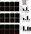
Ingestion of Lactobacillus plantarum by first-instar Drosophila larvae induces cellular ROS generation. Detection of ROS generation following the ingestion of indicated bacteria by germ-free newly emerged gstD1-gfp first-instar larvae for 30 min with the indicated Gram-positive or Gram-negative bacteria isolated from Drosophila midguts (Supplementary Table S1). Germ-free gstD1-gfp embryos were placed in a vial containing sterilized Drosophila growth media inoculated with 1 × 10⁸ cfu of the indicated bacteria. ROS were detected by oxidation of the hydrocyanin ROS-sensitive dye (upper panels), that is present in the larval food. Larvae used also harbour an ROS inducible gstD1-gfp reporter gene (green lower panels). (A′) Cartoon of first-instar midgut. Enterocyte (EC), intestinal stem cell (ISC), luminal contents (LCs). (A″) Tissue orientation control by staining of first-instar midgut stained for DNA. (A′′′) Exploded view of the interface between the ECs and the LC in larvae fed L. plantarum. Numbers of bacteria ingested by larva were quantified and results are presented in Materials and methods.
Figure 2
Figure 2
Ingestion of Lactobacillus plantarum by Drosophila induces cellular ROS generation in the midgut. (A) ROS generation following the ingestion of L. plantarum by germ-free third-instar larvae over 1 h. ROS were detected by oxidation of the hydrocyanin ROS-sensitive dye also included in the media. (B) Microscopic analysis at × 4 magnification of larval midgut dissected from (A). (C) ROS generation in the third-instar midgut following the ingestion of L. plantarum, Bacillus cereus or Erwinia carotovora for up to 4 h. (D) Densitometric analysis of larval midguts described in (C). Results are an average for 5 dissected midguts from each assay. (E) ROS generation in the gstD1-gfp adult Drosophila midgut following the ingestion of L. plantarum, or Erwinia carotovora-GFP for up to 4 h. Note Hydro-Cy3 fluorescence and expression of GFP in enterocytes following the L. plantarum ingestion. Also note GFP fluorescence detected in the midgut following the ingestion of E. carotovora-GFP. (F) ROS generation following the ingestion of L. plantarum in adult Drosophila midguts of the indicated genotypes for 1 h. (G) Densitometric analysis of larval midguts described in (F). Results are an average for five dissected midguts from each assay. All histograms report densitometric analysis (arbitrary units) of hydro-Cy3 oxidation, using the ImageJ software. Ten identically sized areas within an image were measured. n=50. **P<0.01, ***P<0.0001.
Figure 3
Figure 3
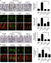
Ingestion of Lactobacillus plantarum induces ROS-dependent cellular proliferation in the Drosophila intestine. (A) EdU-positive cells in the midgut of w1118 germ-free third-instar larvae, or germ-free larvae fed with 1 × 10⁸ cfu L. plantarum for 4 h. Where indicated, the media also contained 1 mM N-acetylcysteine (NAC). (A′) Cartoon of en face third-instar midgut. Enterocyte (EC) in grey, adult midgut progenitors (AMPs) in red. Note some large enterocytes are EdU positive due to endonuclear DNA replication in maturing larval. (B) Number of EdU-positive cells under conditions described in (A) n=20, ***P<0.001. (C) Detection of EdU-positive cells in the midgut of adult conventionally raised, germ-free, or germ-free adult Drosophila following the ingestion of L. plantarum or Erwinia carotovora for 12 h. (D) Number of EdU-positive cells under conditions described in (C) n=20, ***P<0.001. (E) Detection of EdU-positive cells in the midgut of adult where the levels of Nox or Duox are diminished under the enterocyte-specific myoIA-GAL4 driver. Full genotypes myoIA-GAL4;UAS-dnox-RNAi and myoIA-GAL4;UAS-dduox-RNAi, and myoIA-GAL4;UAS-gal4-RNAi. (F) Number of EdU-positive cells in (A). n=10, ***P<0.001.
Figure 4
Figure 4
Contact of cultured cells with lactobacilli induces the generation of cellular reactive oxygen species (ROS). (A) Bacterial-induced ROS in cells contacted by the indicated bacteria for up to 40 min. Caco-2 cells seeded in a 96-well format were preloaded with 100 μM hydro-Cy3, then contacted with 3 × 10⁸/100 μl viable bacteria for the indicated times. Cells were then washed three times with PBS before fluoromometric analysis at 575 nm. (B) Bacterial-induced ROS in cells treated as described in (A) detected by confocal microscopy at 585 nm. In some experiments, 5 mM TEMPOL (a membrane-permeable oxygen radical scavenger) was also included. (C) ROS generation in response to contact of Caco-2 cells as described in (A) by the cecal contents of a conventionally raised BL6, or of germ-free BL6 mice. ROS was detected by fluoromometric analysis at 575 nm.
Figure 5
Figure 5
Ingestion of Lactobacillus induces Nox1-dependent generation of cellular reactive oxygen species (ROS) in murine enterocytes. (A) Detection of ROS generation in the distal small intestine of B6.wild type or B6.Nox1ΔIEC 6-week-old mice at 1 h following the oral gavage feeding of L. rhamnosus GG or E. coli. Mice were administered 5 μl of 200 μM hydro-Cy3 at 15 min before bacterial feeding. (B) Detection of ROS generation in the colon of 6-week-old mice treated as described in (A). In each figure, Haematoxylin and Eosin (H&E) stain is included for tissue orientation and scale. Scale bar=100 μm. Histograms report densitometric analysis (arbitrary units) of hydro-Cy3 oxidation, using the ImageJ software. The densitometry of 10 identically sized areas within an image was measured. Results are an average for three mice for each treatment. n=30. ***P<0.001. Error bars indicate s.e.m.
Figure 6
Figure 6
Ingestion of Lactobacillus induces Nox1-dependent cell proliferation in the murine gut epithelium. (A) Detection and numeration of p-Histone H3 in cells within the colon of 6-week-old w.t. or B6.Nox1ΔIEC mice at 4 h following the oral gavage feeding of HBSS or L. rhamnosus GG. (B) Detection and numeration of EdU-positive cells within the colon of 6-week-old w.t. or B6.Nox1ΔIEC mice at 4 h following the oral gavage feeding of HBSS or L. rhamnosus GG. (C) Detection and numeration of p-Histone H3 in cells within the small intestine of 6-week-old w.t. or B6.Nox1ΔIEC mice at 4 h following the oral gavage feeding of HBSS or L. rhamnosus GG. (D) Detection and numeration of EdU-positive cells within the small intestine of 6-week-old w.t. or B6.Nox1ΔIEC mice at 4 h following the oral gavage feeding of 1 × 10⁸ cfu of HBSS or L. rhamnosus GG. For each numeration, 40 × 20 fields were counted in three mice for each treatment. ***P<0.001.

Comment in

References

    1. Bedard K, Krause KH (2007) The NOX family of ROS-generating NADPH oxidases: physiology and pathophysiology. Physiol Rev 87: 245–313 - PubMed
    1. Buchon N, Broderick NA, Poidevin M, Pradervand S, Lemaitre B (2009) Drosophila intestinal response to bacterial infection: activation of host defense and stem cell proliferation. Cell Host Microbe 5: 200–211 - PubMed
    1. Chakrabarti S, Liehl P, Buchon N, Lemaitre B (2012) Infection-induced host translational blockage inhibits immune responses and epithelial renewal in the Drosophila gut. Cell Host Microbe 12: 60–70 - PubMed
    1. Chen X, Lee KA, Ha EM, Lee KM, Seo YY, Choi HK, Kim HN, Kim MJ, Cho CS, Lee SY, Lee WJ, Yoon J (2011) A specific and sensitive method for detection of hypochlorous acid for the imaging of microbe-induced HOCl production. Chem Commun (Camb) 47: 4373–4375 - PubMed
    1. Coant N, Ben Mkaddem S, Pedruzzi E, Guichard C, Treton X, Ducroc R, Freund JN, Cazals-Hatem D, Bouhnik Y, Woerther PL, Skurnik D, Grodet A, Fay M, Biard D, Lesuffleur T, Deffert C, Moreau R, Groyer A, Krause KH, Daniel F et al. (2010) NADPH oxidase 1 modulates WNT and NOTCH1 signaling to control the fate of proliferative progenitor cells in the colon. Mol Cell Biol 30: 2636–2650 - PMC - PubMed

Publication types

MeSH terms

"V体育2025版" LinkOut - more resources