"V体育官网入口" Skip to main page content
U.S. flag

An official website of the United States government

Dot gov

The . gov means it’s official. Federal government websites often end in . gov or VSports app下载. mil. Before sharing sensitive information, make sure you’re on a federal government site. .

Https

The site is secure. The https:// ensures that you are connecting to the official website and that any information you provide is encrypted and transmitted securely. V体育官网.

. 2013 Jun 11;110(24):9845-50.
doi: 10.1073/pnas.1305472110. Epub 2013 May 22.

A microRNA signature defines chemoresistance in ovarian cancer through modulation of angiogenesis

Affiliations

A microRNA signature defines chemoresistance in ovarian cancer through modulation of angiogenesis

Andrea Vecchione et al. Proc Natl Acad Sci U S A. .

"V体育安卓版" Abstract

Epithelial ovarian cancer is the most lethal gynecologic malignancy; it is highly aggressive and causes almost 125,000 deaths yearly. Despite advances in detection and cytotoxic therapies, a low percentage of patients with advanced stage disease survive 5 y after the initial diagnosis. The high mortality of this disease is mainly caused by resistance to the available therapies. Here, we profiled microRNA (miR) expression in serous epithelial ovarian carcinomas to assess the possibility of a miR signature associated with chemoresistance VSports手机版. We analyzed tumor samples from 198 patients (86 patients as a training set and 112 patients as a validation set) for human miRs. A signature of 23 miRs associated with chemoresistance was generated by array analysis in the training set. Quantitative RT-PCR in the validation set confirmed that three miRs (miR-484, -642, and -217) were able to predict chemoresistance of these tumors. Additional analysis of miR-484 revealed that the sensitive phenotype is caused by a modulation of tumor vasculature through the regulation of the VEGFB and VEGFR2 pathways. We present compelling evidence that three miRs can classify the response to chemotherapy of ovarian cancer patients in a large multicenter cohort and that one of these three miRs is involved in the control of tumor angiogenesis, indicating an option in the treatment of these patients. Our results suggest, in fact, that blockage of VEGF through the use of an anti-VEGFA antibody may not be sufficient to improve survival in ovarian cancer patients unless VEGFB signaling is also blocked. .


Conflict of interest statement

The authors declare no conflict of interest.

Figures
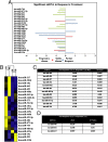
Fig. 1.
Fig. 1.
miR signature classifies responder vs. nonresponder ovarian cancer patients. (A) Analysis of training set using TLDA cards. Significant miRs in the different classes are shown. Values represent miR fold differences between the groups compared with complete response. (B) Centroid analysis of the identified miRs. Blue, down-regulated miRs; yellow, up-regulated miRs. Complete response (CR), partial response (PR), stable disease (SD), and progressive disease (PD) are shown. (C) Significant miRs in the training set redefined in two classes: nonresponder (SD and PD) and responder (CR and PR). (D) Significant miRs in the validation set. RQ represents fold changes in the two groups.
Fig. 2.
Fig. 2.
miR-484 modulates in vivo response to chemotherapy. (A) Tumor volume of mice injected in the right flank with MDAH-2774 control cells and injected in the left flank with MDAH-2774–overexpressing miR-484. The size of the tumors (Left) at day 0 of the CBDCA+Tax treatment and (Right) their increase after 21 d of treatment are shown. (B) In vivo imaging of nude mice injected in the right flank with SKOV-3 control cells and injected in the left flank with SKOV-3–overexpressing miR-484. Images were taken (Left) immediately before the start of the treatment and (Right) after 21 d of CBDCA+Tax treatment. (C) Quantification of in vivo EGFP fluorescence of the experiment described in B at (Left) day 0 and (Right) after 7, 14, and 21 d of treatment. (D) Effects of intratumoral injection of lentivirus-expressing control (scr) or miR-484 in the presence of CBDCA+Tax treatment. (E) Tumor necrosis percentages in scr- and miR-484–transduced tumors are shown. (F) H&E examples of (Left) scr-transduced tumor and (Right) miR-484. Green circles represent the tumor area, and the yellow circle is the area of necrosis. (Scale bar: 0.5 mm.) The significant differences are reported in each graph as evaluated by nonparametric t tests. Differences were considered significant when P < 0.05.
Fig. 3.
Fig. 3.
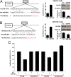
miR-484 directly targets VEGFB and VEGFR2. (A, Left) Alignment of potential miR-484 binding sites in the 3′ UTR of VEGFB, (A, Center) expression of miR-484 in SKOV-3 ovarian cancer cells transfected either with control (scr) or miR-484, (A, Lower Right) Western blot analysis of VEGFB after transfection of miR-484 in SKOV-3 cells, and (A, Upper Right) densitometric ratio between the expression of tubulin and VEGFB. (B, Left) Alignment of potential miR-484 binding sites in the 3′ UTR of VEGFR2, (B, Center) expression of miR-484 in HUVEC cells transfected with control (scr) or miR-484, (B, Lower Right) Western blot analysis of VEGFR2 after transfection of miR-484 in HUVEC cells, and (B, Upper Right) densitometric ratio between the expression of tubulin and VEGFR2. (C) Luciferase assay showing decreased luciferase activity in cells cotransfected with pGL3-VEGFB-3′ UTR or pGL3-VEGFR2-3′ UTR and control or miR-484 oligonucleotides. Mutations in the putative miR-484 binding sites, depicted in red in A for VEGFB and B for VEGFR2, abrogate this effect (Mut). Bars indicate Firefly luciferase activity normalized to Renilla luciferase activity ± SD. Each reporter plasmid was transfected in SKOV-3 cells at least two times (on different days), and each sample was assayed in triplicate.
Fig. 4.
Fig. 4.
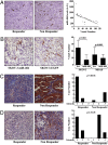
miR-484 levels correlate with vessel density in ovarian cancer samples. (A) Human tumors: CD34 staining (brown) of (Left) responder and (Center) nonresponder tumors showing a higher vascular density with pronounced microvessel formation in the latter. (Right) Regression analysis of miR-484 and vessel number in the same samples. (B) Mouse tumors: CD34 staining (brown) of SKOV-3 cell lines xenograft tumors transduced with (Left) miR-484 or (Center) control (EGFP) showing a higher vascular density in the latter. (Right) Tumor vessel count in mouse xenograft tumors transduced with control (EGFP) or miR-484 in SKOV-3 or MDAH-2774 cells. (C) VEGFB staining in human tumors: responder tumor showing (Left) weak (intensity 1) cytoplasmic VEGFB staining. (Center) Nonresponder tumor showing strong (intensity 3) cytoplasmic staining of VEGFB. (Right) Case number (y axis) and intensity of staining in the different groups (x axis). (D) VEGFR2 staining in human tumors: responder tumor showing (Left) weak (intensity 1) cytoplasmic VEGFR2 staining. (Center) Nonresponder tumor showing strong (intensity 3) cytoplasmic staining of VEGFR2. (Right) Case number (y axis) and intensity of staining in the different groups (x axis). The significant differences are reported in each graph as evaluated by nonparametric t tests. Differences between groups (responder vs. nonresponder) were considered significant when P < 0.05.
Fig. 5.
Fig. 5.
miR-484 is secreted by ovarian cancer cells and targets VEGFR2 in endothelial cells. (A and B) Expression of miR-484 in (A) ovarian cancer cells and (B) their conditioned mediums in cells stably transduced with control or miR-484 vectors. (C) Levels of miR-484 in HUVEC cells cocultured with ovarian cancer-derived cell lines. (D) Confocal microscopy image of cocultured SKOV-3 cells transfected with miR-484–fluorescin-conjugated (green) and HUVEC stained with CD31 antibody–Texas Red-conjugated (red). (E, Left) Western blot analysis of VEGFR2 expression in HUVEC cells cultured in CM from SKOV-3 cells stable transfected with miR-484 or EGFP. (E, Right) Densitometric ratio between the expression of tubulin, and VEGFR2. The significant differences are reported in each graph as evaluated by nonparametric t tests. Differences were considered significant when P < 0.05.

References

    1. Jemal A, et al. Cancer statistics, 2008. CA Cancer J Clin. 2008;58(2):71–96. - PubMed
    1. Bookman MA, et al. Evaluation of new platinum-based treatment regimens in advanced-stage ovarian cancer: A Phase III Trial of the Gynecologic Cancer Intergroup. J Clin Oncol. 2009;27(9):1419–1425. - PMC - PubMed
    1. Bartel DP. MicroRNAs: Genomics, biogenesis, mechanism, and function. Cell. 2004;116(2):281–297. - PubMed
    1. Harfe BD. MicroRNAs in vertebrate development. Curr Opin Genet Dev. 2005;15(4):410–415. - V体育平台登录 - PubMed
    1. Vecchione A, Croce CM. Apoptomirs: Small molecules have gained the license to kill. Endocr Relat Cancer. 2010;17(1):F37–F50. - PubMed

Publication types

MeSH terms

Associated data