<b draggable="ghTcWI"><bdo draggable="KXwunYA"></bdo></b><area dropzone="0QGaq75"></area> Skip to main page content
U.S. flag

An official website of the United States government

Dot gov

The . gov means it’s official. Federal government websites often end in . gov or . mil VSports app下载. Before sharing sensitive information, make sure you’re on a federal government site. .

Https

The site is secure V体育官网. The https:// ensures that you are connecting to the official website and that any information you provide is encrypted and transmitted securely. .

. 2013:2013:865604.
doi: 10.1155/2013/865604. Epub 2013 Feb 26.

Inhibitors of Fatty Acid Synthesis Induce PPAR α -Regulated Fatty Acid β -Oxidative Genes: Synergistic Roles of L-FABP and Glucose

Affiliations

Inhibitors of Fatty Acid Synthesis Induce PPAR α -Regulated Fatty Acid β -Oxidative Genes: Synergistic Roles of L-FABP and Glucose

Huan Huang et al. PPAR Res. 2013.

Abstract

While TOFA (acetyl CoA carboxylase inhibitor) and C75 (fatty acid synthase inhibitor) prevent lipid accumulation by inhibiting fatty acid synthesis, the mechanism of action is not simply accounted for by inhibition of the enzymes alone. Liver fatty acid binding protein (L-FABP), a mediator of long chain fatty acid signaling to peroxisome proliferator-activated receptor- α (PPAR α ) in the nucleus, was found to bind TOFA and its activated CoA thioester, TOFyl-CoA, with high affinity while binding C75 and C75-CoA with lower affinity. Binding of TOFA and C75-CoA significantly altered L-FABP secondary structure. High (20 mM) but not physiological (6 mM) glucose conferred on both TOFA and C75 the ability to induce PPAR α transcription of the fatty acid β -oxidative enzymes CPT1A, CPT2, and ACOX1 in cultured primary hepatocytes from wild-type (WT) mice. However, L-FABP gene ablation abolished the effects of TOFA and C75 in the context of high glucose. These effects were not associated with an increased cellular level of unesterified fatty acids but rather by increased intracellular glucose. These findings suggested that L-FABP may function as an intracellular fatty acid synthesis inhibitor binding protein facilitating TOFA and C75-mediated induction of PPAR α in the context of high glucose at levels similar to those in uncontrolled diabetes. VSports手机版.


Figures

Figure 1
Figure 1
Structural comparisons of the fatty acid synthesis inhibitors TOFA and C75 with natural and fluorescent fatty acids.
Figure 2
Figure 2
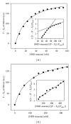
Representative binding curves of stearic acid binding to L-FABP (panel a) and SCP2 (panel b). NBD-stearic acid binding curves were obtained as described in Methods. Briefly, a 2 mL sample of L-FABP (25 nM) or SCP-2 (25 nM) in 10 mM phosphate buffer (pH 7.4) was titrated with small increments of NBD stearate at 24°C (Methods). NBD-stearate fluorescence emission spectra (515–600 nm) were recorded with 490 nm excitation. F: fluorescence intensity of NBD stearate in the presence of proteins (at 530 nm for binding to SCP-2 and at 548 nm for binding to L-FABP) and F 0 being NBD-stearate fluorescence intensity in buffer (at the same wavelength as for F). Insets are double reciprocal plots of the fluorescence binding data in the same panel.
Figure 3
Figure 3
Displacement of L-FABP- and SCP2-bound NBD-stearic acid by TOFA (panels a and b) and C75 (panels c and d). NBD-stearic acid displacement assays were performed as shown in Methods. L-FABP or SCP-2 (25 nM in 10 mM phosphate buffer) was incubated with NBD stearate (40 nM) for 12 min at 24°C to obtain maximal fluorescence, then titrated with increasing amount of ligand. Mean ± SEM, n = 3.
Figure 4
Figure 4
UV spectra and HPLC analysis of TOFA-CoA and C75-CoA. Ultraviolet spectra of TOFyl-CoA and C75-CoA were obtained with a Cary 100 Scan UV-Visible Spectrophotometer (Varian, Inc., Palo Alto, CA, USA) as described in Methods. TOFyl-CoA and C75-CoA were purified by HPLC as previously described [15]. When the final purified TOFyl-CoA and C75-CoA were reapplied to the HPLC column, representative HPLC runs detected only single absorbance peaks at 260 nm for TOFyl-CoA and C75-CoA with retention times of 13 and 9 min, respectively.
Figure 5
Figure 5
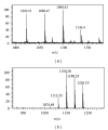
Mass spectral characterization of CoA derivatives of TOFA and C75. HPLC purified TOFYL-CoA (Panel a, m/z = 1074.69) and C75-CoA (Panel b, m/z = 1022.51) were examined by matrix-assisted laser desorption ionization time-of-flight (MALDI-TOF) mass spectrometry utilizing a Shimadzu/Kratos Axima CFR MALDI-TOF mass spectrometer (Columbia, MD, USA) in reflectron mode. Samples were analyzed by the dried-drop method using α-4-hydroxycinnamic acid (Sigma-Aldrich) as the matrix. The instrument was calibrated with angiotensin (m/z = 1046.5) and fibrinopeptide B (m/z = 1570.7). The calibrants were obtained from Sigma-Aldrich. The parent ions for TOFyl-CoA and C75-CoA were obtained at m/z = 1022.51 (a) and m/z = 1074.69 (b), respectively.
Figure 6
Figure 6
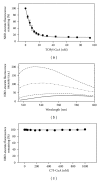
Effect of TOFyl-COA on fluorescence emission characteristics of L-FABP-bound NBD-stearic acid. NBD-stearic acid, bound to L-FABP as in Figure 3, was excited at 490 nm and fluorescence emission spectra obtained before and after addition of TOFyl-CoA (Methods). Panel (a): fluorescence emission spectra of L-FABP- (25 nM) bound NBD stearate (80 nM) without (filled circles) and with (open circles) of TOFyl-CoA (1500 nM). With increasing TOFyl-CoA concentration, the emission maximum of L-FABP-bound NBD stearate shifted to lower wavelength (panel b), and fluorescence intensity increased (panel c).
Figure 7
Figure 7
Effect of TOFyl-CoA and C75-CoA on NBD stearic binding to SCP-2. SCP-2 (25 nM in 10 mM phosphate buffer) was incubated with NBD-stearic acid (40 nM) for 12 min at 24°C to obtain maximal fluorescence. The solution was titrated with increasing amount of ligand (TOFyl-CoA or C75-CoA). TOFyl-CoA displaced SCP2-bound NBD stearate (panel a, with representative spectra in panel b). Panel (b), from top to bottom: shot dash line: NBDS+SCP2; dash-double dot-dash line: NBDS+SCP2+TOFyl-CoA (10 nM); long dashed line: NBDS+SCP2+TOFyl-CoA (100 nM); solid line: NBDS; dotted line: NBDS+TOFyl-CoA (100 nM). C75-CoA did not displace SCP2-bound NBD-stearate (panel c). Panel (a), mean ± SEM, n = 3.
Figure 8
Figure 8
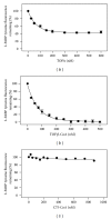
TOFA, TOFyl-CoA, and C75-CoA binding to L-FABP as determined by quenching of intrinsic L-FABP tyrosine quenching. L-FABP tyrosine quenching by TOFA (panel a), TOFyl-CoA (panel b), and C75-CoA (panel c) was determined as described in Methods. Tyrosine fluorescence emission of L-FABP (100 nM) in 10 mM phosphate buffer (pH = 7.4) was monitored by scanning from 290 to 400 nm, with excitation wavelength 280 nm, before and after small increments of added binding ligand. The binding curve was constructed by plotting percentage of tyrosine fluorescence intensity remaining at 305 nm versus concentration of the ligand. Panel (a) and (b), mean ± SEM, n = 3.
Figure 9
Figure 9
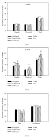
Selective fatty acid synthesis inhibitors and their CoA thioesters alter L-FABP secondary structure determined by circular dichroism (CD). L-FABP (1 μM) was incubated in the absence or presence of 10 μM ligand for 10 min at 25°C. Circular dichroism (CD) spectra were obtained utilizing a JASCO J-815 CD spectrometer (JASCO Analytical Instruments, Easton, MD, USA). Each sample spectrum represented the average of ten scans, and each sample spectrum was baseline corrected. Secondary structure was determined using the CONTIN algorithm as supplied by the instrument manufacturer. Statistical significance of secondary structure differences was determined by one-way ANOVA with the Newman-Keuls posttest (n = 3). *P < 0.05 for L-FABP + ligand versus rat L-FABP (no ligand); **P < 0.01 for rat L-FABP + ligand versus rat L-FABP (no ligand); ***P < 0.001 for rat L-FABP + ligand versus rat L-FABP (no ligand).
Figure 10
Figure 10
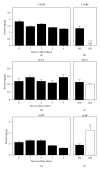
Expression of LCFA and LCFA-CoA binding proteins in cultured mouse primary hepatocytes. Primary hepatocytes from wild-type and L-FABP null mice were isolated from mouse livers and maintained in culture for up to four days as described in Methods. Quantitative western blotting was performed by comparison to a standard curve of known amounts of the respective recombinant L-FABP, SCP-2, or ACBP on the same blot as described in Methods. Quantitative western blots were obtained as a function of increasing time for wild-type hepatocytes in culture: (a) L-FABP; (b) SCP-2; and (c) ACBP. Time 0 = concentration in liver while time of 1–4 days indicates time in culture. For determining the effect of L-FABP gene ablation on expression of these proteins, quantitative western blots of (d) L-FABP; (e) SCP-2; and (f) ACBP were also obtained for hepatocytes from wild-type (WT) and L-FABP null (KO) hepatocytes after three days in culture. Mean ± SEM, n = 3–6.
Figure 11
Figure 11
Expression of key nuclear receptors involved in fatty acid and glucose metabolism as a function of time. Primary hepatocytes were isolated from mouse livers and maintained in culture for up to four days as described in Methods. Representative western blots relative to a housekeeping protein (COX-1) are shown in the inserts. Quantitative analysis of multiple western blots relative to housekeeping protein was shown as black bars for PPARα (Panel a), LXRα (Panel b), SREBP1 (Panel c), and ChREBP (Panel d) as described in Methods. Time 0 = concentration in liver while time of 1–4 days indicates time in culture. Mean ± SEM, n = 3–6.
Figure 12
Figure 12
Effect of TOFA and C75 on CPT1, CPT2, and ACOX1 gene expression in cultured mouse primary hepatocytes isolated from livers of wild-type (WT, L-FABP (+/+)) and null [(L-FABP (−/−)] mice. Hepatocytes isolated from wild-type [WT, L-FABP (+/+)] or gene-ablated [null, L-FABP (−/−)] mice were preincubated for 30 min with 10 μg/mL TOFA or C75 in serum-free culture medium before addition of glucose (6 or 20 mM) as described in Methods. Total RNA was isolated from hepatocytes 5 hr after glucose addition and used for quantitative real-time PCR. The fold change in CPT1A (a, b), CPT2 (c, d), ACOX1 (e, f) mRNA levels was determined relative to internal control housekeeping gene as described in Methods. Values for each genotype were expressed relative to [Alb + 6 mM glucose] within that genotype. Panels (a), (b): CPT1A mRNA fold changes in WT and L-FABP null hepatocytes; (c), (d): CPT2 mRNA fold changes in WT and L-FABP null hepatocytes; (e), (f): ACOX1 mRNA fold changes in WT and L-FABP null hepatocytes. Mean ± SEM, n = 3.
Figure 13
Figure 13
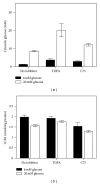
Cytosolic glucose (panel a) and free fatty acid (LCFA, panel b) levels in mouse primary hepatocytes cultured with and without TOFA or C75. Hepatocytes were incubated with TOFA or C75 (10 μg/mL) with 6 or 20 mM glucose (Section 2). Cytosolic glucose level was then determined as in Methods. Briefly, hepatocytes were washed quickly with ice old solution of MgCl2 (100 mM) with 0.1 mM phloretin. Cells were then homogenized with a probe sonicator and, after centrifugation, the supernatant was used for glucose analysis with the Amplite Glucose Quantitation Kit as instructed by the manufacturer. For LCFA determination, hepatocyte homogenate was extracted twice with 1% Triton X-100 in pure chloroform. The organic phase was collected, and the FFA content of each sample was measured with the Free Fatty Acid Quantification Kit from BioVision, Inc. according to manufacturer's instructions using enclosed palmitic acid as standard. Mean ± SEM, n = 3.

"VSports" References

    1. Landree LE, Hanlon AL, Strong DW, et al. C75, a fatty acid synthase inhibitor, modulates AMP-activated protein kinase to alter neuronal energy metabolism. The Journal of Biological Chemistry. 2004;279(5):3817–3827. - "VSports app下载" PubMed
    1. Loftus TM, Jaworsky DE, Frehywot CL, et al. Reduced food intake and body weight in mice treated with fatty acid synthase inhibitors. Science. 2000;288(5475):2379–2381. - PubMed
    1. Tu Y, Thupari JN, Kim E-K, et al. C75 alters central and peripheral gene expression to reduce food intake and increase energy expenditure. Endocrinology. 2005;146(1):486–493. - PubMed
    1. Anderson N, Borlak J. Molecular mechanisms and therapeutic targets in steatosis and steatohepatitis. Pharmacological Reviews. 2008;60(3):311–357. - "VSports最新版本" PubMed
    1. Kneeman JM, Misdraji J, Corey KC. Secondary causes of nonalcoholic liver disease. Therapeutic Advances in Gastroenterology. 2012;5(3):199–207. - PMC - PubMed

VSports注册入口 - LinkOut - more resources