Skip to main page content
U.S. flag

An official website of the United States government

Dot gov

The . gov means it’s official. Federal government websites often end in VSports app下载. gov or . mil. Before sharing sensitive information, make sure you’re on a federal government site. .

Https

The site is secure V体育官网. The https:// ensures that you are connecting to the official website and that any information you provide is encrypted and transmitted securely. .

. 2013 May 17;288(20):14372-14383.
doi: 10.1074/jbc.M113.459602. Epub 2013 Mar 26.

Microsomal triglyceride transfer protein inhibition induces endoplasmic reticulum stress and increases gene transcription via Ire1α/cJun to enhance plasma ALT/AST (VSports)

Affiliations

Microsomal triglyceride transfer protein inhibition induces endoplasmic reticulum stress and increases gene transcription via Ire1α/cJun to enhance plasma ALT/AST

Joby Josekutty et al. J Biol Chem. .

VSports在线直播 - Abstract

Microsomal triglyceride transfer protein (MTP) is a target to reduce plasma lipids because of its indispensable role in triglyceride-rich lipoprotein biosynthesis. MTP inhibition in Western diet fed mice decreased plasma triglycerides/cholesterol, whereas increasing plasma alanine/aspartate aminotransferases (ALT/AST) and hepatic triglycerides/free cholesterol. Free cholesterol accumulated in the endoplasmic reticulum (ER) and mitochondria resulting in ER and oxidative stresses VSports手机版. Mechanistic studies revealed that MTP inhibition increased transcription of the GPT/GOT1 genes through up-regulation of the IRE1α/cJun pathway leading to increased synthesis and release of ALT1/AST1. Thus, transcriptional up-regulation of GPT/GOT1 genes is a major mechanism, in response to ER stress, elevating plasma transaminases. Increases in plasma and tissue transaminases might represent a normal response to stress for survival. .

Keywords: Cardiovascular Disease; Cell Metabolism; Cholesterol; Endoplasmic Reticulum Stress; Lipid Binding Protein; Lipid Metabolism; Lipids; Lipoprotein; Lipoprotein Metabolism; Liver Injury. V体育安卓版.


Figures

FIGURE 1.
FIGURE 1.
Association between hepatic free cholesterol in MTP inhibited mice and increases in plasma ALT/AST. Male C57BL/6J mice were fed a Western diet for 30 days and divided into 4 groups (n = 4/group) and continued to receive Western diet ad libitum for an additional week. During the last week, mice were gavaged daily with DMSO (Control), MTPi (1 mg of BMS212122/kg/day), MTPi + pioglitazone (MTPi + Pio, 25 mg/kg/day), or MTPi + lovastatin (MTPi + Lo, 50 mg/kg/day). Data are representative of three experiments. A–I, hepatic tissue was used to measure MTP activity and lipids (A–D). Plasma was used to quantify lipids and transaminases (E–I). Comparisons with Controls and MTPi are designated with * and #, respectively. Values are mean ± S.D.; *, #, p < 0.05; **, ##, p < 0.01; ***, ###, p < 0.001. J and K, values for hepatic free cholesterol were plotted against plasma ALT (J) and AST (K) levels from 6 independent experiments. A linear regression and Pearson correlation was performed. The black line represents the linear regression with the red dots adjacent to the line indicate the 95% confidence interval. The r2 and significance of correlation were determined after the analysis with a Pearson correlation test. L, representative Oil Red O-stained sections of livers.
FIGURE 2.
FIGURE 2.
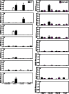
MTP inhibition increases free cholesterol in different organelles and induces oxidative and ER stress. A–D, male C57BL/6J mice fed a Western diet were gavaged with DMSO (Control, n = 4), MTPi (n = 5), or MTPi + pioglitazone (MTPi + Pio, n = 5). Data are representative of 3 different experiments. Subcellular liver fractions were utilized to measure MTP activity (A), triglycerides (B), free cholesterol (C), and total GSH (D). E, Western blot analysis of ER stress response proteins. F, mRNA levels of hepatic ER stress effectors were measured by quantitative RT-PCR and normalized to ARPp0 mRNA. Comparisons with Control groups are designated with *. Comparisons with MTPi groups are designated with #. Values are mean ± S.D.; *, #, p < 0.05; **, ##, p < 0.01; ***, ###, p < 0.001.
FIGURE 3.
FIGURE 3.
MTP inhibitor does not increase cytokine levels or cell death. A–C, C57BL/6J (n = 6) mice were fed Western diet for 37 days. DMSO (Control) or MTPi was administered in the last 7 days. A, ELISA was used to assay plasma cytokines (SABiosciences). Plasma samples from 2 animals were pooled. Therefore, data are average and S.D. of 3 determinations. B, Western blot analysis of proteins associated with inflammation. C, protein bands were quantified. D, lactate dehydrogenase (LDH) was assessed from plasma samples obtained from animals as described in the legend to Fig. 2. E, different caspase activities were measured in the livers of mice from Fig. 2. F, C57BL/6J mice on chow diet were injected intraperitoneally with 1.5 mg/kg of tunicamycin. Livers were collected after 24 h and used to measure caspase activities. G, Huh-7 cells on coverslips were treated with either 1 μm MTPi or staurosporine, stained with propidium iodide (PI), and photographed. Data are representative of three experiments. H–K, Huh-7 cells (n = 3) were treated with 0.5, 1, or 2 μm MTPi or staurosporine (STS) for 24 h. Media were used to measure ALT/AST (H and I). Cells were fixed, stained with PI, and analyzed by flow cytometry. Percent of dead cells (J) is plotted. Flow diagrams (K) illustrate sub-diploid/non-viable and diploid/viable cell counts. Data are representative of three experiments. Comparisons with Controls and MTPi are designated with * and #, respectively. Values are mean ± S.D.; *, #, p < 0.05; **, ##, p < 0.01; ***, ###, p < 0.001.
FIGURE 4.
FIGURE 4.
MTPi increases transcription of GPT and GOT1. A, Huh-7 cells (n = 3) were treated with 1 μm MTPi for 48 h and ALT1/2 (GPT/GPT2) and AST1/2 (GOT1/GOT2) mRNA levels were measured. B and C, Huh-7 cells were treated with or without 1 μm MTPi for 48 h. Cells then received actinomycin D (10 μg/ml) for the indicated times. Cell lysates were used to quantify mRNA levels and ratios at time 0 were normalized to 100%. Percent change with time is plotted. D, Huh-7 cells (n = 3) were transfected with plasmids (1 μg) expressing firefly luciferase under the control of GPT and GOT1 promoters for 24 h, and then treated with 1 μm MTPi or 1 μm tunicamycin for 24 h. Luminescence was determined in cell lysates (n = 3 per group). Data are representative of three experiments. Values are mean ± S.D. *, p < 0.05; **, p < 0.01; ***, p < 0.001 compared with DMSO treatment (Control).
FIGURE 5.
FIGURE 5.
MTP inhibition increases synthesis of ALT1 and AST1. A, Huh-7 cells treated with 1 μm MTPi for 24 h were continuously labeled with 150 μCi of 35S-Promix for the indicated times. Different proteins were immunoprecipitated using appropriate antibodies from cell lysates and media. Representative data are from 4 experiments. B–G, quantifications of proteins shown in panel A. H, Huh-7 cells were treated with or without 1 μm MTPi for 24 h, pulse labeled with 300 μCi of 35S-Promix labeling mix for 1 h, and chased with 1% FBS containing DMEM with/without MTPi in the presence of 1 mm cold l-methionine/l-cysteine for the indicated times. Proteins were immunoprecipitated from cell lysates and media using specific antibodies. Data are representative of five experiments. I–P, quantifications of proteins shown in panel H. Values at the beginning of the chase time 0 (lysate time 0) were normalized to 100%. Values at different times in cell lysates and media represent a percentage of the values at time 0. AU, absorbance unit.
FIGURE 6.
FIGURE 6.
MTPi induced elevations in plasma ALT/AST are IRE1α dependent. A, siRNA (100 pmol) targeting human IRE1α, PERK, and ATF-6 were transfected for 24 h into Huh-7 cells and then treated with or without 1 μm MTPi for an additional 24 h. mRNA were extracted to measure different transcripts by qRT-PCR. Data were normalized to ARPp0 mRNA (n = 3). Data are representative of three experiments. B–I, 14 days after the onset of Western diet feeding, Ire1αfl/fl animals were divided into 2 groups (n = 4/group) and injected intravenously with either AAV2/8.TBG.PI.luciferase.RBG (Ire1αfl/fl) or AAV2/8.TBG.PI.Cre.RBG (L-Ire1α−/−). Beginning at day 30, animals were gavaged daily with or without MTPi for 7 days. Data are representative of two experiments. Hepatic mRNA was used to measure gene expression (B). Liver homogenates were used to measure MTP activity and lipids (C–E). Plasma samples were used to quantify lipids (F and G) and transaminases (H and I). Comparisons with Ire1αfl/fl animals are designated with *. Comparisons with Ire1αfl/fl + MTPi-treated animals are designated with #. Values are mean ± S.D. *, #, p < 0.05
FIGURE 7.
FIGURE 7.
IRE1α mediates transcriptional activation of GPT and GOT1 through cJun. A, proposed schematic model of MTPi-mediated elevations in plasma/media ALT1 and AST1. Cellular free cholesterol accumulation after MTPi treatment induces ER stress. Autophosphorylation of IRE1α leads to splicing of XBP-1. Furthermore, phosphorylated IRE1α interacts with Traf-2 and Ask1 to activate JNK, leading to phosphorylation of the cJun transcription factor. In the nucleus, a schematic diagram shows conserved cis regions in GPT and GOT1 promoters for the binding of XBP-1, cJun, p53, and NF-κB identified after aligning promoter sequences from different species. P1, P2, P3, and P4 denote different promoter regions amplified using primers (→ and ←) described in supplemental Table S1. B, Western blot analysis of XBP-1(s) and p-cJun in liver homogenates of Ire1αfl/fl and L-Ire1α−/− mice treated or not with MTPi in Fig. 5. C, quantification of blots in panel B. D–G, Huh7 cells (n = 3) were transfected with 100 pmol of siRNA targeting cJun or XBP-1 for 24 h. Cells were then treated with/without 1 μm MTPi for 24 h. Lysates (D and E) and media (F and G) were harvested for Western blot analysis and ALT/AST activity measurements, respectively. Comparisons with MTPi-treated siControl cells are designated with * and #, respectively. Values are mean ± S.D. *, #, p < 0.05; **, ##, p < 0.01; ***, ###, p < 0.001.
FIGURE 8.
FIGURE 8.
cJun binds to the GPT and GOT1 promoters in MTP inhibited cells. A–G, Huh-7 cells treated with or without 1 μm MTPi were utilized for ChIP using control IgG, anti-polymerase II, anti-p-cJun, and anti-XBP-1 antibodies. Quantitative PCR was performed to amplify different promoter regions and data are represented as a percent of input DNA. Regions that showed significant amplifications are shown. Comparisons with controls are designated with *. Values are mean ± S.D., n = 3. *, p < 0.05; **, p < 0.01; ***, p < 0.001. H–N, siControl or si-cJun (100 pmol) were transfected into Huh-7 cells for 24 h. Cells were treated with 1 μm MTPi for an additional 24 h. ChIP was performed utilizing antibodies targeting XBP-1, p-cJun, NF-κB, and p53. Positive association of transcription factors was ascertained when a minimum of 0.1% input DNA was amplified. Comparisons with siControl-treated cells are designated with *. Values are mean ± S.D. *, p < 0.05; **, p < 0.01; ***, p < 0.001; n = 3 per group.

References

    1. Grundy S. M., Cleeman J. I., Merz C. N., Brewer H. B., Jr., Clark L. T., Hunninghake D. B., Pasternak R. C., Smith S. C., Jr., Stone N. J., and Coordinating Committee of the National Cholesterol Education Program (2004) Implications of recent clinical trials for the National Cholesterol Education Program Adult Treatment Panel III Guidelines. J. Am. Coll. Cardiol. 44, 720–732 - PubMed
    1. LaRosa J. C., Grundy S. M., Waters D. D., Shear C., Barter P., Fruchart J.-C., Gotto A. M., Greten H., Kastelein J. J., Shepherd J., Wenger N. K., and Treating to New Targents (TNT) Investigators (2005) Intensive lipid lowering with atorvastatin in patients with stable coronary disease. N. Engl. J. Med. 352, 1425–1435 - PubMed
    1. Hussain M. M., Rava P., Walsh M., Rana M., Iqbal J. (2012) Multiple functions of microsomal triglyceride transfer protein. Nutr. Metab. 9, 14 - PMC - PubMed
    1. Cuchel M., Bloedon L. T., Szapary P. O., Kolansky D. M., Wolfe M. L., Sarkis A., Millar J. S., Ikewaki K., Siegelman E. S., Gregg R. E., Rader D. J. (2007) Inhibition of microsomal triglyceride transfer protein in familial hypercholesterolemia. N. Engl. J. Med. 356, 148–156 - PubMed
    1. Liu R., Pan X., Whitington P. F. (2009) Increased hepatic expression is a major determinant of serum alanine aminotransferase elevation in mice with nonalcoholic steatohepatitis. Liver Int. 29, 337–343 - "V体育ios版" PubMed

Publication types

MeSH terms

LinkOut - more resources