Skip to main page content (V体育平台登录)
U.S. flag

An official website of the United States government

Dot gov

The . gov means it’s official. Federal government websites often end in . gov or . mil. Before sharing sensitive information, make sure you’re on a federal government site VSports app下载. .

Https

The site is secure. The https:// ensures that you are connecting to the official website and that any information you provide is encrypted and transmitted securely. V体育官网.

. 2013 Feb 1;304(3):G241-56.
doi: 10.1152/ajpgi.00334.2012. Epub 2012 Dec 13.

VSports - Impact of L-FABP and glucose on polyunsaturated fatty acid induction of PPARα-regulated β-oxidative enzymes

Affiliations

Impact of L-FABP and glucose on polyunsaturated fatty acid induction of PPARα-regulated β-oxidative enzymes

Anca D Petrescu et al. Am J Physiol Gastrointest Liver Physiol. .

"V体育2025版" Abstract

Liver fatty acid binding protein (L-FABP) is the major soluble protein that binds very-long-chain n-3 polyunsaturated fatty acids (n-3 PUFAs) in hepatocytes. However, nothing is known about L-FABP's role in n-3 PUFA-mediated peroxisome proliferator activated receptor-α (PPARα) transcription of proteins involved in long-chain fatty acid (LCFA) β-oxidation. This issue was addressed in cultured primary hepatocytes from wild-type, L-FABP-null, and PPARα-null mice with these major findings: 1) PUFA-mediated increase in the expression of PPARα-regulated LCFA β-oxidative enzymes, LCFA/LCFA-CoA binding proteins (L-FABP, ACBP), and PPARα itself was L-FABP dependent; 2) PPARα transcription, robustly potentiated by high glucose but not maltose, a sugar not taken up, correlated with higher protein levels of these LCFA β-oxidative enzymes and with increased LCFA β-oxidation; and 3) high glucose altered the potency of n-3 relative to n-6 PUFA. This was not due to a direct effect of glucose on PPARα transcriptional activity nor indirectly through de novo fatty acid synthesis from glucose. Synergism was also not due to glucose impacting other signaling pathways, since it was observed only in hepatocytes expressing both L-FABP and PPARα. Ablation of L-FABP or PPARα as well as treatment with MK886 (PPARα inhibitor) abolished/reduced PUFA-mediated PPARα transcription of these genes, especially at high glucose. Finally, the PUFA-enhanced L-FABP distribution into nuclei with high glucose augmentation of the L-FABP/PPARα interaction reveals not only the importance of L-FABP for PUFA induction of PPARα target genes in fatty acid β-oxidation but also the significance of a high glucose enhancement effect in diabetes. VSports手机版.


Figures

Fig. 1.
Fig. 1.
Glucose enhanced upregulation of peroxisome proliferator activated receptor-α (PPARα)-regulated gene transcription by polyunsaturated fatty acids (PUFA) in wild-type (WT) mouse hepatocytes. As indicated by the vertical bars from left to right, WT mouse hepatocytes were cultured for 6 h in serum-free medium containing glucose (6, 11, 20, or 30 mM) or (6 mM glucose + 14 mM maltose). Culture medium was supplemented with either fatty acid-free albumin (Alb, 40 μM) or Alb complexed with 200 μM arachidonic acid (AA), eicosapentaenoic acid (EPA), or docosahexaenoic acid (DHA) as described in materials and methods. The fold change in CPT1A (A), CPT2 (B), and ACOX1 (C) mRNA levels was determined relative to internal control housekeeping gene as described in materials and methods. Values were expressed relative to Alb + 6 mM glucose. Values are means ± SE, n = 3–4. *P < 0.05 vs. Alb alone at the same glucose concentration; #P < 0.05 vs. 6 mM glucose concentration within each treatment group.
Fig. 2.
Fig. 2.
Effect of PUFA and high glucose on PPARα transcription of proteins in LCFA uptake/transport [liver fatty acid binding protein (L-FABP), acyl-CoA binding protein (ACBP)] and nuclear regulation (PPARα, HNF4α, HNF1α) in WT mouse hepatocytes. Total RNA was isolated from WT mouse hepatocytes treated with Alb only (40 μM) or PUFA (AA, EPA, or DHA, 200 μM) and Alb with 6 or 20 mM glucose for 6 h. The RNA was analyzed by quantitative PCR for changes in L-FABP mRNA (A), ACBP mRNA (B), PPARα mRNA (C), HNF4α mRNA (D), and HNF1α mRNA (E). Values are means ± SE, n = 4, P < 0.05. *Significant difference between lipid and Alb at constant concentration of glucose; #significant difference between 6 and 20 mM glucose for the same lipid.
Fig. 3.
Fig. 3.
L-FABP gene ablation markedly reduced high glucose potentiation of PUFA-mediated PPARα transcription of genes in LCFA β-oxidation, transport (ACBP), and PPARα. As indicated by the vertical bars from left to right, L-FABP-null hepatocytes were cultured for 6 h in serum-free medium containing glucose (6, 11, 20 or 30 mM) supplemented with either fatty acid-free Alb (40 μM) or Alb complexed with 200 μM AA, EPA, or DHA as described in materials and methods. Total RNA was isolated and relative fold change in the expression level of PPARα-regulated genes was determined by quantitative PCR: CPT1A mRNA (A), CPT2 mRNA (B), ACOX1 mRNA (C), ACBP mRNA (D), and PPARα mRNA (E). Values for each genotype were expressed relative to Alb + 6 mM glucose within that genotype. Values are means ± SE, n = 3–4. *P < 0.05 vs. Alb alone at the same glucose concentration; #P < 0.05 vs. 6 mM glucose concentration within each treatment group.
Fig. 4.
Fig. 4.
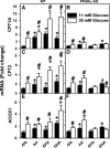
PPARα gene ablation markedly reduced both PUFA-mediated PPARα transcription of fatty acid β-oxidative genes as well as potentiation by high glucose. Primary hepatocytes from WT (A, C, and E) and PPARα-knockout (PPARα-KO; B, D, and F) mice were isolated and cultured with serum-free medium containing 11 or 30 mM glucose plus 40 μM fatty acid-free Alb or Alb complexed with 200 μM of AA, EPA, or DHA as described in materials and methods. CPT1A mRNA (A and B), CPT2 mRNA (C and D), and ACOX1 mRNA (E and F) levels were then measured relative to an internal housekeeping gene control as described in materials and methods. Values for each genotype were then expressed relative to Alb + 6 mM glucose treatment within that genotype. Values are means ± SE, n = 3–4. *P < 0.05 vs. Alb at the same glucose concentration; #P < 0.05 for 30 mM vs. 11 mM glucose within each treatment group (i.e., Alb, AA, EPA, or DHA).
Fig. 5.
Fig. 5.
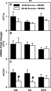
PPARα inhibitor MK886 prevented stimulation of fatty acid β-oxidation gene transcription by high glucose in WT mouse hepatocytes. CPT1A (A), CPT2 (B), and ACOX1 (C) mRNA levels were measured in hepatocytes cultured with serum-free medium containing glucose (6 or 20 mM) and Alb (40 μM) or Alb complexed with 200 μM of AA or EPA. Values are means ± SE, n = 3–4. *P < 0.05 vs. Alb at the same glucose concentration. #P < 0.05 for 20 mM vs. 6 mM glucose within each lipid treatment group (i.e., Alb, AA, EPA).
Fig. 6.
Fig. 6.
Western blots for CPT2 and ACOX1: effect of PUFA and high glucose. Hepatocytes from WT L-FABP (+/+) mice were treated with Alb only (Alb, 40 μM) as negative control and Alb in complex with PUFA (AA, EPA, DHA, 200 μM) in medium containing either 6 or 20 mM glucose for 24 h followed by Western blotting (see materials and methods). A: Adobe Photoshop (Adobe Systems, San Jose, CA) and CorelDraw X5 (Corel, Ottawa, ON, Canada) were used to crop and compile the cropped images from Western blots for CPT2, ACOX1, and β-actin (as loading control). B: relative quantitative determination of CPT2 protein level vs. treatments as determined by use of Scion Image. C: quantitative analysis of ACOX1 protein level as a function of treatments; values are means ± SE, n = 4. *P < 0.05 vs. Alb alone at the same glucose concentration; #P < 0.05 vs. 6 mM glucose concentration within each PUFA treatment group.
Fig. 7.
Fig. 7.
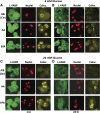
Impact of PUFA on L-FABP distribution to hepatocyte nuclei. Primary hepatocytes from WT mice were cultured with glucose (6 or 20 mM) and Alb (40 μM) or Alb in complex with 200 μM AA or EPA as described in materials and methods. After 1 and 24 h incubation, medium was removed and hepatocytes were fixed, labeled with FITC-anti-L-FABP and TO-PRO nuclear stain, and simultaneously imaged through separate photomultipliers (to detect FITC and TO-PRO) by confocal microscopy as described in materials and methods and analyzed similarly as was described earlier (35, 38, 86). Representative confocal fluorescence images of L-FABP (green, first column), nuclei (red, second column), and colocalized pixels (yellow, third column) of hepatocytes are shown for 6 mM glucose (A and B) or 20 mM glucose (C and D) after incubation for 1 h (A and C) or 24 h (B and D). Ctr, control; Coloc., colocalized.
Fig. 8.
Fig. 8.
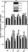
PUFA enhanced nuclear distribution of L-FABP in cultured mouse hepatocytes, as demonstrated by quantitative analysis of confocal images. WT mouse primary hepatocytes were isolated, cultured, and processed for confocal microscopy as described in the legend to Fig. 7 except that the incubation times were 0.5, 1, 4, or 24 h. Images of multiple hepatocytes were analyzed with Image J software as described in materials and methods. Graphs represent analysis of images of hepatocytes cultured in 6 mM (A) and 20 mM (B) glucose. The L-FABP fluorescence intensity per unit surface area was measured within as well as outside the nucleus area, as determined by the nuclear stain, and the ratio of nuclear L-FABP to cytoplasmic L-FABP was determined. Values are means ± SE, n = 40 cells. *P < 0.05 vs. Alb control at the same time point; #P < 0.05 vs. the identical lipid treatment at 0.5 h.

"VSports app下载" References

    1. Akiyama TE, Ward JM, Gonzalez FJ. Regulation of liver fatty acid binding protein gene by hepatocyte nuclear factors 1α (HNF1α). J Biol Chem 275: 27117–27122,2000 - PubMed
    1. Aoyama T, Peters JM, Iritani N, Nakajima T, Furihata K, Hashimoto T, Gonzalez FJ. Altered constitutive expression of fatty acid metabolizing enzymes in mice lacking PPARα. J Biol Chem 273: 5678–5684,1998 - PubMed
    1. Atshaves BP, Martin GG, Hostetler HA, McIntosh AL, Kier AB, Schroeder F. Liver fatty acid binding protein (L-FABP) and dietary obesity. J Nutr Biochem 21: 1015–1032,2010 - PMC - PubMed
    1. Atshaves BP, McIntosh AL, Kier AB, Schroeder F. High dietary fat exacerbates weight gain and obesity in female liver fatty acid binding protein gene ablated mice. Lipids 45: 97–110,2010 - PMC - PubMed
    1. Atshaves BP, McIntosh AL, Lyuksyutova OI, Zipfel WR, Webb WW, Schroeder F. Liver fatty acid binding protein gene ablation inhibits branched-chain fatty acid metabolism in cultured primary hepatocytes. J Biol Chem 279: 30954–30965,2004 - PubMed (VSports最新版本)

Publication types

MeSH terms

LinkOut - more resources (V体育平台登录)