"VSports最新版本" Skip to main page content
U.S. flag

An official website of the United States government

Dot gov

The . gov means it’s official. Federal government websites often end in . gov or . mil VSports app下载. Before sharing sensitive information, make sure you’re on a federal government site. .

Https

The site is secure. The https:// ensures that you are connecting to the official website and that any information you provide is encrypted and transmitted securely V体育官网. .

. 2012;8(8):e1002866.
doi: 10.1371/journal.ppat.1002866. Epub 2012 Aug 16.

Global metabolic profiling of infection by an oncogenic virus: KSHV induces and requires lipogenesis for survival of latent infection

Affiliations

V体育平台登录 - Global metabolic profiling of infection by an oncogenic virus: KSHV induces and requires lipogenesis for survival of latent infection

V体育2025版 - Tracie Delgado et al. PLoS Pathog. 2012.

"VSports app下载" Abstract

Like cancer cells, virally infected cells have dramatically altered metabolic requirements. We analyzed global metabolic changes induced by latent infection with an oncogenic virus, Kaposi's Sarcoma-associated herpesvirus (KSHV). KSHV is the etiologic agent of Kaposi's Sarcoma (KS), the most common tumor of AIDS patients. Approximately one-third of the nearly 200 measured metabolites were altered following latent infection of endothelial cells by KSHV, including many metabolites of anabolic pathways common to most cancer cells. KSHV induced pathways that are commonly altered in cancer cells including glycolysis, the pentose phosphate pathway, amino acid production and fatty acid synthesis. Interestingly, over half of the detectable long chain fatty acids detected in our screen were significantly increased by latent KSHV infection VSports手机版. KSHV infection leads to the elevation of metabolites involved in the synthesis of fatty acids, not degradation from phospholipids, and leads to increased lipid droplet organelle formation in the infected cells. Fatty acid synthesis is required for the survival of latently infected endothelial cells, as inhibition of key enzymes in this pathway led to apoptosis of infected cells. Addition of palmitic acid to latently infected cells treated with a fatty acid synthesis inhibitor protected the cells from death indicating that the products of this pathway are essential. Our metabolomic analysis of KSHV-infected cells provides insight as to how oncogenic viruses can induce metabolic alterations common to cancer cells. Furthermore, this analysis raises the possibility that metabolic pathways may provide novel therapeutic targets for the inhibition of latent KSHV infection and ultimately KS tumors. .


VSports注册入口 - Conflict of interest statement

The authors have declared that no competing interests exist.

Figures
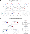
Figure 1
Figure 1. KSHV infection of endothelial cells induces fatty acid production through de novo synthesis.
Relative levels of metabolites from mock- (control) and KSHV-infected (infected) TIME cells are shown at 48 hpi (light blue and pink respectively) and 96 hpi (dark blue and red respectively). A) Box and whisker plots showing relative levels of fatty acid metabolites significantly altered by KSHV infection. B) Box and whisker plots of metabolites that differentiate production of long chain fatty acids (LCFAs) by synthesis or degradation of phospholipids indicating that increased fatty acid metabolites in KSHV infected cells come from increased synthesis.
Figure 2
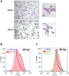
Figure 2. KSHV-infected cells induce the formation of lipid droplet organelles.
A) 48 hpi, TIME cells were fixed and stained with Oil-Red-O, a specific stain for lipid droplets, and hematoxylin, to stain cell nuclei. Cells were visualized by bright field microscopy. B) Mock- and KSHV- infected TIME cells were harvested at 48 hpi, fixed and stained with Nile Red, a specific fluorescent stain for lipid droplets. Staining was analyzed by flow cytometry. C) Mock-, KSHV- and UV-irradiated-KSHV- infected TIME cells were harvested at 96 hpi and stained as in B for lipid droplets.
Figure 3
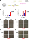
Figure 3. Fatty acid synthesis inhibitors selectively induce cell death in KSHV latently infected endothelial cells.
A) Schematic of lipogenesis pathway and where specific inhibitors block the pathway. At 48 hpi, mock- and KSHV-infected TIME cells were treated with B) 0, 10, or 20 µg/mL of TOFA and incubated for an additional 48 hours or C) 0, 4 or 8 µg/mL C75 and incubated for an additional 24 hours. Cell death rates were then determined by a trypan blue exclusion assay. Cell death rate (%) is # of dead cells/# of total cells. 48 hpi, TIME cells (D & E) or 1° hDMVECs (F & G) were treated with 0 µg/mL or 20 µg/mL TOFA for 36 hours (C & E) or 0 µg/mL or 4 µg/mL C75 for 24 hours (D & F). Cells were then treated with Image-It Dead Green viability stain and visualized on an inverted fluorescent microscope.
Figure 4
Figure 4. Fatty acid synthesis inhibitors selectively induce apoptosis in KSHV latently infected cells.
48 hpi, mock- (M) and KSHV- (K) infected cells were treated with A) 20 µg/mL TOFA for 36 hours or B) 4 µg/mL C75 for 24 hours and then harvested forWestern blot analysis with antibodies that recognize the cleaved PARP or cleaved caspase-3.
Figure 5
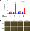
Figure 5. Palmitic Acid rescues TOFA induced cell death in KSHV latently infected endothelial cells.
A) At 48 hpi, mock- and KSHV-infected TIME cells were treated with DMSO (control) or 20 µg/mL of TOFA, in the presence or absence of 24 µM Palmitic Acid, and incubated for an additional 48 hours. Cell death rates were then determined by a trypan blue exclusion assay. Cell death rate (%) is # of dead cells/# of total cells. B) At 48 hpi, TIME cells were treated as in panel A. Cells were then treated with Image-It Dead Green viability stain and visualized on an inverted fluorescent microscope.

References

    1. Warburg O (1956) On respiratory impairment in cancer cells. Science 124: 269–270. - PubMed
    1. Coleman MC, Asbury CR, Daniels D, Du J, Aykin-Burns N, et al. (2008) 2-deoxy-D-glucose causes cytotoxicity, oxidative stress, and radiosensitization in pancreatic cancer. Free Radic Biol Med 44: 322–331. - V体育平台登录 - PubMed
    1. Aft RL, Zhang FW, Gius D (2002) Evaluation of 2-deoxy-D-glucose as a chemotherapeutic agent: mechanism of cell death. Br J Cancer 87: 805–812. - PMC - PubMed
    1. Liu H, Hu YP, Savaraj N, Priebe W, Lampidis TJ (2001) Hypersensitization of tumor cells to glycolytic inhibitors. Biochemistry (Mosc) 40: 5542–5547. - PubMed
    1. Shrivastava V, Mishra AK, Dwarakanath BS, Ravindranath T (2006) Enhancement of radionuclide induced cytotoxicity by 2-deoxy-D-glucose in human tumor cell lines. J Cancer Res Ther 2: 57–64. - PubMed

Publication types

MeSH terms