Skip to main page content
U.S. flag

An official website of the United States government

Dot gov

The . gov means it’s official. Federal government websites often end in . gov or VSports app下载. mil. Before sharing sensitive information, make sure you’re on a federal government site. .

Https

The site is secure. The https:// ensures that you are connecting to the official website and that any information you provide is encrypted and transmitted securely. V体育官网.

. 2011 Sep 30;286(39):33845-53.
doi: 10.1074/jbc.M111.274159. Epub 2011 Aug 3.

Human Mre11/human Rad50/Nbs1 and DNA ligase IIIalpha/XRCC1 protein complexes act together in an alternative nonhomologous end joining pathway

Affiliations

Human Mre11/human Rad50/Nbs1 and DNA ligase IIIalpha/XRCC1 protein complexes act together in an alternative nonhomologous end joining pathway

Julie Della-Maria et al. J Biol Chem. .

Abstract

Recent studies have implicated a poorly defined alternative pathway of nonhomologous end joining (alt-NHEJ) in the generation of large deletions and chromosomal translocations that are frequently observed in cancer cells. Here, we describe an interaction between two factors, hMre11/hRad50/Nbs1 (MRN) and DNA ligase IIIα/XRCC1, that have been linked with alt-NHEJ. Expression of DNA ligase IIIα and the association between MRN and DNA ligase IIIα/XRCC1 are altered in cell lines defective in the major NHEJ pathway. Most notably, DNA damage induced the association of these factors in DNA ligase IV-deficient cells. MRN interacts with DNA ligase IIIα/XRCC1, stimulating intermolecular ligation, and together these proteins join incompatible DNA ends in a reaction that mimics alt-NHEJ. Thus, our results provide novel mechanistic insights into the alt-NHEJ pathway that not only contributes to genome instability in cancer cells but may also be a therapeutic target VSports手机版. .


Figures (VSports注册入口)

FIGURE 1.
FIGURE 1.
DNA damage induced association of DNA ligase IIIα/XRCC1 and MRN in DNA ligase IV-deficient cells. Nuclear extracts were prepared from wild-type human pre-B cell line, Nalm6 and its lig4-null derivative, N114P2 (A) or wild-type human lymphoblastoid cell line, NC10, and DNA ligase IV mutant lymphoblastoid cell line, LB2304 (B), that had been either mock treated (0) or treated with γ-irradiation (IR, 10 grays). Left, proteins in the nuclear extracts (Input 1:5, 20 μg) were detected by immunoblotting with the indicated antibodies. Right, nuclear extracts (100 μg) were incubated with hMre11 antibody (IP: α-Mre11), and proteins in the immunoprecipitates were detected by immunoblotting.
FIGURE 2.
FIGURE 2.
Effect of DNA PK activity on DNA damage-dependent dissociation of DNA ligase IIIα and MRN. Nuclear extracts were prepared from DNA-PK-deficient, MO59J, and DNA PK-proficient, MO59K, glioblastoma cells (A, 45 μg) or HeLa cells (60 μg) (B) that had been either mock-treated (0) or treated with γ-irradiation (IR, 10 grays). Where indicated, cells were incubated without (−LY294) or with 200 μm LY294,002 (+LY294) for 30 min prior to irradiation. Left, proteins in the nuclear extracts (Input 1:5) were detected by immunoblotting with the indicated antibodies. Right, nuclear extracts were incubated with hMre11 antibody (IP: α-Mre11), and proteins in the immunoprecipitates were detected by immunoblotting.
FIGURE 3.
FIGURE 3.
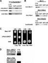
Mapping the interacting regions of MRN and DNA ligase IIIα. A and B, pulldown assays performed using glutathione beads liganded by either GST (2 μg) or GST-DNA ligase IIIβ (GST-LigIII, 2 μg) and purified human MRN (3 nm), purified hMre11, hMre11/hRad50, or hMre11/Nbs1 (3 nm each) as indicated. Binding was detected by immunoblotting using the indicated antibodies. C, interactions of full-length Nbs1 and truncated derivatives with DNA ligase IIIα and β using the yeast two-hybrid assay. D, binding of labeled in vitro translated derivatives of Nbs1 lacking either the BRCT (upper) or FHA domain (lower) to glutathione beads liganded by either GST (2 μg) or GST-DNA ligase IIIβ (GST-LigIII, 2 μg).
FIGURE 4.
FIGURE 4.
MRN stimulates intermolecular ligation by DNA ligase IIIα/XRCC1. A, Coomassie Blue-stained gel of purified DNA ligase IIIα/XRCC1 (150 ng). B, linear 580-bp DNA molecules with cohesive four-nucleotide 3′ overhangs (lane 1, 2 pmol) incubated with: DNA ligase IIIα/XRCC1 (400 fmol) and no addition (lane 2), 400 fmol of human MRN (lane 3), 800 fmol of human MRN (lane 4), 1.2 pmol of human MRN (lane 5). C, graphic representation of the DNA joining assay shown in B. The results of three independent experiments after quantification with the Quantity One software (Bio-Rad) are expressed as increase in DNA joining compared with reactions with DNA ligase IIIα/XRCC1 alone. Error bars indicate S.D. D, linear 580-bp DNA molecules with cohesive four-nucleotide 3′ overhangs (lane 1, 1.2 pmol) incubated with DNA ligase IIIα/XRCC1 (400 fmol) and no addition (lane 2), 400 fmol of human MRN and yeast MRX (lanes 3 and 6, respectively), 800 fmol of human MRN and yeast MRX (lanes 4 and 7, respectively). E, graphic representation of the DNA joining assay shown in D. The results of three independent experiments after quantification with the Quantity One software (Bio-Rad) are expressed as increase in DNA joining compared with reactions with DNA ligase IIIα/XRCC1 alone. Error bars indicate S.D.
FIGURE 5.
FIGURE 5.
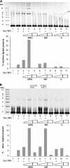
MRN stimulates compatible and incompatible DNA end ligation by DNA ligase IIIα/XRCC1. A, linear plasmid DNA (lane 1, 16 fmol) with compatible ends (PstI) was incubated with 1.4 fmol of DNA ligase IIIα/XRCC1 in the presence of no addition (lane 2), 312.5 fmol of MRN (lane 3), 625 fmol of MRN (lane 4), 312.5 fmol of MR (lane 6), 625 fmol of MR (lane 7), 425 fmol of Nbs1 (lane 9), 850 fmol of Nbs1 (lane 10). Lane 5, 625 fmol of MRN; lane 8, 625 fmol of MR, and lane 11, 850 fmol of Nbs1. DNA bands were quantified using ImageQuant TL version 2005 software, and the amount of multimeric ligated products was calculated as a percentage of the total DNA in the reaction. B, linear plasmid DNA (lane 1, 20 fmol) with incompatible, nonpalindromic ends (BglI and EcoNI) was incubated with 180 fmol of DNA ligase IIIα/XRCC1 in the presence of no addition (lane 2), 625 fmol of MRN (lane 3), 1.25 pmol of MRN (lane 4), 625 fmol of MR (lane 6), 1.25 pmol of MR (lane 7), 850 fmol of Nbs1 (lane 9), 1.7 pmol of Nbs1 (lane 10). Lane 5, 1.25 pmol of MRN; lane 8, 1.25 pmol of MR; and lane 11, 1.7 pmol of Nbs1. DNA bands were quantified, and the percentage of ligation product was calculated as a percentage of the total DNA in the reaction.
FIGURE 6.
FIGURE 6.
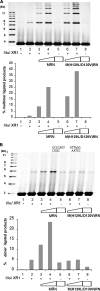
hMre11 nuclease activity is not required for stimulating compatible DNA end ligation but enhances incompatible DNA end ligation. A, linear plasmid DNA (lane 1, 16 fmol) with compatible ends (PstI) was incubated with 1.4 fmol of DNA ligase IIIα/XRCC1 in the presence of no addition (lane 2), 312.5 fmol of wild-type MRN (lane 3), 625 fmol of wild-type MRN (lane 4), 312.5 fmol of nuclease-deficient MRN (lane 6), 625 fmol of nuclease-deficient MRN (lane 7). Lane 5, 625 fmol of wild-type MRN; lane 8, 625 fmol of nuclease-deficient MRN. DNA bands were quantified using ImageQuant TL version 2005 software, and the amount of multimeric ligated products was calculated as a percentage of the total DNA in the reaction. B, linear plasmid DNA (lane 1, 20 fmol) with incompatible, nonpalindromic ends (BglI and EcoNI) was incubated with 180 fmol of DNA ligase IIIα/XRCC1 in the presence of no addition (lane 2), 625 fmol of wild-type MRN (lane 3), 1.25 pmol of wild-type MRN (lane 4), 625 fmol of nuclease-deficient MRN (lane 6), 1.25 pmol of nuclease-deficient MRN (lane 7). Lane 5, 1.25 pmol of wild-type MRN; lane 8, 1.25 pmol of nuclease-deficient MRN. DNA bands were quantified, and the percentage of ligation product was calculated as a percentage of the total DNA in the reaction.
FIGURE 7.
FIGURE 7.
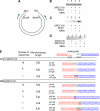
Joining of incompatible DNA ends by MRN and DNA ligase IIIα/XRCC1: microhomology-mediated joining of DNA ends with incompatible 5′ overhangs. A, circular plasmid DNA was digested with either KpnI (left) and PstI (right) to generate noncomplementary 3′ overhangs or EcoRI (left) and BamHI (right), to generate noncomplementary 5′ overhangs. Joining of the incompatible DNA ends was detected by the PCR using the indicated primers. B, DNA substrate (0.8 pmol) with noncomplementary 3′ overhangs was incubated with 0.01 pmol of DNA ligase IIIα/XRCC1 alone or in combination with equimolar amounts of hMre11 and MRN as indicated. C, DNA substrate (0.8 pmol) with noncomplementary 5′ overhangs was incubated with 0.01 pmol of DNA ligase IIIα/XRCC1 alone or in combination with equimolar amounts of hMre11 and MRN as indicated. D, DNA substrate (0.8 pmol) with noncomplementary 5′ overhangs was incubated with 0.01 pmol of either DNA ligase IIIα/XRCC1 or DNA ligase IV/XRCC4 in the absence or presence of an equimolar amount of MRN. E, sequences of the junctions generated by the joining of incompatible 5′ ends by DNA ligase IIIα/XRCC1 in combination with either hMre11 or MRN are shown. Bolded residues indicate microhomologies.

V体育平台登录 - References

    1. Venkitaraman A. R. (2002) Cell 108, 171–182 - PubMed
    1. Ferguson D. O., Sekiguchi J. M., Chang S., Frank K. M., Gao Y., DePinho R. A., Alt F. W. (2000) Proc. Natl. Acad. Sci. U.S.A. 97, 6630–6633 - PMC - PubMed
    1. Sharpless N. E., Ferguson D. O., O'Hagan R. C., Castrillon D. H., Lee C., Farazi P. A., Alson S., Fleming J., Morton C. C., Frank K., Chin L., Alt F. W., DePinho R. A. (2001) Mol. Cell 8, 1187–1196 - PubMed
    1. Lieber M. R. (2008) J. Biol. Chem. 283, 1–5 - PubMed
    1. Weterings E., Chen D. J. (2008) Cell Res. 18, 114–124 - PubMed

"VSports注册入口" Publication types

"V体育平台登录" MeSH terms

Substances