Skip to main page content (VSports app下载)
U.S. flag

An official website of the United States government

Dot gov

The . gov means it’s official. Federal government websites often end in VSports app下载. gov or . mil. Before sharing sensitive information, make sure you’re on a federal government site. .

Https

The site is secure. The https:// ensures that you are connecting to the official website and that any information you provide is encrypted and transmitted securely. V体育官网.

. 2011 Sep 22;118(12):3290-300.
doi: 10.1182/blood-2011-02-336552. Epub 2011 Jul 25.

Activation of p38 MAPK in CD4 T cells controls IL-17 production and autoimmune encephalomyelitis

Affiliations

Activation of p38 MAPK in CD4 T cells controls IL-17 production and autoimmune encephalomyelitis

Rajkumar Noubade et al. Blood. .

Abstract

Although several transcription factors have been shown to be critical for the induction and maintenance of IL-17 expression by CD4 Th cells, less is known about the role of nontranscriptional mechanisms. Here we show that the p38 MAPK signaling pathway is essential for in vitro and in vivo IL-17 production by regulating IL-17 synthesis in CD4 T cells through the activation of the eukaryotic translation initiation factor 4E/MAPK-interacting kinase (eIF-4E/MNK) pathway. We also show that p38 MAPK activation is required for the development and progression of both chronic and relapsing-remitting forms of experimental allergic encephalomyelitis (EAE), the principal autoimmune model of multiple sclerosis. Furthermore, we show that regulation of p38 MAPK activity specifically in T cells is sufficient to modulate EAE severity. Thus, mechanisms other than the regulation of gene expression also contribute to Th17 cell effector functions and, potentially, to the pathogenesis of other Th17 cell-mediated diseases. VSports手机版.


Figures

Figure 1
Figure 1
p38 MAPK regulates IL-17 production by in vitro–generated Th17 cells. FACS-sorted CD4 T cells from WT B6 mice in vitro differentiated into Th17 cells in the presence of different concentrations of SB203580 (A) or BIRB796 (B) for 72 hours. IL-17 production by Th17-polarized FACS-sorted CD4 T cells from WT B6 or MKK3−/−MKK6+/− mice (C) or total CD4 T cells from WT B10.BR and dn-p38-Tg (D) or MKK6 Tg (E) differentiated into Th17 cells. IL-17 levels in the supernatants were assessed by ELISA. The significance of differences observed in panels A and B was determined by linear regression analysis (A, P < .0001; B, P = .0001). The significance of the differences observed in panels C-E was determined using the Student t test (*** ≤ 0.001).
Figure 2
Figure 2
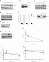
p38 MAPK controls IL-17 production at the posttranscriptional level. (A) FACS-sorted CD4 T cells from WT B6 mice were differentiated into Th17 cells in the absence or presence of SB203580 (5μM) for the indicated periods of time. The phosphorylation of STAT3 at Ser727 (P-Ser) or at Tyr705 (P-Tyr) was examined by Western blot analysis. Total STAT3 (Total) is also shown. FACS-sorted CD4 T cells from WT mice were activated under Th17 conditions in the absence (control) or presence of SB203580 for 48h. Relative Rorc (B) and Il17 (C) mRNA levels were examined by quantitative real-time PCR using β2-microglobulin as the endogenous control. (D) Relative Il17 mRNA levels in FACS-sorted CD4 T cells from WT B6 and MKK3−/−MKK6+/− mice activated under Th17 conditions for 48h. (E) Intracellular staining for IL-17 and IFNγ in WT total CD4 T cells differentiated into Th17 cells in the absence (control) or presence of SB203580 for 72 hours.
Figure 3
Figure 3
p38 MAPK regulates IL-17 via eIF-4E. (A) CD4 T cells from WT B6 mice were activated under Th17 conditions in the absence (−) or presence (+) of SB203580 (SB) for the indicated periods of time and phosphorylation of eIF-4E at Ser209 (P-eIF-4E) was examined by Western blot analysis. Actin is shown as a loading control throughout. (B) B6 CD4 T cells were activated as in panel A in the presence or absence of BIRB796 (BIRB) for 48 hours, and phosphorylation of eIF-4E at Ser209 was examined by Western blot analysis. (C) CD4 T cells from WT B6 and MKK3−/−MKK6+/− (KO) mice were activated as in panel A and phosphorylation of eIF-4E was examined by Western blot analysis. (D-E) Phosphorylation of eIF-4E in CD4 T cells from WT B10.BR and MKK6-Tg mice before activation (E) or 24 and 48 hours on activation (D) as described in panel A was examined by Western blot analysis. A longer exposure was used for detection of P-eIF-4E in panel E. (F) Relative levels of Il17, c-myc, and Il2 mRNA present in the immunoprecipitates obtained with an anti-eIF-4E Ab or a control IgG using whole-cell lysates from CD4 T cells activated under Th17 conditions for 3 days. Analyses were performed by quantitative real-time PCR and mRNA values for each gene are relative to the levels detected in the control IgG immunoprecipitates. The presence of eIF-4E in the input lysate and the immunoprecipitates was determined by Western blot analysis (right panel). (G) CD4 T cells from WT B6 mice were activated under Th17 conditions in the absence (−) or the presence (+) of the MNK inhibitor CGP57380 (CGP) for the indicated periods of time. Phospho-eIF-4E was examined by Western blot analysis. (H) CD4 T cells from WT B6 mice were differentiated into Th17 cells in the presence of the indicated amounts of CGP57380, and the levels of IL-17 in the culture supernatants after 72 hours were quantified by ELISA. (I) Relative Il17 mRNA levels in CD4 T cells activated as in panel H were assessed after 48 hours by quantitative real-time PCR using β2-microglobulin as the endogenous control. (J) IL-2 production in CD4 T cells activated as in panel H was quantified by ELISA. The significance of differences observed in panels H through J was determined by linear regression analysis (H, P < .0001; I, P = .23; J, P = .27).
Figure 4
Figure 4
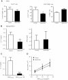
p38 MAPK controls IL-17 production by Th17 cells in vivo. (A) Mononuclear cells were isolated from the CNS of 2× MOG35-55-CFA immunized mice on day 35 after immunization, stimulated with PMA/ionomycin for 4 hours in the presence of brefeldin A, stained, and analyzed by flow cytometry. (Left panel) Cells were gated on CD45 and the frequency of CD4+TCRβ+ cells was determined. (Right panels) Cells were gated on CD4 and TCRβ and the frequency of IL-17+ and IFNγ+ cells was determined. (B) Spleen and draining lymph node (DLN) cells from 2× MOG35-55-CFA immunized mice treated with either carrier (n = 7) or the SB203580 (n = 8) were harvested on day 21 after immunization and stimulated with MOG35-55 (50 μg/mL) for 72 hours and IL-17 and IFNγ in the supernatants was quantified by ELISA. (C) Spleen and DLN cells from 2× MOG35-55-CFA immunized mice (n = 10) were harvested on day 10 after immunization and stimulated with MOG35-55 in the presence or absence of SB203580 (SB; 5μM) for 72 hours. IL-17 production was quantified by ELISA. (D) Cells obtained as in panel C were stimulated with the indicated amounts of MOG35-55 in the presence or the absence of SB203580 and proliferation was assessed by [3H]-thymidine incorporation. The significance of differences observed in panels A through C was determined using the Student t test (* ≤ 0.05; *** ≤ 0.001), and by 2-way ANOVA for panel D (SB, P = .31; MOG35-55, P = .003; and interaction, P = .95).
Figure 5
Figure 5
p38 MAPK blockade ameliorates EAE. (A) Clinical course of EAE in B6 mice immunized with 2× MOG35-55-CFA treated with either carrier or SB203580. (B) Clinical course of EAE in B6 mice immunized with 2× MOG35-55-CFA treated with either carrier or SB203580 starting on day 0 (first arrow), treatment terminated on day 31 (arrow with cross), and retreated from day 41 through day 60 (second arrow). (C) Clinical course of EAE in B6 mice immunized with 2× MOG35-55-CFA and randomly selected for treatment with either carrier or SB203580 on reaching a clinical score ≥ 1. (D) Remitting-relapsing EAE course in SJL mice immunized with 2× PLP135-151-CFA. After the initial episode of EAE and after 2 days of remission defined as a score of 0 for a minimum of 2 days, mice were treated with either carrier or SB203580 (arrow). The significance of the differences between the clinical courses of disease in panels A and B (day 0-day 40), and panel C was calculated by regression analysis and best-fit curves are shown. In pane D, 2-way ANOVA followed by Bonferroni posthoc comparisons was used to determine significant differences in disease severity for individual days (*P = .01, **P = .001).
Figure 6
Figure 6
Modulation of p38 MAPK activity specifically in T cells influences EAE susceptibility and severity. (A) Clinical course of EAE in WT B10.BR and dn-p38-Tg mice immunized with 1× MOG97-114-CFA + PTX. (B) Clinical course of EAE in WT and MKK6-Tg mice immunized with 1× MOG97-114-CFA + PTX. (A-B) Significance of the differences in the clinical course of disease was determined by regression analysis and the best-fit curves are shown. (C-D) EAE was induced in WT (n = 29) and MKK6-Tg (n = 8) mice by immunization with 1× PLP190-209-CFA + PTX. The incidence (C) and the cumulative disease score (CDS; D) are shown. The significance of the differences was determined using the independent samples t test within SPSS (Levene test for equality of variances and t test for equality of means; ***≤ 0.001).

V体育2025版 - References

    1. Zhu J, Paul WE. CD4 T cells: fates, functions, and faults. Blood. 2008;112(5):1557–1569. - VSports app下载 - PMC - PubMed
    1. Dong C. TH17 cells in development: an updated view of their molecular identity and genetic programming. Nat Rev Immunol. 2008;8(5):337–348. - PubMed
    1. Awasthi A, Kuchroo VK. Th17 cells: from precursors to players in inflammation and infection. Int Immunol. 2009;21(5):489–498. - PMC (V体育安卓版) - PubMed
    1. Kroenke MA, Carlson TJ, Andjelkovic AV, Segal BM. IL-12- and IL-23-modulated T cells induce distinct types of EAE based on histology, CNS chemokine profile, and response to cytokine inhibition. J Exp Med. 2008;205(7):1535–1541. - PMC - PubMed
    1. Rengarajan J, Mowen KA, McBride KD, Smith ED, Singh H, Glimcher LH. Interferon regulatory factor 4 (IRF4) interacts with NFATc2 to modulate interleukin 4 gene expression. J Exp Med. 2002;195(8):1003–1012. - PMC - PubMed

Publication types (V体育安卓版)

"VSports app下载" MeSH terms

"V体育官网入口" Substances