VSports在线直播 - Skip to main page content
U.S. flag

An official website of the United States government

Dot gov

The . gov means it’s official. Federal government websites often end in VSports app下载. gov or . mil. Before sharing sensitive information, make sure you’re on a federal government site. .

Https

The site is secure V体育官网. The https:// ensures that you are connecting to the official website and that any information you provide is encrypted and transmitted securely. .

. 2011 Mar 10;471(7337):245-8.
doi: 10.1038/nature09794.

Crucial role for DNA ligase III in mitochondria but not in Xrcc1-dependent repair

Affiliations

Crucial role for DNA ligase III in mitochondria but not in Xrcc1-dependent repair

Deniz Simsek et al. Nature. .

Abstract

Mammalian cells have three ATP-dependent DNA ligases, which are required for DNA replication and repair. Homologues of ligase I (Lig1) and ligase IV (Lig4) are ubiquitous in Eukarya, whereas ligase III (Lig3), which has nuclear and mitochondrial forms, appears to be restricted to vertebrates. Lig3 is implicated in various DNA repair pathways with its partner protein Xrcc1 (ref. 1). Deletion of Lig3 results in early embryonic lethality in mice, as well as apparent cellular lethality, which has precluded definitive characterization of Lig3 function. Here we used pre-emptive complementation to determine the viability requirement for Lig3 in mammalian cells and its requirement in DNA repair. Various forms of Lig3 were introduced stably into mouse embryonic stem (mES) cells containing a conditional allele of Lig3 that could be deleted with Cre recombinase. With this approach, we find that the mitochondrial, but not nuclear, Lig3 is required for cellular viability VSports手机版. Although the catalytic function of Lig3 is required, the zinc finger (ZnF) and BRCA1 carboxy (C)-terminal-related (BRCT) domains of Lig3 are not. Remarkably, the viability requirement for Lig3 can be circumvented by targeting Lig1 to the mitochondria or expressing Chlorella virus DNA ligase, the minimal eukaryal nick-sealing enzyme, or Escherichia coli LigA, an NAD(+)-dependent ligase. Lig3-null cells are not sensitive to several DNA-damaging agents that sensitize Xrcc1-deficient cells. Our results establish a role for Lig3 in mitochondria, but distinguish it from its interacting protein Xrcc1. .


Conflict of interest statement

Competing financial interests The authors declare no competing financial interests.

Figures

Figure 1
Figure 1. Mitochondrial Lig3 activity is critical for cellular viability
(a) Pre-emptive complementation strategy for Lig3 deletion. Lig3KO/KO clones were identified by their lack of growth in G418 (neo–). PCR confirmed the genotype, as indicated. (b) Mitochondrial Lig3 activity is critical for cellular viability. n, number of independently-derived Lig3 transgenic cell lines analyzed; Total, total number of post-Cre colonies analyzed by replica plating with and without G418; G418S, G418 sensitive; %, ratio of G418S to Total. (c) Lig3 proteins tested for pre-emptive complementation. MLS, mitochondrial leader sequence; ZnF, zinc finger; BRCT, BRCA1 C-terminal related domain; DBD, DNA-binding domain; CC, catalytic core; *M88T, mutation of the nuclear translation initiation site. (d) Western blot analysis showing the loss of endogenous Lig3 in Lig3KO/KO clones stably expressing GFP-tagged Lig3 or MtLig3. Lig3 is 105 kDa, whereas the GFP fusions are ~135 kDa. neo+, Lig3KO/cKOneo+; KO, Lig3KO/KO; α-tub, α-tubulin.
Figure 2
Figure 2. Mitochondrial DNA ligase activity can be provided by a variety of DNA ligases
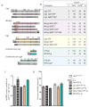
(a) DNA ligases tested for pre-emptive complementation of Lig3KO/KO cells. ΔZnF, deletion of Lig3 amino acids 89 to 258; ΔBRCT, deletion of Lig3 amino acids 934 to 1009; ΔNLS, deletion of Lig1 amino acids 135 to147; NES, nuclear export signal from MAPKK; Mt, presence of Lig3 MLS; NLS, nuclear localization signal; HhH, helix-hairpin-helix. (b) Mitochondrial DNA ligase activity can be provided by a variety of DNA ligases. ChVLig and EcoLigA presumably enter mitochondria without the requirement for an MLS. (c) Cells expressing exogenous DNA ligases are competent to replicate and maintain mtDNA. mtDNA copy number was quantified by qPCR by amplifying a 117 bp fragment from mtDNA. Values are presented relative to levels of mtDNA in Lig3KO/KO; Lig3 GFP Tg cells. Data represent the mean of two biological repeats each determined twice by qPCR +/- SEM. (d) Cells expressing exogenous DNA ligases showed similar capacities for mtDNA repair after oxidative damage. Cells were treated with 175 μM H2O2 for 15 min and allowed to recover for 1.5 h. To measure repair, a 10 kb mtDNA fragment was amplified following damage and quantified by qPCR. Values were normalized to the amplification of a 117 bp mtDNA fragment. Percent repair is the amount of damage remaining after 1.5 h recovery divided by the initial damage. There was no significant difference between cell lines expressing wild-type Lig3 (parental cells and transgene rescued cells) and the other ligase forms. For cells expressing MitLig1-ΔNLS, and MtChVLig two transgenic cell lines were analyzed. Data represent the mean of 2-4 determinations on multiple clones with each qPCR performed twice +/- SEM.
Figure 3
Figure 3. Loss of Lig3 is not associated with sensitivity to several DNA damaging agents or with increased sister-chromatid exchange
(a) Western blot analysis showing the loss of endogenous Lig3 protein in Lig3KO/KO cells with the indicated transgenes. (b-f) Sensitivity of Lig3KO/KO cell lines to the indicated DNA damaging agents was measured using colony formation assays. Brca1–/– and Ercc1–/– cells are only shown on graphs when they are sensitive. For each cell line and agent, n = 4 and error bars = SEM; error bars in some cases are smaller than the symbol. (g) SCE analysis. The range of SCEs was between 5 and 21 per metaphase for each cell line. For cells expressing MtLig1, two transgenic cell lines were analyzed. The differences between the cell lines are not significant using a two-tailed unpaired t-test. Values are presented with 1 SD from the mean.

References

    1. Ellenberger T, Tomkinson AE. Eukaryotic DNA ligases: structural and functional insights. Annu Rev Biochem. 2008;77:313–338. - "VSports注册入口" PMC - PubMed
    1. Puebla-Osorio N, Lacey DB, Alt FW, Zhu C. Early embryonic lethality due to targeted inactivation of DNA ligase III. Mol Cell Biol. 2006;26:3935–3941. - PMC - PubMed
    1. Ho CK, Van Etten JL, Shuman S. Characterization of an ATP-dependent DNA ligase encoded by Chlorella virus PBCV-1. J Virol. 1997;71:1931–1937. - PMC - PubMed
    1. Thompson LH, Brookman KW, Jones NJ, Allen SA, Carrano AV. Molecular cloning of the human XRCC1 gene, which corrects defective DNA strand break repair and sister chromatid exchange. Mol Cell Biol. 1990;10:6160–6171. - PMC - PubMed
    1. Tebbs RS, et al. Requirement for the Xrcc1 DNA base excision repair gene during early mouse development. Dev Biol. 1999;208:513–529. - PubMed

Publication types

MeSH terms (V体育2025版)