Skip to main page content
U.S. flag

An official website of the United States government

Dot gov

The . gov means it’s official VSports app下载. Federal government websites often end in . gov or . mil. Before sharing sensitive information, make sure you’re on a federal government site. .

Https

The site is secure V体育官网. The https:// ensures that you are connecting to the official website and that any information you provide is encrypted and transmitted securely. .

. 2011 Jan;47(1):8-15.
doi: 10.1016/j.oraloncology.2010.10.011. Epub 2010 Dec 15.

p53-Reactivating small molecules induce apoptosis and enhance chemotherapeutic cytotoxicity in head and neck squamous cell carcinoma

Affiliations

p53-Reactivating small molecules induce apoptosis and enhance chemotherapeutic cytotoxicity in head and neck squamous cell carcinoma (V体育官网入口)

VSports app下载 - Jong-Lyel Roh et al. Oral Oncol. 2011 Jan.

Abstract

We evaluate whether p53-reactivating (p53RA) small molecules induce p53-dependent apoptosis in head and neck squamous cell carcinoma (HNSCC), a question that has not been previously addressed in head and neck cancer. PRIMA-1, CP-31398, RITA, and nutlin-3 were tested in four human HNSCC cell lines differing in TP53 status. Cell growth, viability, cell cycle progression, and apoptosis after treatment with p53RA small molecules individually or in combination with chemotherapeutic agents were assessed. Prominent p53 reactivation was observed in mutant TP53-bearing tumor cell lines treated with PRIMA-1 or CP-31398, and in wild-type TP53-bearing cell lines treated with nutlin-3 VSports手机版. Cell-cycle arrest and apoptosis induced by p53RA small molecules were associated with upregulation of p21 and BAX, and cleavage of caspase-3. Nutlin-3 showed maximal growth suppression in tumor cells showing MDM2-dependent p53 degradation. High-dose treatment with p53RA small molecules also induced apoptosis in cell lines independent of p53 or MDM2 expression. In combination therapy, p53RA small molecules enhanced the anti-tumor activity of cisplatin, 5-fluorouracil, paclitaxel, and erlotinib against HNSCC cells. The p53RA small molecules effectively restored p53 tumor-suppressive function in HNSCCs with mutant or wild-type TP53. The p53RA agents may be clinically useful against HNSCC, in combination with chemotherapeutic drugs. .


V体育平台登录 - Conflict of interest statement

Conflicts of Interest Statement: None declared.

Figures

Figure 1
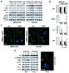
Figure 1
p53 expression and mutation status in the HNSCC cell lines used in this study. A, Western blot analysis of p53 expression before and after stimulation with CDDP. β-actin was used as a loading control. B, immunofluorescence staining of p53 in untreated cells. DAPI-stained nuclei and intracellular p53 are shown. C, TP53 mutation status determined by direct sequencing of exons 2–11.
Figure 2
Figure 2
Differential response of HNSCC cell lines to p53RA small molecules. A, growth-inhibitory effects examined by MTT assays after incubating cells for 96 h with increasing doses of small molecules. Graphs were drawn from mean values ± SD from three different experiments. B, colony forming assays. The number of colonies was counted 10–14 days after cells treated with the indicated concentrations of the agents for 72 h were plated in drug-free media. Error bars represent SD. C, growth suppression tested by MTT assays after incubating OKF6/TERT1 and HDF for 96 h with 5–10 µM p53RA small molecules. D, Western blot analysis of p53, MDM2, and p21WAF1 after treatment with p53RA agents for 24 h. Normalized ratios of p53 or p21waf1:β-actin to control (Ct) were determined by densitometry. P, PRIMA-1 (5 µM); C, CP-31398 (5 µM); R, RITA (10 µM); N, nutlin-3 (10 µM).
Figure 3
Figure 3
Cell cycle and apoptosis assays after treatment with p53RA small molecules. A, cell-cycle changes examined by PI staining of cell lines after treatment for 24 h. B, Annexin-V binding fraction representing an apoptotic population of cells induced by treatment for 48 h. The figures are the representative of three independent experiments.
Figure 4
Figure 4
Activation of the p53 pathway by treatment with p53RA small molecules. A, Western blot analysis after treatment of two selected cell lines with different concentrations of small molecules for different times. B, changes in mRNA expression measured by real-time quantitative RT-PCR, shown as fold-reduction relative to DMSO-treated cells. P-values versus untreated cells were calculated using a two-tailed Mann-Whitney test. C, immunofluorescence staining of p53 after PRIMA-1 or nutlin-3 treatment for 8 or 24 h. Accumulation of p53 was seen in nuclei (counterstained with DAPI). D, Western blot and fluoromicroscopic examinations after treatment with RITA for 24 h. Cell viability was examined by trypan blue exclusion assay after treatment with RITA or nutlin-3 for 96 h.
Figure 5
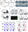
Figure 5
Combined effects of p53RA small molecules and chemotherapeutic agents on HNSCC cell lines. A, MTT assays of cells incubated for 96 h with increasing concentrations of CDDP in combination with 2.5 µM PRIMA-1 or nutlin-3. B, phase-contrast images of two cell lines treated with p53RA or CDDP alone or in combination for 48 h. C, growth inhibition relative to control, assayed by MTT after treatment with individual p53RA and chemotherapeutic agents alone or in combination for 96 h. D–F, Western blot, immunofluorescence (p53), and real-time quantitative RT-PCR analyses of the expression of p53 pathway and proapoptotic genes in two selected cell lines after individual or combined treatment for 24 h. G, cell-cycle analysis of three cell lines treated with p53RA agents and CDDP, alone and in combination, for 48 h. The tabulated data are the representative of three independent experiments. H, apoptosis induction in JHU-O28 cell was increased by combined treatment with nutlin-3 and CDDP for 48 h compared with either agent alone.

References

    1. Levine AJ. p53, the cellular gatekeeper for growth and division. Cell. 1997;88(3):323–331. - PubMed
    1. Vousden KH, Lu X., q Live or let die: the cell's response to p53. Nat Rev Cancer. 2002;2(8):594–604. - PubMed
    1. Vousden KH, Prives C. Blinded by the Light: The Growing Complexity of p53. Cell. 2009;137(3):413–431. - PubMed
    1. Green DR, Kroemer G. Cytoplasmic functions of the tumour suppressor p53. Nature. 2009;458(7242):1127–1130. - PMC (VSports手机版) - PubMed
    1. Olivier M, Eeles R, Hollstein M, Khan MA, Harris CC, Hainaut P. The IARC TP53 database: new online mutation analysis and recommendations to users. Hum Mutat. 2002;19(6):607–614. - V体育平台登录 - PubMed

Publication types

"VSports手机版" MeSH terms

LinkOut - more resources