Skip to main page content
U.S. flag

An official website of the United States government

Dot gov

The . gov means it’s official. Federal government websites often end in . gov or . mil. Before sharing sensitive information, make sure you’re on a federal government site. VSports app下载.

Https

The site is secure. The https:// ensures that you are connecting to the official website and that any information you provide is encrypted and transmitted securely V体育官网. .

. 2010 Jun;24(6):1914-24.
doi: 10.1096/fj.09-149765. Epub 2010 Feb 2.

Palmitoylation of ketogenic enzyme HMGCS2 enhances its interaction with PPARalpha and transcription at the Hmgcs2 PPRE

Affiliations

Palmitoylation of ketogenic enzyme HMGCS2 enhances its interaction with PPARalpha and transcription at the Hmgcs2 PPRE

Morris A Kostiuk et al. FASEB J. 2010 Jun.

Abstract

Excessive liver production of ketone bodies is one of many metabolic complications that can arise from diabetes, and in severe untreated cases, it can result in ketoacidosis, coma, and death. Mitochondrial HMG-CoA synthase (HMGCS2), the rate-limiting enzyme in ketogenesis, has been shown to interact with PPARalpha and act as a coactivator to up-regulate transcription from the PPRE of its own gene. Although protein palmitoylation is typically a cytosolic process that promotes membrane association, we recently identified 21 palmitoylated proteins in rat liver mitochondria, including HMGCS2 VSports手机版. Herein, our data support a mechanism whereby palmitate is first added onto HMGCS2 active site Cys166 and then transacylated to Cys305. Palmitoylation promotes the HMGCS2/PPARalpha interaction, resulting in transcriptional activation from the Hmgcs2 PPRE. These results, together with the fact that 8 of the 21 palmitoylated mitochondrial proteins that we previously identified have nuclear receptor interacting motifs, demonstrate a novel--and perhaps ubiquitous--role for palmitoylation as a modulator of transcription. .


Figures
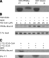
Figure 1.
Figure 1.
Level of interaction between HMGCS2 and PPARα increases in a palmitoyl-CoA concentration-dependent manner. A) In vitro transcribed and translated [35S]HMGCS2 was incubated with 1 μg of GS/PPARα prebound to glutathione Sepharose beads or GST Sepharose alone as shown, followed by SDS-PAGE. Fluorogram of the dried gel representing 10% of the flow through (FT) or the bound protein (B) is shown. B) One milligram of purified HMGCS2-His6 was incubated with various concentrations of palmitoyl-CoA or palmitate or the combination of 30 min of pretreatment in 10 mM NEM followed by palmitoyl-CoA as shown for 30 min. One hundred nanograms of HMGCS2 (10% of the reaction) was incubated with immobilized GSTPPARα followed by SDS PAGE and Western blot analysis with anti-His5 antibody. C) One microgram of purified HMGCS2-His6 was incubated with 25 μM [125I]-iodopalmitoyl-CoA (lanes 1 and 3) or 25 μM [125I]-iodopalmitate (lanes 2 and 4) following a 30-min preincubation with buffer alone (lanes 1 and 2) or 10 mM NEM (lanes 3 and 4). Ten percent of these reactions (100 ng protein) were incubated with immobilized GST-PPARα, followed by SDS PAGE gel. Autoradiography of the dried gels from the bound fractions and 5% of eluted fractions is shown.
Figure 2.
Figure 2.
Palmitoylation of HMGCS2 and its interaction with PPARα are specific to acylation of HMGCS2 by long-chain fatty acids. A) One microgram of HMGCS2-His6 was incubated with 50 μM acyl-CoAs of varying acyl-chain lengths as shown, and 10% of the reaction (100 ng HMGCS2) was incubated with 1 μg GST-PPARα immobilized on glutathione-Sepharose beads followed by SDS PAGE and Western blot analysis with anti-His5 antibody. Results from Western blots of the bound protein and 5% of the protein loaded in the binding reaction are shown. B) One microgram of HMGCS2-His6 was incubated in reactions containing 50 μM succinyl-CoA, acetoacetyl-CoA, or 50 μM of acyl-CoAs of varying acyl-chain lengths as shown for 30 min, followed by reaction with 100 μM [125I]-iodopalmitoyl-CoA for 30 min. Following SDS-PAGE, the dried Coomassie-stained gels were subjected to autoradiography.
Figure 3.
Figure 3.
HMGCS2 is palmitoylated on cysteines 166 and 305. A) MALDI mass spectra excerpt showing tryptic peptides containing Cys305 (sequence: QAGSDRPFTLDDLQYMIFHTPFCK) from wild-type HMGCS2-His6 after S-acylation with azidopalmitoyl-CoA (I), palmitoyl-CoA (II), or control (no palmitoylation; III). Signals at m/z 2887 ([M+H]+) and m/z 2909 ([M+Na]+) stem from a tryptic peptide containing no cysteines (sequence: GTHMENVYDFYKPNLASEYPIVDGK). B–D) MALDI MS/MS spectra (with MS spectra excerpts of precursor ions) of chymotryptic peptides of wild-type HMGCS2-His6 after S-acylation with palmitoyl-CoA. B) Palmitoylated peptide containing Cys305. C). Signal from palmitoylated peptide containing Cys166. D) Signal from carbamidomethylated peptide containing Cys166.
Figure 4.
Figure 4.
Palmitoylation of Cys 305 requires the active site Cys 166. A) One milligram of HMGCS2-His6 wild-type or mutant as shown was incubated with or without azidopalmitoyl-CoA or NEM, followed by reaction with phosphine-biotin. Acylation was detected by protein blotting with neutravidin-HRP/ECL, and corresponding protein was visualized by Coomassie blue staining of the membrane. B) One milligram of HMGCS2-His6 wild-type or mutant as shown was labeled using [125I]-iodopalmitoyl-CoA in the presence or absence of NEM, followed by SDS-PAGE. Acylation was detected by autoradiography and corresponding protein by Coomassie blue staining. C) MALDI MS/MS spectrum (with MS spectra excerpt of precursor ions) of azido-palmitoylated chymotryptic peptide containing Cys166 from the HMGCS2-His6 Cys305Ser mutant. For this mutant, a strong signal is observed compared to the wild type shown in figure 3C. D, E) Palmitoylation, reduction, alkylation with NEM for wild-type, and NEM-d5 for Cys166Ser were done before mixing of the two proteins and tryptic digestion after mixing. D) Tryptic peptides containing Cys305. Only the peptide originating from the Cys166Ser mutant is clearly observable (m/z=2960), further supporting our findings that S-palmitoylation of Cys305 is more enhanced in the wild-type protein compared to the Cys166Ser mutant. E) Signals for the tryptic peptides containing Cys454 (which is not involved in palmitoylation), confirming that both wild-type and Cys166Ser are present in approximately similar amounts.
Figure 5.
Figure 5.
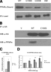
Palmitoylation of HMGCS2 stimulates the HMGCS2/PPARα interaction and transcriptional up-regulation at the Hmgcs2 PPRE. A) One milligram of purified HMGCS2-His6 and the Cys to Ser mutants as shown was reacted with 25 mM palmitoyl-CoA for 30 min, and 10% of the reaction (100 ng HMGCS2) was incubated with 1 mg GST-PPARα immobilized on glutathione-Sepharose beads, followed by SDS PAGE and Western blot analysis with anti-His5 antibody. B) COS 7 cells transfected with PPARα and HA-tagged wild-type or C166S HMGCS2 were immunoprecipitated with an anti-HA antibody, followed by SDS-PAGE and transfer to a PVDF membrane, and probed with anti-HA and anti-PPARα. C, D) COS 7 cells were cotransfected with pGL3HSPPRE and pSG5PPARα with or without increasing amounts of pcDNAHMGCS2-WT or pcDNA-HMGCS2-C166,305S, and resulting luciferase activity was measured. C) Stimulation of the transcriptional up-regulation of the reporter vector by PPARα normalized to the transfection with the reporter vector alone set to 1. D) Typical increases of luciferase activity in cells transfected with increasing amounts of pcDNA-HMGCS2-WT or pcDNA-HMGCS2-C166,305S normalized as fold over cells transfected with PPARα alone set to 1 (n=4). Error bars = se.
Figure 6.
Figure 6.
Models of the HMGCS2-PPARα palmitoylation-dependent interaction and nuclear localization, showing the 4 possible PPARα-binding scenarios leading to nuclear localization of HMGCS2. A) We postulate that processing of the leader sequence and palmitoylation both occur in the mitochondria prior to the interaction with PPARα. Once palmitoylated, HMGCS2 exits the mitochondria, albeit via an unknown mechanism, the nuclear entry could occur either directly or following the binding to PPARα. B) We postulate that palmitoylation of HMGCS2 occurs in the cytosol and results in direct nuclear translocation or results in the binding of HMGCS2 to PPARα prior to nuclear import. HMGCS2 (HS) shown in yellow, PPARα (P) shown in blue, RXRα (RXR) shown in green; grey structures represent ribosomes.

References

    1. Cullingford T E. The ketogenic diet; fatty acids, fatty acid-activated receptors and neurological disorders. Prostaglandins Leukot Essent Fatty Acids. 2004;70:253–264. - PubMed
    1. Fukao T, Lopaschuk G D, Mitchell G A. Pathways and control of ketone body metabolism: on the fringe of lipid biochemistry. Prostaglandins Leukot Essent Fatty Acids. 2004;70:243–251. - VSports - PubMed
    1. Hegardt F G. Mitochondrial 3-hydroxy-3-methylglutaryl-CoA synthase: a control enzyme in ketogenesis. Biochem J. 1999;338:569–582. - PMC - PubMed
    1. Eledrisi M S, Alshanti M S, Shah M F, Brolosy B, Jaha N. Overview of the diagnosis and management of diabetic ketoacidosis. Am J Med Sci. 2006;331:243–251. - PubMed
    1. Wolfsdorf J, Craig M E, Daneman D, Dunger D, Edge J, Lee W R, Rosenbloom A, Sperling M A, Hanas R. Diabetic ketoacidosis. Pediatr Diabetes. 2007;8:28–43. - PubMed

Publication types

MeSH terms

LinkOut - more resources