Skip to main page content
U.S. flag

An official website of the United States government

Dot gov

The . gov means it’s official. Federal government websites often end in VSports app下载. gov or . mil. Before sharing sensitive information, make sure you’re on a federal government site. .

Https

The site is secure. The https:// ensures that you are connecting to the official website and that any information you provide is encrypted and transmitted securely V体育官网. .

. 2010 Jul 15;127(2):313-20.
doi: 10.1002/ijc.25049.

V体育官网入口 - MicroRNA-34a is an important component of PRIMA-1-induced apoptotic network in human lung cancer cells

Affiliations

"VSports最新版本" MicroRNA-34a is an important component of PRIMA-1-induced apoptotic network in human lung cancer cells

Wenrui Duan et al. Int J Cancer. .

Abstract

Restoration of p53 function in tumor cells would be an attractive strategy for lung cancer therapy because p53 mutations are found in more than 50% of lung cancers. The small molecule PRIMA-1 has been shown to restore the tumor suppression function of p53 and to induce apoptosis in human tumor cells. The mechanism of apoptosis induced by PRIMA-1 remains unclear. We investigated the effects of PRIMA-1 in apoptosis with Western immunoblot analysis, TaqMan microRNA real-time PCR, cell viability analysis and flow cytometry using human lung cancer cell lines containing mutant (H211 and H1155), wild-type (A549) or null (H1299) p53. PRIMA-1 induced massive apoptosis in the H211 and H1155 cells, but was less toxic to the A549 and H1299 cells VSports手机版. Western immunoblot analysis showed cleavage of PARP in H211 and H1155 cells but not in A549 and H1299 cells following treatment with PRIMA-1. In addition, p53 protein was also phosphorylated in H211 and H1155 cells. TaqMan microRNA assay showed that the expression of microRNA-34a was increased in the H211 and H1155 cells posttreatment. Knockdown microRNA-34a decreased the rate of apoptosis caused by PRIMA-1. The above results suggest that microRNA-34a is one of the important components of PRIMA-1-induced apoptotic network in the cancer cells harboring mutant p53. .


Figures

Figure 1
Figure 1
Analysis of cell viability post PRIMA-1 treatment. Cells were treated with the PRIMA-1 (100 μM), and the MTT assay was performed to determine cell viability 24 hr posttreatment. Each treatment was repeated in quadruplicate. An averaged absorbance of blank values (with no cells) was subtracted from all absorbance to yield corrected absorbance. The relative absorbance of each sample was calculated by comparing the average of corrected absorbance with an average of corrected untreated control. Each value presented in this figure was an average value obtained from 4 measurements.
Figure 2
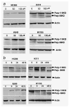
Figure 2
Western immunoblot analysis of PARP cleavage induced by PRIMA-1 in lung cancer cell lines. (a) Human lung cancer cells A549 (wild-type p53), H1299 (p53 null), H211 (mutant p53, 248Q) and H1155 (mutant p53, 273H) were treated with PRIMA-1 at a dose of 50 or 100 μM. Cells were harvested 24 hr posttreatment. A total of 100 μg cell lysate each sample was subjected for Western immunoblot analysis. Western immunoblot membrane was probed with antibodies against full length and cleaved PARP. (b) Human lung cancer cells were treated with PRIMA-1 (100 μM). Cells were harvested and lysed at 6, 24 and 48 hr posttreatment. Western immunoblot analysis was used to analyze the cleavage of PARP protein in the lung cancer cells at indicated time point. Membrane was probed with antibodies against full length and cleaved PARP. Loading control was β-actin. The cleaved PARP was detected in the H211 and H1155 cells after treatment with PRIMA-1.
Figure 3
Figure 3
Analysis of p53 phosphorylation in lung cancer cell lines. Human lung cancer cells A549, H1299, H211 and H1155 were treated with PRIMA-1 (100 μM) and then harvested at 6, 24 and 48 hr posttreatment. Cell lysates were subjected to SDS-PAGE and probed with anti-p53 and anti-phospho-p53 antibodies against Ser15, respectively. PRIMA-1 exposure led to increase phosphorylation of p53 at Ser15. Loading control was β-actin.
Figure 4
Figure 4
PRIMA-1 upregulates expression of microRNA-34a. (a) TaqMan microRNA real-time PCR analysis was used to evaluate the expression of microRNA-34a, b and c. Cells were treated with the PRIMA-1 (100 μM), and cells were harvested at 6, 24 and 48 hr posttreatment. Real-time PCR was carried out in a 96-well plate using an Applied Biosystems 7900HT Sequence Detection System. The relative quantitative method was used to determine expression of the microRNA-34 family members. The averaged microRNA-34 family member from the control (Ctr) group of each cell line was used as calibrator. For each cell line, the averaged values obtained from other time points were compared with the calibrator. Each value presented in this figure was an average value obtained from 3 amplifications. (b) Western blot analysis was used to confirm the knocking down of p53. (c) Expression levels of microRNA-34a were estimated with TaqMan microRNA real-time PCR in the anti-p53 siRNA transfected and control cells. The error bars show standard deviation. * indicates p < 0.05, 2-tailed t-test.
Figure 5
Figure 5
Knockdown microRNA-34a reduces PRIMA-1-induced apoptosis. H211 and H1155 cells were transfected with miRIDIAN hairpin inhibitor to inhibit expression of microRNA-34a or negative control. The transfected cells were treated with PRIMA-1 (25 μM for H211 and 100 μM for H1155) 24 hr posttransfection. After an additional incubation of 24 hr, the transfected and PRIMA-1-treated cells were harvested. TaqMan microRNA real-time PCR was used to confirm the specific knockdown of microRNA-34a in the H211 and H1155cells (a). The transfected and PRIMA-1-treated cells were then stained with Annexin V-FITC apoptosis kit and analyzed by fluorescence activated cell sorting (b). The apoptosis was also evaluated by Western blot analysis of PARP cleavage (c). Each value presented in this figure was an average value obtained from 3 independent analyses. The error bars show standard deviation. * indicates p < 0.05, 2-tailed t-test.

References

    1. Levine AJ. p53, the cellular gatekeeper for growth and division. Cell. 1997;88:323–31. - "VSports在线直播" PubMed
    1. Vogelstein B, Lane D, Levine AJ. Surfing the p53 network. Nature. 2000;408:307–10. - PubMed
    1. El-Hizawi S, Lagowski JP, Kulesz-Martin M, Albor A. Induction of gene amplification as a gain-of-function phenotype of mutant p53 proteins. Cancer Res. 2002;62:3264–70. - V体育2025版 - PubMed
    1. Olivier M, Eeles R, Hollstein M, Khan MA, Harris CC, Hainaut P. The IARC TP53 database: new online mutation analysis and recommendations to users. Hum Mutat. 2002;19:607–14. - "VSports app下载" PubMed
    1. Mitsudomi T, Steinberg SM, Nau MM, Carbone D, D’Amico D, Bodner S, Oie HK, Linnoila RI, Mulshine JL, Minna JD, Gazdar AF. p53 gene mutations in non-small-cell lung cancer cell lines and their correlation with the presence of ras mutations and clinical features. Oncogene. 1992;7:171–80. - VSports在线直播 - PubMed

VSports最新版本 - Publication types

MeSH terms (VSports注册入口)