Skip to main page content
U.S. flag

An official website of the United States government

Dot gov

The . gov means it’s official. Federal government websites often end in . gov or . mil. Before sharing sensitive information, make sure you’re on a federal government site VSports app下载. .

Https

The site is secure. The https:// ensures that you are connecting to the official website and that any information you provide is encrypted and transmitted securely. V体育官网.

. 2009 Apr;119(4):772-87.
doi: 10.1172/JCI33950. Epub 2009 Mar 16.

WNT1-inducible signaling protein-1 mediates pulmonary fibrosis in mice and is upregulated in humans with idiopathic pulmonary fibrosis

Affiliations

WNT1-inducible signaling protein-1 mediates pulmonary fibrosis in mice and is upregulated in humans with idiopathic pulmonary fibrosis

Melanie Königshoff et al. J Clin Invest. 2009 Apr.

V体育官网 - Abstract

Idiopathic pulmonary fibrosis (IPF) is characterized by distorted lung architecture and loss of respiratory function. Enhanced (myo)fibroblast activation, ECM deposition, and alveolar epithelial type II (ATII) cell dysfunction contribute to IPF pathogenesis. However, the molecular pathways linking ATII cell dysfunction with the development of fibrosis are poorly understood VSports手机版. Here, we demonstrate, in a mouse model of pulmonary fibrosis, increased proliferation and altered expression of components of the WNT/beta-catenin signaling pathway in ATII cells. Further analysis revealed that expression of WNT1-inducible signaling protein-1 (WISP1), which is encoded by a WNT target gene, was increased in ATII cells in both a mouse model of pulmonary fibrosis and patients with IPF. Treatment of mouse primary ATII cells with recombinant WISP1 led to increased proliferation and epithelial-mesenchymal transition (EMT), while treatment of mouse and human lung fibroblasts with recombinant WISP1 enhanced deposition of ECM components. In the mouse model of pulmonary fibrosis, neutralizing mAbs specific for WISP1 reduced the expression of genes characteristic of fibrosis and reversed the expression of genes associated with EMT. More importantly, these changes in gene expression were associated with marked attenuation of lung fibrosis, including decreased collagen deposition and improved lung function and survival. Our study thus identifies WISP1 as a key regulator of ATII cell hyperplasia and plasticity as well as a potential therapeutic target for attenuation of pulmonary fibrosis. .


Figures

Figure 1
Figure 1. Enhanced proliferative capacity of ATII cells in experimental lung fibrosis.
The purity (A) and phenotype (B) of ATII cell isolations from saline- or bleomycin-treated (Bleo) mice, 14 days after instillation, was analyzed by immunofluorescence staining. ATII cells were fixed directly after isolation (cytocentrifuge preparations) and stained with antibodies against the ATII cell marker SPC (original magnification, ×40, scale bar: 10 μm) or the (myo)fibroblast marker α-SMA (insets, magnification, ×10) (A) or fixed after 24 hours of attachment and subsequently stained with antibodies against SPC, TJP1, or ECAD (original magnification, ×40) (B). (C) Double immunostaining for panCK (green) and Ki67 (red) was performed in primary ATII cells from saline- or bleomycin-treated mice, 14 days after instillation (original magnification, ×40). Nuclei were visualized by DAPI staining (insets; original magnification, ×40). All stainings are representative of at least 3 independent experiments. (D) ATII cell proliferation was analyzed in primary cells isolated from mice 7 or 14 days after instillation with bleomycin by [3H]thymidine incorporation. Data are presented as fold change in [3H]thymidine incorporation compared with saline-instilled controls (n = 10 per group). (E) mRNA levels of the proliferation markers Ki67, Ccng1, and Ccnb2 were analyzed by qRT-PCR using primary ATII cells and plotted as log-fold increase (ΔΔCt) of mRNA levels in bleomycin- versus saline-treated mice, 14 days after instillation (n = 6 each). Results are presented as mean ± SEM; *P < 0.05, **P < 0.02.
Figure 2
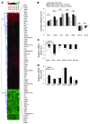
Figure 2. Increased mRNA expression of Wisp1 and WNT signaling components in ATII cells isolated from bleomycin-treated mice.
(A) ATII cell gene expression profiles were analyzed by whole genome expression analysis using RNA from freshly isolated ATII cells from saline- or bleomycin-treated mouse lungs 14 days after administration. Red and green indicate increased and decreased gene expression levels, respectively, in ATII cells isolated from bleomycin- versus saline-treated mice. Columns represent individual samples, including dye-swap experiments. Selected genes are represented in rows. Detailed description of the whole genome expression analysis is given in the Supplemental Data. (B) Confirmation of microarray results was performed for selected genes in freshly isolated ATII cells (n = 6), as well as in ATII cells 72 hours after isolation (n = 3) by qRT-PCR, as indicated. The following genes were analyzed: secreted frizzled-related protein 1 (Sfrp1), inhibin beta A (Inhba), found in inflammatory zone 1 (Fizz1), secreted phosphoprotein 1 (Spp1), Wisp1, cadherin 16 (Cdh16), and potassium voltage-gated channel subfamily E member 2 (Kcne2). Results are presented as mean ± SEM; **P < 0.02 for all bars, compared with ATII cells isolated from saline-treated mice. (C and D) The mRNA levels of the WNT target gene Mmp7, the WNT ligands Wnt1, Wnt2, Wnt3a, Wnt7b, and Wnt10b (C), the receptors frizzled 1 (Fzd1), Fzd2, and Fzd4, and the intracellular signal transducers Ctnnb1, Gsk3b, and Tcf4 (D) were assessed in ATII cells isolated from bleomycin- and saline-treated mice (n = 6 each) by qRT-PCR. Results are presented as mean ± SEM. *P < 0.05, **P < 0.02.
Figure 3
Figure 3. Increased epithelial expression of WNT/β-catenin signaling components in experimental lung fibrosis.
Immunohistochemical staining for CTNNB1, GSK-3β, and WNT1 was performed on whole-lung sections of saline- or bleomycin-treated mice 7 or 14 days after bleomycin application, as indicated. The arrows indicate distinct alveolar epithelial cells. Stainings are representative of 2 independent experiments using at least 3 different bleomycin- or saline-treated lung tissues.
Figure 4
Figure 4. Activation of WNT/β-catenin signaling in vivo during experimental lung fibrosis.
TOPGAL reporter mice were treated orotracheally with WNT3A or bleomycin, as described in detail in Methods. Supplemental Figure 3 depicts the treatment scheme. (A) X-gal staining of β-gal activity in lungs from WNT3A- and vehicle-treated mice (top row) or bleomycin- and saline-treated mice (bottom row). Pictures are representative of at least 2 independent experiments using at least 4 different lung tissues for each condition. (B) X-gal, SPC, and clara cell–specific protein (CCSP) protein expression in serial whole-lung sections from bleomycin-treated TOPGAL reporter mouse was assessed by immunohistochemistry. (C) Primary ATII cells were stimulated with WNT3A (100 ng/ml), and the mRNA levels of Ctgf, Wisp1, and Ccnd1 were analyzed by qRT-PCR (n = 4 for each) at the indicated time points and plotted as log-fold increase (ΔΔCt) of mRNA levels in WNT-stimulated versus unstimulated cells. Results are presented as mean ± SEM; *P < 0.05, **P < 0.02.
Figure 6
Figure 6. Increased WISP1 expression in ATII cells in vitro and in vivo in IPF.
(A) mRNA levels of the CCN family members were analyzed by qRT-PCR using lung homogenates derived from donor or IPF lung explants (n = 10 each). (B) mRNA levels of WISP1 and CTGF were analyzed by qRT-PCR in microdissected septae from donor or IPF lungs (n = 5 each). Results in A and B are plotted as relative mRNA levels (ΔCt) and presented as mean ± SEM. (C) mRNA levels of WISP1 (white bars) and CTGF (black bars) were determined by qRT-PCR in primary human ATII cells (n = 4) or fibroblasts (n = 3) isolated from donor or IPF lung tissue. Results are plotted as log-fold increase (ΔΔCt) of mRNA levels in IPF-derived versus donor-derived cells and presented as mean ± SEM. (D) WISP1 protein expression in sections from control or IPF lung specimens was assessed by immunohistochemistry. Arrows indicate extracellular WISP1 staining. WISP1 and phospho–histone H3 (Phospho H3) protein expression in serial whole-lung sections from IPF patients was assessed by immunohistochemistry (bottom row). Data are representative of at least 2 independent experiments using at least 4 different lung tissues from IPF specimens. (E) WISP1 protein expression was determined in total protein lysates from donor or IPF lung tissue using Western blot analysis. Lamin A/C was used as a loading control. Data are representative of at least 2 independent experiments using 6 different lung tissues for donor and IPF specimens. (F) mRNA levels of WISP1 were analyzed by qRT-PCR using lung homogenates derived from IPF (n = 6), nonspecific interstitial pneumonia (NSIP; n = 4), or chronic obstructive pulmonary disease (COPD; n = 6) lung explants. Results are plotted as log-fold increase (ΔΔCt), compared with control lungs (transplant donor), and are presented as mean ± SEM. *P < 0.05, **P < 0.02.
Figure 5
Figure 5. Increased WISP1 expression in ATII cells in vitro and in vivo in experimental lung fibrosis.
(A) Time-course analysis of CCN family member gene expression was performed using qRT-PCR of lung homogenates harvested 7, 14, or 21 days after administration of bleomycin. Respective mRNA levels were plotted as log-fold change (ΔΔCt) of mRNA levels in bleomycin- versus time-matched saline-treated mice (n = 4) and are presented as mean ± SEM. (B) WISP1 protein expression was assessed using immunohistochemical staining of whole-lung sections of bleomycin- or saline-treated mice 14 days after application (upper panels) and Western blot analysis in total protein lysates (lower panels). Recombinant mouse WISP1 protein served as a positive control; β-actin served as a loading control. Data are representative of at least 2 independent experiments using 6 (Saline) or 5 (Bleo) different lung tissues each. (C) The mRNA levels of all CCN family members were determined by qRT-PCR in primary ATII cells (black bars, n = 6) or primary mouse fibroblasts (mFb; white bars, n = 4) isolated from the lungs of saline- or bleomycin-treated mice 14 days after administration. Results are plotted as log-fold change (ΔΔCt) of mRNA levels in bleomycin-derived versus saline-derived cells and are presented as mean ± SEM. (D) WISP1 protein expression was assessed using double immunostaining for ECAD (green) and WISP1 (red) of primary ATII cells from saline- or bleomycin-treated mice, respectively (original magnification, ×40). Nuclei were visualized by DAPI staining (inset; original magnification, ×40). Data are representative of at least 3 independent experiments.*P < 0.05, **P < 0.02.
Figure 7
Figure 7. Increased ATII cell proliferation in response to WISP1.
(A) The effects of WISP1 (1 μg/ml), CTGF (2.5 ng/ml), or keratinocyte growth factor (KGF; 10 ng/ml) on primary mouse ATII cell proliferation were assessed by [3H]thymidine incorporation and presented as relative proliferation, compared with unstimulated ATII cells isolated from saline-treated mice (control [Ctr]) (n = 10); **P < 0.02. (B) The effect of WISP1 (1 μg/ml, 24 hours) on the proliferation of primary ATII cells was assessed by coimmunostaining of Ki67 (red) and TJP1 (green) (original magnification, ×40). Nuclei were visualized by DAPI staining (blue). (C) The effects of neutralizing α-WISP1 antibodies (20 μg/ml α-WISP1) or preimmune serum (IgG control), each applied 30 minutes prior to the addition of WISP1, were analyzed by [3H]thymidine incorporation. Data are presented as relative proliferation, compared with unstimulated ATII cells isolated from saline-treated mice (n = 5); **P < 0.02 versus control; #P < 0.02 versus WISP1 stimulation; ##P < 0.02 versus control Bleo. (D) Proliferation of ATII cells subjected to scrambled (Scr) or Wisp1 siRNA (siW1) treatment (150 nM each) was assessed by cell counting 24 hours after treatment. Data are presented as mean ± SEM; P < 0.02, Bleo versus saline; P < 0.02, siRNA versus scrambled. The efficiency and specificity of WISP1 knockdown by siRNA treatment were investigated by qRT-PCR and Western blot analysis (see Supplemental Figure 5, B and C).
Figure 8
Figure 8. EMT of ATII cells in response to WISP1.
(A) Primary mouse ATII cells were stimulated with WISP1 (1 μg/ml, 12 hours), and the mRNA levels of the EMT marker genes Tjp1, Cdh1, Ocln, Fsp1, Vim, and Acta2 were analyzed by qRT-PCR (n = 5 for each). (B) Primary ATII cells were stimulated with WISP1 (1 μg/ml, 12 hours) in the absence or presence of neutralizing α-WISP1 antibodies or preimmune serum (IgG control). EMT was assessed by immunofluorescence detection of α-SMA expression (left panels, original magnification, ×10) and colocalization of α-SMA (green) and TJP1 (red) (middle and right panels; original magnification, ×40). Nuclei were visualized by DAPI staining. (C) The migration of ATII cells in response to WISP1 was determined in a Boyden chamber assay; TGF-β1 (2 ng/ml) was used as a positive control. Data are presented as the mean ± SEM of 2 independent experiments performed in triplicate. (D) Primary ATII cells were stimulated with WISP1 (1 μg/ml, 12 hours), and the mRNA levels of the metalloproteinases Mmp2, Mmp7, and Mmp9 and the profibrotic marker genes Pai1 and Spp1 were analyzed by qRT-PCR (n = 5 for each) and plotted as log-fold increase (ΔΔCt) of mRNA levels in WISP1-stimulated versus unstimulated cells. (E) The mRNA levels of the EMT marker genes Tjp1, Cdh1, Ocln, Fsp1, Vim, and Acta2 were determined by qRT-PCR in primary ATII cells isolated from saline- or bleomycin-treated mice 14 days after administration (n = 6). All qRT-PCR results are presented as mean ± SEM. **P < 0.02, *P < 0.05.
Figure 9
Figure 9. Enhanced ECM deposition and myofibroblast marker expression by fibroblasts in response to WISP1.
(A) NIH 3T3 fibroblasts were stimulated with WISP1 (1 μg/ml; 6 or 12 hours, as indicated), and the mRNA levels of the ECM components Col1a1, Col1a2, fibronectin (Fn1), and the (myo)fibroblast activation markers Fsp1 and Acta2 were analyzed by qRT-PCR (n = 4). Results are plotted as log-fold increase (ΔΔCt) of mRNA levels in WISP1-stimulated versus unstimulated cells and presented as mean ± SEM. (B) NIH 3T3 fibroblasts were stimulated with WISP1 (1 μg/ml) or TGF-β1 (2 ng/ml) for 24 hours, and total collagen content was quantified using the Sircol collagen assay (n = 6). (C) Fibroblast collagen expression and localization after WISP1 stimulation for 24 hours were assessed by immunofluorescence detection of type I collagen 1 (red). Nuclei were visualized by DAPI staining (blue). Data are representative for at least 3 independent experiments. Original magnification, ×40. (D) Human lung fibroblasts were stimulated with WISP1 (1 μg/ml; 6 or 12 hours, as indicated), and the mRNA levels of the ECM components Col1a1, fibronectin (Fn1), the (myo)fibroblast activation markers Fsp1, Acta2, tenascin C (Tnc), and the tissue inhibitor of matrix metalloproteinases 1 (Timp1) were analyzed by qRT-PCR (n = 3) as described in A. (E) Human lung fibroblasts were stimulated with WISP1 (1 μg/ml) or TGF-β1 (2 ng/ml) for 24 hours, and total collagen content was quantified using the Sircol collagen assay (n = 3).*P < 0.05, **P < 0.02.
Figure 10
Figure 10. WISP1 neutralization in vivo leads to the attenuation of lung fibrosis.
(A) Mice were subjected to saline or bleomycin instillation, as described above, and treated either with neutralizing α-WISP1 antibodies or preimmune serum (IgG control) by orotracheal application as described in detail in Methods. Lungs were processed 14 days after bleomycin application for immunohistochemical analysis and stained for type I collagen. (B) Total collagen content in lung homogenates was quantified using the Sircol collagen assay. Results are derived from whole lungs harvested 14 days after saline, bleomycin, bleomycin plus preimmune serum (IgG control), or bleomycin plus neutralizing α-WISP1 antibody instillation by orotracheal application (n = 5 each). Results are presented as mean ± SEM; **P < 0.02, #P < 0.02 versus Bleo + IgG treatment. (C) Indicated lung sections were used for immunohistochemical analysis and stained with α-SMA. Pictures are representative of at least 2 independent experiments using at least 4 different lung tissues for each condition.
Figure 11
Figure 11. WISP1 inhibition in vivo leads to decreased profibrotic marker gene expression and increased survival in lung fibrosis.
(A) Indicated lung sections were used for immunohistochemical analysis and stained with H&E. (B and C) mRNA levels of the profibrotic marker genes Col1a1, Spp1, Mmp7, Pai1, and Ctgf (B) and the EMT marker genes Tjp1, Cdh1, Ocln, Fsp1, Vim, and Acta2 (C) were analyzed by qRT-PCR (n = 5 each). Results are plotted as log-fold change (ΔΔCt) of mRNA levels in lung specimens 14 days after bleomycin instillation treated with neutralizing α-WISP1 antibodies, compared with lungs treated with preimmune serum (IgG control). Results are presented as mean ± SEM. See Supplemental Figure 9, A and B, for a comparison of all treatment groups. (D) Lung compliance measurements were obtained from mice instilled with saline, bleomycin, bleomycin plus IgG control, or bleomycin plus α-WISP1 antibody (n = 10 for each), 14 days after initial exposure to bleomycin. (E) The survival of mice subjected to neutralizing α-WISP1 or preimmune serum (IgG control) instillations (n = 18 for each) was monitored. Days of antibody instillations are indicated on the x axis. *P < 0.05, **P < 0.02; #P < 0.02 versus Bleo + IgG treatment.
Figure 12
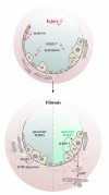
Figure 12. The role of WISP1 in lung fibrosis.
A proposed model depicting the role of WISP1 in lung fibrosis is shown. Initial injury leads to increased WISP1 expression by hyperplastic ATII cells, which sustains ATII cell hyperplasia. Fibrogenesis is then promoted via autocrine (lower right) or paracrine (lower left) effects on ATII cell mediator release and EMT and/or fibroblast ECM synthesis, respectively.

References

    1. Martinez F.J., et al. The clinical course of patients with idiopathic pulmonary fibrosis. Ann. Intern. Med. 2005;142:963–967. doi: 10.1001/archinte.142.5.963. - DOI - PubMed
    1. American Thoracic Society and European Respiratory Society. . American Thoracic Society/European Respiratory Society International Multidisciplinary Consensus Classification of the Idiopathic Interstitial Pneumonias. Am. J. Respir. Crit. Care Med. 2002;165:277–304. - PubMed
    1. Selman M., King T.E., Pardo A. Idiopathic pulmonary fibrosis: prevailing and evolving hypotheses about its pathogenesis and implications for therapy. Ann. Intern. Med. 2001;134:136–151. - PubMed (VSports手机版)
    1. Thannickal V.J., Toews G.B., White E.S., Lynch J.P., 3rd, Martinez F.J. Mechanisms of pulmonary fibrosis. Annu. Rev. Med. 2004;55:395–417. doi: 10.1146/annurev.med.55.091902.103810. - V体育官网入口 - DOI - PubMed
    1. Noble P.W., Homer R.J. Back to the future: historical perspective on the pathogenesis of idiopathic pulmonary fibrosis. Am. J. Respir. Cell Mol. Biol. 2005;33:113–120. doi: 10.1165/rcmb.F301. - V体育2025版 - DOI - PubMed

Publication types

VSports最新版本 - MeSH terms

VSports注册入口 - Substances