"V体育官网入口" Skip to main page content
U.S. flag

An official website of the United States government

Dot gov

The . gov means it’s official. Federal government websites often end in . gov or . mil. Before sharing sensitive information, make sure you’re on a federal government site. VSports app下载.

Https

The site is secure V体育官网. The https:// ensures that you are connecting to the official website and that any information you provide is encrypted and transmitted securely. .

. 2008 Sep;118(9):3065-74.
doi: 10.1172/JCI34739.

Inhibition of mTORC1 leads to MAPK pathway activation through a PI3K-dependent feedback loop in human cancer (V体育ios版)

Affiliations

VSports注册入口 - Inhibition of mTORC1 leads to MAPK pathway activation through a PI3K-dependent feedback loop in human cancer

Arkaitz Carracedo (VSports手机版) et al. J Clin Invest. 2008 Sep.

Abstract

Numerous studies have established a causal link between aberrant mammalian target of rapamycin (mTOR) activation and tumorigenesis, indicating that mTOR inhibition may have therapeutic potential VSports手机版. In this study, we show that rapamycin and its analogs activate the MAPK pathway in human cancer, in what represents a novel mTORC1-MAPK feedback loop. We found that tumor samples from patients with biopsy-accessible solid tumors of advanced disease treated with RAD001, a rapamycin derivative, showed an administration schedule-dependent increase in activation of the MAPK pathway. RAD001 treatment also led to MAPK activation in a mouse model of prostate cancer. We further show that rapamycin-induced MAPK activation occurs in both normal cells and cancer cells lines and that this feedback loop depends on an S6K-PI3K-Ras pathway. Significantly, pharmacological inhibition of the MAPK pathway enhanced the antitumoral effect of mTORC1 inhibition by rapamycin in cancer cells in vitro and in a xenograft mouse model. Taken together, our findings identify MAPK activation as a consequence of mTORC1 inhibition and underscore the potential of a combined therapeutic approach with mTORC1 and MAPK inhibitors, currently employed as single agents in the clinic, for the treatment of human cancers. .


Figures
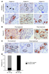
Figure 2
Figure 2. Phosphorylation of ERK in human cancer specimens upon RAD001 treatment.
(AC) p-ERK immunostaining in breast cancer patients before and after the treatment with RAD001. Tumor cells are indicated by arrows or circles. Original magnification, ×40. (D) Percentage of patients with MAPK activation after RAD001 treatment, as measured by ERK phosphorylation, in the 2 different administration schedules.
Figure 1
Figure 1. Schematic representation of the administration schedules in the clinical trial with RAD001.
Patients included in the trial were subjected to a first surgery for the obtention of the tumor biopsy (pre-treatment). Later, daily or weekly RAD001 was administered for 4 weeks and the post-treatment biopsy obtained for analysis.
Figure 3
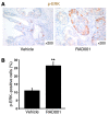
Figure 3. RAD001 treatment leads to MAPK activation in Pten-null prostate conditional mice.
(A) p-ERK immunostaining in Pten-null mouse prostates treated with vehicle or RAD001 for 4 weeks. A representative area is shown. n = 3 mice per group. (B) Quantification of p-ERK staining in hot spots (high–p-ERK areas). **P < 0.01 compared with vehicle treatment. Error bars indicate SD.
Figure 4
Figure 4. Rapamycin activates Ras-Raf1-MEK-ERK in vitro.
(A) Effect of rapamycin treatment (20 nM, 24 h) on ERK, AKT, and RpS6 (S6) phosphorylation in MEFs with different genetic modifications (n = 3). p-S6, phosphorylated RpS6. (B) ERK, AKT, and RpS6 phosphorylation status in SV40-immortalized MEFs upon acute mTOR genetic deletion. (C) Raf1, ERK, and RpS6 phosphorylation status in MCF7 cells upon rapamycin treatment (20 nM, 24 h; n = 3). (D) Effect of rapamycin (20 nM, 24 h) and/or MEK inhibitor UO126 (10 μM, 24 h) on ERK and RpS6 phosphorylation in MCF7 cells (n = 3). (E) ERK and RpS6 phosphorylation in MCF7 transfected for 24 hours with an empty vector (mock) or a dominant-negative form of Ras (RasN17) and treated with rapamycin (20 nM, 24 h; n = 3). Numbers indicate the ratio of the phosphorylated protein related to total protein levels.
Figure 5
Figure 5. Rapamycin-induced MAPK activation is downstream of S6K/IRS-1/PI3K.
(A) ERK phosphorylation in MCF7 transfected for 24 hours with wild-type S6K1 (HAS6K1wt) or a rapamycin-insensitive constitutively active form of S6K1 (HAS6K1 E389 D3E) and treated with rapamycin (20 nM, 24 h; n = 3). (B) ERK and RpS6 phosphorylation status in starved (24 h, 0.1% FBS) T24 cells treated with vehicle or rapamycin (20 nM, 24 h) and stimulated with insulin (200 nM, 15 min) or IGF-1 (100 ng/ml, 15 min; n = 3). (C) ERK and RpS6 phosphorylation in Tsc2-null p53-null MEFs 24 hours after transfection with empty vector or 0.5 or 4 μg of Tsc2-expressing vector (n = 3). Asterisk indicates non-specific band. HSP90 was used as a loading control. (D) Effect of rapamycin (20 nM, 24 h) and/or PI3K inhibitor LY294002 (10 μM, 24 h) on AKT, ERK, and RpS6 phosphorylation in MCF7 cells (n = 3). (E) AKT, ERK, and RpS6 phosphorylation status in starved (24 h, 0.1% FBS) MCF7 cells preincubated with DMSO or wortmannin (500 nM, 45 min) and stimulated with insulin (100, 200, and 400 nM; n = 3). Numbers indicate the ratio of phosphorylated protein related to total protein levels.
Figure 6
Figure 6. MAPK pathway pharmacological blockade increases rapamycin-mediated growth inhibition in vitro.
(A and B) Effect of rapamycin (Rapa; 20 nM), UO126 (10 μM), or the combination of both on cell growth (A) and BrdU incorporation (B; after 24 hours of treatment; n = 4). **P < 0.01 compared with vehicle-treated cells; #P < 0.05 and ##P > 0.01 compared with rapamycin-treated cells; $P < 0.05 and $$P < 0.01 compared with UO126-treated cells. Error bars indicate SD.
Figure 7
Figure 7. Antitumoral action of combined mTORC1 and MAPK inhibition in vivo.
(A) Growth curves of MCF7 tumor xenografts induced in Nude immunocompromised mice. Bar graph shows the tumor growth of RAD001-, PD0325901-, or RAD001+PD0325901-treated tumors at day 10 compared with the size at the beginning of the treatment. A representative picture of the tumors at the time of harvesting (day 10) is shown; n = 4. (B) Weight of tumors subjected to each treatment at the time of the harvest (day 10); n = 4. (CF) Representative immunostaining of p-RpS6 (C), p-ERK (D), H&E and TUNEL (central inset; E), and Ki-67 (F) of tumors from each experimental group; n = 4. Insets show representative positive staining. Original magnification, ×200 (CF), ×800 (insets). Arrows indicate apoptotic areas; asterisk indicate nonproliferative areas. C, RAD001+PD0325901; P, PD0325901; R, RAD001; V, vehicle. (G) Quantification of the effect of the different treatments on apoptosis/necrosis (% of apoptotic/necrotic area; n = 3 tumors quantified per group) and proliferation (Ki-67 positivity; >350 total cells counted per tumor; n = 3). **P < 0.01 compared with vehicle-treated mice; #P < 0.05 and ##P < 0.01 compared with RAD001-treated mice; $P < 0.05 and $$P < 0.01 compared with PD0325901-treated mice. Error bars indicate SD.
Figure 8
Figure 8. Schematic representation of the pathway described in the study.
Briefly, mTORC1 activation leads to PI3K and MAPK inhibition through a negative feedback loop stemming from S6K1 (left panel), while treatment with mTORC1 inhibitors results in a hyperactive RTK/IRS-1/PI3K pathway, increasing the signal toward the Ras-Raf1-MEK1/2-ERK pathway (middle panel). Therefore, the combination of MEK and mTORC1 inhibitors may provide a therapeutic benefit in the treatment of certain cancers through the abrogation of the feedback described in this study (right panel). Activated proteins are represented with blue circles and inhibition status with red squares.

Comment in

  • J Clin Invest. 118:3003.

References

    1. Wullschleger S., Loewith R., Hall M.N. TOR signaling in growth and metabolism. Cell. 2006;124:471–484. doi: 10.1016/j.cell.2006.01.016. - DOI - PubMed
    1. Guertin D.A., Sabatini D.M. Defining the role of mTOR in cancer. Cancer Cell. 2007;12:9–22. doi: 10.1016/j.ccr.2007.05.008. - DOI - PubMed
    1. Ballif B.A., et al. Quantitative phosphorylation profiling of the ERK/p90 ribosomal S6 kinase-signaling cassette and its targets, the tuberous sclerosis tumor suppressors. Proc. Natl. Acad. Sci. U. S. A. 2005;102:667–672. doi: 10.1073/pnas.0409143102. - DOI - PMC - PubMed
    1. Inoki K., Li Y., Zhu T., Wu J., Guan K.L. TSC2 is phosphorylated and inhibited by Akt and suppresses mTOR signalling. Nat. Cell Biol. 2002;4:648–657. doi: 10.1038/ncb839. - DOI - PubMed
    1. Ma L., Chen Z., Erdjument-Bromage H., Tempst P., Pandolfi P.P. Phosphorylation and functional inactivation of TSC2 by Erk implications for tuberous sclerosis and cancer pathogenesis. Cell. 2005;121:179–193. doi: 10.1016/j.cell.2005.02.031. - DOI - PubMed

Publication types

MeSH terms (V体育2025版)