Skip to main page content
U.S. flag

An official website of the United States government

Dot gov

The VSports app下载. gov means it’s official. Federal government websites often end in . gov or . mil. Before sharing sensitive information, make sure you’re on a federal government site. .

Https

The site is secure V体育官网. The https:// ensures that you are connecting to the official website and that any information you provide is encrypted and transmitted securely. .

. 2007 Nov 15;21(22):2908-22.
doi: 10.1101/gad.1586007.

"V体育ios版" Nuclear accumulation of cyclin D1 during S phase inhibits Cul4-dependent Cdt1 proteolysis and triggers p53-dependent DNA rereplication

Affiliations

Nuclear accumulation of cyclin D1 during S phase inhibits Cul4-dependent Cdt1 proteolysis and triggers p53-dependent DNA rereplication (V体育2025版)

Priya Aggarwal et al. Genes Dev. .

"VSports" Abstract

Deregulation of cyclin D1 occurs in numerous human cancers through mutations, alternative splicing, and gene amplification VSports手机版. Although cancer-derived cyclin D1 mutants are potent oncogenes in vitro and in vivo, the mechanisms whereby they contribute to neoplasia are poorly understood. We now provide evidence derived from both mouse models and human cancer-derived cells revealing that nuclear accumulation of catalytically active mutant cyclin D1/CDK4 complexes triggers DNA rereplication, resulting from Cdt1 stabilization, which in turn triggers the DNA damage checkpoint and p53-dependent apoptosis. Loss of p53 through mutations or targeted deletion results in increased genomic instability and neoplastic growth. Collectively, the data presented reveal mechanistic insights into how uncoupling of critical cell cycle regulatory events will perturb DNA replication fidelity, thereby contributing to neoplastic transformation. .


Figures

Figure 1.
Figure 1.
Malignant D1T286A lymphocytes are aneuploid. (A) Representative SKY analysis of tumors derived from D1T286A. (B) Quantification of the chromosome number per metaphase spread for nontransgenic (purple; 200/11; spreads per number of mice), premalignant D1T286A transgenic (blue; 100/9), and malignant D1T286A transgenic (light blue; 219/9) mice splenocytes.
Figure 2.
Figure 2.
Cdt1 overexpression in D1T286A-expressing splenocytes. (A) Cdt1 and Cul4A/4B levels in tissues or tumors derived from nontransgenic or D1T286A mice were assessed by immunoblot analysis. (B,C) Cdt1, Cul4A, Cul4B, and Cdt2 mRNA levels from nontransgenic spleen and D1T286A transgenic tumors were assessed by real-time RT–PCR using the Applied Biosystems 7900HT detection. (D) The Cdt1 half-life in nontransgenic or transgenic splenocytes was determined following brief culture in RPMI-1640 complete media. Cells were treated with cycloheximide (100 μg/mL), harvested at the indicated intervals post-treatment, and subjected to Western analysis for Cdt1 and βActin. Cdt1 levels are expressed as an average of three independent experiments; bars represent standard error between experiments.
Figure 3.
Figure 3.
Acute inhibition of Cdt1 proteolysis by the cyclin D1T286A/CDK4 kinase. (A) HeLa cells transfected with the indicated plasmids along with myc-Cdt1 were treated with nocodazole for 16–18 h, after which cells were shaken off and replated in two plates: The first plate, in complete media, was harvested 8 h after release (G1 phase); in the second dish, HU was added and cells were harvested 14 h after shake-off (S phase). Levels of Cdt1, cyclin A (marker for S phase), cyclin D1, β-Actin, and GFP were determined by immunoblot. (B) Cul4A/4B and Cdt2 mRNA levels determined by real-time RT–PCR from HeLa cells synchronized in either G1 or S phase (as in A) following transfection with vectors encoding D1T286A along with either wild-type or kinase-dead CDK4. Target mRNAs were normalized to actin and expressed as fold change between G1 and S phase. (C,D) HeLa cells were transfected with the indicated plasmids and synchronized in G1 or S phase. Lysates prepared from these cells were analyzed by immunoblot with antibodies directed toward the proteins indicated to the right. (E,F) Western analysis of soluble lysates prepared from TE3, TE7 (which express endogenous cyclin D1P287A), and KYSE-520 human esophageal cancer cell lines for cyclin D1, Cul4B, βActin, and Cdt1.
Figure 4.
Figure 4.
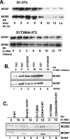
Cyclin D1-dependent stabilization of Cdt1 promotes reloading of MCM during S phase. (A) NIH-3T3 cell lines overexpressing cyclin D1 or D1T286A were synchronized with HU for 24 h and released in complete medium lacking HU. Chromatin-bound proteins were analyzed by Western analysis for MCM3 and MCM7 at the indicated intervals following replating in media without HU. (B) TE3, TE7, and KYSE-520 cell lines were synchronized at the G1/S boundary with HU for 24 h. Chromatin-bound proteins were assessed at 0 and 10 h post-HU release. (C) HeLa cells transfected with the indicated plasmids synchronized at the G1/S boundary with HU for 24 h. Chromatin-bound proteins were assessed at 0 and 10 h post-HU.
Figure 5.
Figure 5.
Cdt1 cooperates with the D1T286A-dependent kinase to induce p53-dependent DNA rereplication. (A) H1299 cells transfected with indicated cyclin expression vectors, and CDK4 along with GFP and Cdt1 where indicated, were stained with propidium iodide and analyzed by flow cytometry following gating on the GFP-positive cell population. (B) H1299 cells were transfected with plasmids encoding Cdt1, Cdt1 along with D1T286A and CDK4, or empty vector and treated with nocodazole (G2–M block) 10 h after transfection. Fifteen hours following nocodazole, cells were harvested by mitotic shake-off, and cell cycle profile was assessed by propidium iodide staining. The percentage of cells in a representative experiment showing >4N DNA content is represented graphically. (C) NIH-3T3 cells were transfected with the indicated expression plasmids with or without p53V143A and analyzed as in A. Graphic quantification of the percentage of >4N cells (rereplication). (D) Reintroduction of Cul4A, but not Skp2, inhibits D1T286A-dependent rereplication.
Figure 6.
Figure 6.
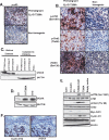
Expression of D1T286A in splenic lymphocytes induces a DNA damage response. (A) IHC of premalignant D1T286A transgenic (top) and age-matched nontransgenic (bottom) spleens for p-p53 (S-15). (B) IHC of p-ATM (S1981) (top panel), p-Chk2 (T68) (middle panel), and γ-H2AX (S139) (bottom panel) in premalignant D1T286A transgenic (left panel) and age-matched nontransgenic (right panel) mice. (C) Accumulation of γH2AX in histone extracts prepared from D1T286A transgenic tumor. β-Actin was used as a loading control for cytoplasmic extract lysates. (D) Lysates prepared from U2OS cells, transfected as indicated, were subjected to immunoblot for γH2AX and βActin. (E) Lysates prepared from HeLa cells transfected with the indicated plasmids were subjected to immunoblot for p-ATM, total ATM, p-Chk2, total Chk2, GFP (transfection control), and β-Actin. (F) Representative IHC for cyclin D1b and γH2AX from esophageal adenocarcinoma.
Figure 7.
Figure 7.
Targeted deletion of p53 accelerates B-cell lymphoma development. (A) Survival curves representing nontransgenic (black line; n-25), D1T286A (blue line; n = 36), p53+/− (red line; 28), D1T286A/p53+/− (green line; n = 44), p53−/− (yellow line; n = 5), and D1T286A/p53−/− (purple line; n = 12) cohorts over a 24-mo period. (B) Representative histology of p53+/− spleen at the magnification of 4× (panel a) and 40× (panel b), a tumor-burdened p53+/− D1T286A spleen at 4× (panel c) and 40× (panel d), or a D1T286A tumor-burdened spleen at 4× (panel e) and 40× (panel f) stained with hematoxylin and eosin. (C) Immunohistochemical staining of D1T286A/p53+/− spleens for B220, cyclin D1, CD3, and Ki-67. (D) Splenocytes from a cohort of age-matched p53+/− (a,c) or D1T286A/p53+/− tumor-burdened (panels b,d) mice were analyzed by flow cytometry with antibodies specific for B220 and CD19 (panels a,b) or surface IgM and IgD (panels c,d).
Figure 8.
Figure 8.
Deletion of p53 decreases D1T286A-dependent apoptosis and accelerates the accumulation of aneuploid cells. (A) Cdt1 levels in tumors derived from D1T286A/p53+/− mice. (B) Cdt1 levels in nontransgenic spleens or from premalignant versus malignant spleen derived from either Eμ-D1T286A or Eμ-D1T286A/p53+/− mice. (C) Representative SKY of tumors derived fromD1T286A/p53+/− mice. (D) Quantification of TUNEL-positive cells in premalignant D1T286A, D1T286A/p53+/−, or age-matched nontransgenic control. (E) Quantification of the chromosome number per metaphase spread for nontransgenic (purple), premalignant D1T286A transgenic (blue), premalignant p53+/− (light blue), and premalignant D1T286A/p53+/− (red) mice. One-hundred to 200 metaphase spreads from multiple mice of each genotype were analyzed. (F) Representative cell cycle profile of p53+/− (top) and D1T286A/p53+/− (bottom) splenocytes.

References

    1. Aktas H., Cai H., Cooper G.M., Cai H., Cooper G.M., Cooper G.M. Ras links growth factor signaling to the cell cycle machinery via regulation of cyclin D1 and the Cdk inhibitor p27KIP1. Mol. Cell. Biol. 1997;17:3850–3857. - PMC - PubMed
    1. Albanese C., Johnson J., Watanabe G., Eklund N., Vu D., Arnold A., Pestell R.G., Johnson J., Watanabe G., Eklund N., Vu D., Arnold A., Pestell R.G., Watanabe G., Eklund N., Vu D., Arnold A., Pestell R.G., Eklund N., Vu D., Arnold A., Pestell R.G., Vu D., Arnold A., Pestell R.G., Arnold A., Pestell R.G., Pestell R.G. Transforming p21ras mutants and c-Ets-2 activate the cyclin D1 promoter through distinguishable regions. J. Biol. Chem. 1995;270:23589–23597. - PubMed
    1. Alt J.R., Cleveland J.L., Hannink M., Diehl J.A., Cleveland J.L., Hannink M., Diehl J.A., Hannink M., Diehl J.A., Diehl J.A. Phosphorylation-dependent regulation of cyclin D1 nuclear export and cyclin D1-dependent cellular transformation. Genes & Dev. 2000;14:3102–3114. - VSports注册入口 - PMC - PubMed
    1. Bani-Hani K., Martin I.G., Hardie L.J., Mapstone N., Briggs J.A., Forman D., Wild C.P., Martin I.G., Hardie L.J., Mapstone N., Briggs J.A., Forman D., Wild C.P., Hardie L.J., Mapstone N., Briggs J.A., Forman D., Wild C.P., Mapstone N., Briggs J.A., Forman D., Wild C.P., Briggs J.A., Forman D., Wild C.P., Forman D., Wild C.P., Wild C.P. Prospective study of cyclin D1 overexpression in Barrett’s esophagus: Association with increased risk of adenocarcinoma. J. Natl. Cancer Inst. 2000;92:1316–1321. - "V体育平台登录" PubMed
    1. Barnes D.M., Gillett C.E., Gillett C.E. Cyclin D1 in breast cancer. Breast Cancer Res. Treat. 1998;52:1–15. - PubMed (VSports最新版本)

Publication types

MeSH terms