Skip to main page content (V体育平台登录)
U.S. flag

An official website of the United States government

Dot gov

The . gov means it’s official. Federal government websites often end in . gov or VSports app下载. mil. Before sharing sensitive information, make sure you’re on a federal government site. .

Https

The site is secure V体育官网. The https:// ensures that you are connecting to the official website and that any information you provide is encrypted and transmitted securely. .

. 2004 Apr;24(8):3505-13.
doi: 10.1128/MCB.24.8.3505-3513.2004.

Dual roles for the Notch target gene Hes-1 in the differentiation of 3T3-L1 preadipocytes

Affiliations

Dual roles for the Notch target gene Hes-1 in the differentiation of 3T3-L1 preadipocytes

David A Ross et al. Mol Cell Biol. 2004 Apr.

Abstract

The process of adipogenesis involves a complex program of gene expression that includes down-regulation of the gene encoding Hes-1, a target of the Notch signaling pathway. To determine if Notch signaling affects adipogenesis, we exposed 3T3-L1 preadipocytes to the Notch ligand Jagged1 and found that differentiation was significantly reduced. This effect could be mimicked by constitutive expression of Hes-1 VSports手机版. The block was associated with a complete loss of C/EBPalpha and peroxisome proliferator-activated receptor gamma (PPARgamma) induction and could be overcome by retroviral expression of either C/EBPalpha or PPARgamma2. Surprisingly, small interfering RNA (siRNA)-mediated reduction of Hes-1 mRNA in 3T3-L1 cells also inhibited differentiation, suggesting an additional, obligatory role for Hes-1 in adipogenesis. This role may be related to our observation that both Notch signaling and Hes-1 down-regulate transcription of the gene encoding DLK/Pref-1, a protein known to inhibit differentiation of 3T3-L1 cells. The results presented in this study establish a new target downstream of the Notch-Hes-1 pathway and suggest a dual role for Hes-1 in adipocyte development. .


Figures
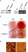
FIG. 1.
FIG. 1.
Constitutive Notch signaling inhibits differentiation of 3T3-L1 cells. (A and B) Analysis of NICD and Mre11 (loading control) protein by Western immunoblotting (A) and Hes-1 and hypoxanthine phosphoribosyltransferase (HPRT; loading control) RNA by RT-PCR (B) in 3T3-L1 cells cultured on control plates or plates containing immobilized Jagged1. RT, reverse transcriptase. (C) Effect of Jagged1 on 3T3-L1 cell differentiation. Cells were transferred to either DM or GM and stained with Oil Red O after 7 days. (D) Micrograph of Oil Red O-stained 3T3-L1 cells from control plates (left) and Jagged1 plates (right).
FIG. 2.
FIG. 2.
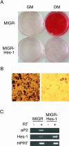
Constitutive expression of Hes-1 inhibits differentiation of 3T3-L1 cells. (A) 3T3-L1 cell populations transduced with control (MIGR) or Hes-1 (MIGR-Hes-1) retroviruses were induced to differentiate in DM or maintained in GM and stained with Oil Red O after 7 days. (B) Micrograph of MIGR (left) and MIGR-Hes-1 (right) cells cultured in DM for 7 days. (C) RT-PCR analyses of aP2, Hes-1, and hypoxanthine phosphoribosyltransferase (HPRT; loading control) RNAs in the indicated cells grown in DM after 7 days. RT, reverse transcriptase.
FIG. 3.
FIG. 3.
PPARγ and C/EBPα are not induced in 3T3-L1 cells harboring the MIGR-Hes-1 virus. Control (MIGR) and Hes-1-expressing (MIGR-Hes-1) cells were grown to confluence (day −2) and transferred to DM (day 0), and the expression of PPARγ, C/EBPα, SREBP-1 (ADD-1), C/EBPβ, and Cdk4 (loading control) proteins was determined after the indicated number of days. PPARγ and C/EBPα are each expressed as two isoforms. Asterisk, position of a nonspecific cross-reacting protein.
FIG. 4.
FIG. 4.
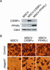
Constitutive expression of C/EBPα or PPARγ2 allows differentiation of 3T3-L1 cells undergoing Notch signaling. (A) Analyses of C/EBPα, PPARγ, and Cdk4 (loading control) proteins in 3T3-L1 cells transduced with MSCV, MSCV-C/EBPα, or MSCV-PPARγ2. Asterisk, position of a nonspecific cross-reacting protein. (B) Micrographs of Oil Red O-stained 3T3-L1 cells transduced with the indicated retroviruses and induced to differentiate on control plates and those containing immobilized Jagged1.
FIG. 5.
FIG. 5.
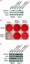
Constitutive expression of C/EBPα or PPARγ2 allows differentiation of 3T3-L1 cells expressing Hes-1. (A) Analyses of C/EBPα, PPARγ, and Cdk4 (loading control) proteins in 3T3-L1 cells transduced with MIGR or MIGR-Hes-1 and with MSCV, MSCV-C/EBPα, or MSCV-PPARγ2, as indicated. Asterisk, position of a nonspecific cross-reacting protein. (B) MIGR and MIGR-Hes-1 3T3-L1 cells were transduced individually with MSCV, MSCV-C/EBPα, or MSCV-PPARγ2, and puromycin-resistant populations were induced to differentiate and stained with Oil Red O after 7 days. (C) Cells induced to differentiate were evaluated for expression of PPARγ, aP2, Hes-1, and hypoxanthine phosphoribosyltransferase (HPRT; control) RNAs by RT-PCR.
FIG. 6.
FIG. 6.
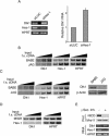
Reduced Hes-1 expression inhibits differentiation of 3T3-L1 cells. (A) 3T3-L1 cells harboring the pSIREN-siLUC (siLUC) or pSIREN-siHES-1 (siHES-1) retroviruses were analyzed for expression of Hes-1 and HPRT (control) RNA by semiquantitative RT-PCR. The amount of input reverse-transcribed first-strand cDNA (input f.s. cDNA) is represented by black triangles. The bar graph represents the relative levels of Hes-1 mRNA in the pSIREN-siLUC (siLUC) or pSIREN-siHES-1 (siHES-1) 3T3-L1 cells. Hes-1 levels are expressed relative to those in siLUC cells and normalized with hypoxanthine phosphoribosyltransferase (HPRT). (B) 3T3-L1 cells containing control (siLUC) or siHes-1 viruses were transferred to DM or maintained in GM and stained with Oil Red O after 7 days. (C) Cells in panel B were assessed for expression of aP2 and HPRT (control) RNA by RT-PCR. RT, reverse transcriptase.
FIG. 7.
FIG. 7.
DLK1 is a Notch/Hes-1 target. (A) 3T3-L1 cells containing low levels of Hes-1 express high levels of Dlk1. 3T3-L1 cells transduced with pSIREN-siLUC (siLUC) or pSIREN-siHES-1 (siHES-1) were assessed for expression of Dlk1, Hes-1, and hypoxanthine phosphoribosyltransferase (HPRT; control) RNA by RT-PCR. The bar graph represents the relative levels of Dlk1 RNA in the siLUC and siHES-1 cells normalized to HPRT, as measured by semiquantitative RT-PCR. (B) Ligand-mediated activation of Notch reduces the level of Dlk1 RNA in 3T3-L1 cells. 3T3-L1 cells were cocultured with control (BABE) or Jagged1 (JAG)-expressing NIH 3T3 cells. Total RNA from the 3T3-L1 cells was assessed for expression of Dlk1, Hes-1, and HPRT (control) RNAs by semiquantitative RT-PCR. Increasing amounts of input reverse-transcribed first-strand cDNA (input f.s. cDNA) are represented by black triangles. (C) Ligand-mediated activation of Notch reduces the level of Dlk1 in NIH 3T3 cells. NIH 3T3 cells were cocultured with control (BABE) or Jagged1 (JAG)-expressing cells and analyzed as described for panel B (left). NIH 3T3 cells were also evaluated for the expression of Dlk1 and β-actin (control) protein by Western immunoblotting (right). (D) NIH 3T3 cells expressing Hes-1 contain reduced amounts of Dlk1. NIH 3T3 cells harboring control (MIGR) or Hes-1-expressing (MIGR-Hes-1) retroviruses were assessed for Dlk1 and HPRT (control) RNA by semiquantitative RT-PCR as for panel B. (E) SUP-T1 cells treated with a γ-secretase inhibitor have increased levels of Dlk1. SUP-T1 T-ALL cells were treated with γ-secretase inhibitor X or DMSO and assessed for expression of NICD and Mre11 (control) protein by Western immunoblotting and for Dlk1, Hes-1, and HPRT (control) RNA by RT-PCR.
FIG. 8.
FIG. 8.
The Dlk1 promoter is inhibited by NICD and by Hes-1. (A) NIH 3T3 cells harboring either a control (MIGR) or NICD-expressing (MIGR-NICD) retrovirus were transfected with the reporter plasmids pGL2-Pro, CSL-Luc, DLK-1400, and DLK-191. Luciferase values are expressed relative to a control Renilla luciferase reporter. (B) Activity of the same reporters was determined in cells containing a Hes-1-expressing virus (MIGR-Hes-1). Mean values and standard errors of the means were determined from at least three individual experiments.
FIG. 9.
FIG. 9.
Model depicting the role of Notch/Hes-1 signaling in differentiating 3T3-L1 cells.

References

    1. Aster, J. C., E. S. Robertson, R. P. Hasserjian, J. R. Turner, E. Kieff, and J. Sklar. 1997. Oncogenic forms of NOTCH1 lacking either the primary binding site for RBP-Jκ or nuclear localization sequences retain the ability to associate with RBP-Jκ and activate transcription. J. Biol. Chem. 272:11336-11343. - PubMed
    1. Berezovska, O., C. Jack, P. McLean, J. C. Aster, C. Hicks, W. Xia, M. S. Wolfe, W. T. Kimberly, G. Weinmaster, D. J. Selkoe, and B. T. Hyman. 2000. Aspartate mutations in presenilin and gamma-secretase inhibitors both impair notch1 proteolysis and nuclear translocation with relative preservation of notch1 signaling. J. Neurochem. 75:583-593. - PubMed
    1. Brou, C., F. Logeat, N. Gupta, C. Bessia, O. LeBail, J. R. Doedens, A. Cumano, P. Roux, R. A. Black, and A. Israel. 2000. A novel proteolytic cleavage involved in Notch signaling: the role of the disintegrin-metalloprotease TACE. Mol. Cell 5:207-216. - V体育官网入口 - PubMed
    1. Castella, P., S. Sawai, K. Nakao, J. A. Wagner, and M. Caudy. 2000. HES-1 repression of differentiation and proliferation in PC12 cells: role for the helix 3-helix 4 domain in transcription repression. Mol. Cell. Biol. 20:6170-6183. - PMC - PubMed
    1. De Strooper, B., W. Annaert, P. Cupers, P. Saftig, K. Craessaerts, J. S. Mumm, E. H. Schroeter, V. Schrijvers, M. S. Wolfe, W. J. Ray, A. Goate, and R. Kopan. 1999. A presenilin-1-dependent gamma-secretase-like protease mediates release of Notch intracellular domain. Nature 398:518-522. - PubMed

"V体育安卓版" Publication types

MeSH terms

Substances