Skip to main page content
U.S. flag

An official website of the United States government

Dot gov

The VSports app下载. gov means it’s official. Federal government websites often end in . gov or . mil. Before sharing sensitive information, make sure you’re on a federal government site. .

Https

The site is secure. The https:// ensures that you are connecting to the official website and that any information you provide is encrypted and transmitted securely V体育官网. .

. 2003 Oct;163(4):1653-62.
doi: 10.1016/S0002-9440(10)63522-5.

Role of fibroblast growth factor type 1 and 2 in carbon tetrachloride-induced hepatic injury and fibrogenesis

Affiliations

"VSports在线直播" Role of fibroblast growth factor type 1 and 2 in carbon tetrachloride-induced hepatic injury and fibrogenesis

Chundong Yu et al. Am J Pathol. 2003 Oct.

Abstract

Genomic ablation of hepatocyte-specific fibroblast growth factor receptor (FGFR)4 in mice revealed a role of FGF signaling in cholesterol and bile acid metabolism and hepatolobular restoration in response to injury without effect on liver development or hepatocyte proliferation. Although the potential role of all 23 FGF polypeptides in the liver is still unclear, the most widely studied prototypes, FGF1 and FGF2, are present and have been implicated in liver cell growth and function in vitro VSports手机版. To determine whether FGF1 and FGF2 play a role in response to injury and fibrosis, we examined the impact of both acute and chronic exposure to carbon tetrachloride (CCl(4)) in the livers of FGF1- and FGF2-deficient mice. After acute CCl(4) exposure, FGF1(-/-)FGF2(-/-) mice exhibited an accelerated release of serum alanine aminotransferase similar to FGFR4 deficiency, but no effect on overall hepatolobular restoration or bile acid metabolism. FGF1(-/-)FGF2(-/-) mice exhibited a normal increase in alpha-smooth muscle actin and desmin associated with activation and migration of hepatic stellate cells to damage, but a reduced level of hepatic stellate cell-derived matrix collagen alpha1(I) synthesis. Liver fibrosis resulting from chronic CCl(4) exposure was markedly decreased in the livers of FGF1/FGF2-deficient mice. These results suggest an agonist role for FGF1 and FGF2 in specifically insult-induced liver matrix deposition and hepatic fibrogenesis and a potential target for the prevention of hepatic fibrosis. .


Figures

Figure 1.
Figure 1.
Accelerated injury in FGF1(−/−)FGF2(−/−) livers after acute CCl4 treatment. A: Plasma ALT levels after acute CCl4 treatment. ALT activity was measured using the GP-Transaminase kit (no. 505-P; Sigma, St. Louis, MO) as described. Values were the mean ± SD (n = 4 mice). Significance of differences between FGF1(−/−)FGF2(−/−) and WT mice at 24, 38, and 48 hours was P < 0.05. B: Hepatocyte DNA synthesis after acute CCl4 treatment. DNA synthesis was assessed by BrdU incorporation as described in Materials and Methods. Values were the mean ± SD (n = 4 mice). Significance of differences between FGF1(−/−)FGF2(−/−) and WT mice at 38 and 48 hours was P < 0.02. C: Liver-to-total body weight ratio after acute CCl4 treatment. Livers were excised and weighed as described. Values were the mean ± SD (n = 4 mice). Significance of the difference between FGF1(−/−)FGF2(−/−) and WT livers was P > 0.05 at all time points.
Figure 2.
Figure 2.
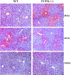
Histology of livers from FGF1(−/−)FGF2(−/−) and WT mice after acute CCl4 treatment. Paraffin-embedded sections were prepared and stained with H&E as described in Materials and Methods. Note the severe centrizonal injury and cellular debris at 38 hours after CCl4 in FGF1(−/−)FGF2(−/−) livers relative to WT. Original magnifications, ×150.
Figure 3.
Figure 3.
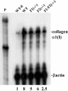
Decreased induction of collagen α1(I) expression in FGF1(−/−)FGF2(−/−) livers after acute CCl4 treatment. Total liver RNA was isolated from three mice in each group at the indicated times after acute CCl4 treatment, pooled before assay by RNase protection, and relative intensity of the collagen α1(I) mRNA was quantitated and normalized as described in Materials and Methods. The signal in untreated WT livers was assigned a value of 1 and all other treatments expressed as a fold change over untreated wild type. P, labeled RNA probe. The displayed autoradiograph is representative of two independent reproductions.
Figure 4.
Figure 4.
Both FGF1 and FGF2 contribute to induction of collagen α1(I) in livers after acute CCl4 administration. Collagen α1(I) mRNA was determined and quantitated as described in Figure 3 ▶ using 50 μg of pooled total RNA from three livers of mice with the indicated phenotype at 48 hours after CCl4 treatment, except the indicated untreated WT (WT-0) control. The displayed autoradiograph is representative of three independent reproductions.
Figure 5.
Figure 5.
Lack of difference in α-SMA and desmin expression in FGF1(−/−)FGF2(−/−) and WT livers after acute CCl4 treatment. A: α-SMA and desmin expression in liver tissue extracts. Livers from mice of the indicated phenotype at the indicated times after acute CCl4 treatment were excised; the extract from three livers in each treatment group was pooled and analyzed by immunoblot (see Materials and Methods). The displayed autoradiograph is representative of three independent reproductions. B: Immunochemical analysis of activated HSCs expressing α-SMA in tissue sections. Livers at 72 hours after CCl4 treatment were processed and tissue sections prepared as described in Figure 2 ▶ . Original magnifications, ×100.
Figure 6.
Figure 6.
Normal activation of TGF-β1 expression in FGF1(−/−)FGF2(−/−) livers after acute CCl4 treatment. TGF-β1 mRNA was determined by RPA as described in Materials and Methods using 50 μg of pooled total RNA isolated from three livers. The intensity of TGF-β1 bands was quantitated as in Figure 3 ▶ and expressed relative to untreated WT assigned a value of 1. The displayed autoradiograph is representative of two independent reproductions.
Figure 7.
Figure 7.
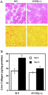
Decreased fibrosis in FGF1(−/−)FGF2(−/−) livers after chronic CCl4 administration. A: The indicated type mice were subjected to chronic CCl4 treatment for 21 days as described in Materials and Methods. Livers were excised; tissue sections were prepared and stained with H&E (top) or Sirius Red (bottom). B: Quantitative analysis of collagen in liver sections. Total liver collagen in sections of WT or FGF1(−/−)FGF2(−/−) mice was quantitated as described in Materials and Methods. Values are the mean ± SD (n = 4 mice). Significance of differences between FGF1(−/−)FGF2(−/−) and WT mice at day 21 was P < 0.02. Original magnifications, ×60 (A).

References

    1. McKeehan WL, Wang F, Kan M: The heparan sulfate-fibroblast growth factor family: diversity of structure and function. Prog Nucleic Acid Res Mol Biol 1998, 59:135-176 - "VSports注册入口" PubMed
    1. Powers CJ, McLeskey SW, Wellstein A: Fibroblast growth factors, their receptors and signaling. Endocr Relat Cancer 2000, 7:165-197 - PubMed (V体育2025版)
    1. Ornitz DM, Itoh N: Fibroblast growth factors. Genome Biol 2001, 2:3005.1-3005.12 - PMC - PubMed
    1. Abraham JA, Mergia A, Whang JL, Tumolo A, Friedman J, Hjerrild KA, Gospodarowicz D, Fiddes JC: Nucleotide sequence of a bovine clone encoding the angiogenic protein, basic fibroblast growth factor. Science 1986, 233:545-548 - V体育官网入口 - PubMed
    1. Jaye M, Howk R, Burgess W, Ricca GA, Chiu IM, Ravera MW, O’Brien SJ, Modi WS, Maciag T, Drohan WN: Human endothelial cell growth factor: cloning, nucleotide sequence, and chromosome localization. Science 1986, 233:541-545 - PubMed

V体育2025版 - Publication types

MeSH terms