Skip to main page content
U.S. flag

An official website of the United States government

Dot gov

The . gov means it’s official. Federal government websites often end in VSports app下载. gov or . mil. Before sharing sensitive information, make sure you’re on a federal government site. .

Https

The site is secure. The https:// ensures that you are connecting to the official website and that any information you provide is encrypted and transmitted securely V体育官网. .

. 2001 Nov;108(9):1369-78.
doi: 10.1172/JCI12373.

V体育ios版 - DDR2 receptor promotes MMP-2-mediated proliferation and invasion by hepatic stellate cells

Affiliations

DDR2 receptor promotes MMP-2-mediated proliferation and invasion by hepatic stellate cells

E Olaso et al. J Clin Invest. 2001 Nov.

Abstract

Type I collagen provokes activation of hepatic stellate cells during liver injury through mechanisms that have been unclear VSports手机版. Here, we tested the role of the discoidin domain tyrosine kinase receptor 2 (DDR2), which signals in response to type I collagen, in this pathway. DDR2 mRNA and protein are induced in stellate cells activated by primary culture or in vivo during liver injury. The receptor becomes tyrosine phosphorylated in response to either endogenous or exogenous type I collagen, whereas its expression is downregulated during cellular quiescence induced by growth on Matrigel. We developed stellate cell lines stably overexpressing either wild-type DDR2, a constitutively active chimeric DDR2 receptor (Fc-DDR2), a truncated receptor expressing the extracellular domain, or a kinase-dead DDR2 Cells overexpressing DDR2 showed enhanced proliferation and invasion through Matrigel, activities that were directly related to increased expression of active matrix metalloproteinase 2 (MMP-2). These data show that DDR2 is induced during stellate cell activation and implicate the phosphorylated receptor as a mediator of MMP-2 release and growth stimulation in response to type I collagen. Moreover, type I collagen-dependent upregulation of DDR2 expression establishes a positive feedback loop in activated stellate cells, leading to further proliferation and enhanced invasive activity. .


Figures

Figure 1
Figure 1
DDR2 is induced during stellate cell activation in vitro. Stellate cells isolated from normal rat livers were maintained in monolayer culture for up to 8 days, and DDR2 mRNA and protein were analyzed. (a) Northern blot analysis performed with 10 μg mRNA from primary cultures of stellate cells hybridized to DDR2 and GAPDH cDNAs. (b) Summary histogram showing the induction of DDR2 during stellate cell activation by in vitro culture based on data from a. Results are expressed as a ratio of DDR2 mRNA versus GAPDH mRNA. (c) Stellate cells were cultured for 0, 2, and 8 days, lysed in RIPA buffer, immunoprecipitated, immunoblotted, and probed with the polyclonal anti-DDR2 Ab R2-JM. COS-1 cells were transfected with full-length DDR2 (+) or empty vector (–). (d) Lysates were also analyzed by Western blot for tubulin as a control for protein loading.
Figure 2
Figure 2
DDR2 is induced in stellate cells during liver injury. (a) Northern blot analysis of DDR2 mRNA expression in rat stellate cells isolated after administration of a single dose of CCl4 or after bile duct ligation. (b) Summary histogram demonstrating the induction of DDR2 during liver injury. Results are expressed in the histograms as the ratio of DDR2 mRNA versus GAPDH mRNA. (c) Total liver extracts from rats treated 4 weeks with CCl4 or corn oil vehicle alone were lysed in RIPA buffer. DDR2 was immunoprecipitated with the R2-JM Ab, followed by Western blot analysis with an anti-phosphotyrosine Ab (4G10) or the anti-DDR2 Ab R2-JM.
Figure 3
Figure 3
Type I collagen stimulates DDR2 phosphorylation in stellate cells. HSC-T6 stellate cells were stimulated with 10 μg/ml type I collagen for increasing intervals and lysed in RIPA buffer. DDR2 was immunoprecipitated with the anti-DDR2 Ab R2-JM, followed by Western blot analysis with an antiphosphotyrosine Ab (4G10) or anti-DDR2 (R2-JM).
Figure 4
Figure 4
Endogenous collagen synthesis is sufficient for DDR2 phosphorylation. HSC-T6 stellate cells were cultured for 48 hours in the presence of the proline analogue cis-OH-Pro to reduce collagen synthesis. (a) Levels of type I collagen were analyzed with a Western blot of cell lysates using a polyclonal Ab. (b) Cell lysates were immunoprecipitated with the polyclonal anti-DDR2 Ab R2-JM, followed by analysis of Western blotting with an antiphosphotyrosine Ab (4G10) or anti-DDR2 (R2-JM).
Figure 5
Figure 5
Extracellular matrix regulates DDR2 expression. Levels of DDR2 mRNA (a) and protein (b) expressed by immortalized HSCs (HSC-T6) were analyzed after 3 days of growth on either Matrigel or type I collagen gels. Tubulin or GAPDH mRNA expression levels were assessed, respectively, as loading controls. (c) In primary stellate cells activated by growth in primary culture on plastic, transfer to Matrigel led to diminished DDR2 expression after 48 hours in culture, as assessed by Western blot analysis.
Figure 6
Figure 6
DDR2 promotes HSC proliferation. HSC-T6 cells were infected with a retrovirus containing a bi-cistronic cDNA expressing either a truncated form of the receptor lacking the kinase domain (ec-DDR2), a kinase-dead mutant (kd-DDR2), GFP (control), or a constitutively active DDR2 receptor (Fc-DDR2). Infected cells were recovered by cell sorting for GFP fluorescence using FACS. (a) Retroviral infection with kd-DDR2 inhibits the phosphorylation of the endogenous DDR2 receptor in response to a 30-minute exposure to 10 μg/ml collagen type I. Proliferation rates were analyzed by measuring (b) [3H]-thymidine incorporation or (c) cell number and expressed as relative values versus control cells.
Figure 7
Figure 7
DDR2 promotes MMP-2 expression and activity in stellate cells. Serum-free supernatants from HSC-T6 cells expressing a kinase-dead mutant of the receptor (kd-DDR2), GFP alone (control), or the constitutively active Fc-DDR2 were analyzed in triplicate experiments for (a) MMP-2 protein using Western blots and (b) MMP-2 activity by gelatin zymography. Relative expression was quantitated by the NIH Image software and shown beneath each lane.
Figure 8
Figure 8
Increased TIMP-2 or the MMP inhibitor GM6001 are required to inhibit proliferation of stellate cells overexpressing DDR2. (a) HSC-T6 cells expressing GFP alone (control) or the constitutively active Fc-DDR2 were cultured for 48 hours in DMEM with 1% FCS in the presence of recombinant TIMP-2 or GM6001, 10 μM (GM). Incorporation of [3H]-thymidine during the last 6 hours of the experiment was measured. (b) TIMP-2 and MT1-MMP expression were analyzed by Western blots in the serum-free supernatants, or in the lysates from HSC-T6 cells expressing GFP alone (control), or Fc-DDR2.
Figure 9
Figure 9
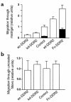
DDR2 promotes invasion into basement membrane matrix by stellate cells and is inhibited by GM6001. (a) A representative experiment of HSC-T6 cells infected with the ec-DDR2, kd-DDR2, GFP alone (control), wt-DDR2, or the constitutively active Fc-DDR2 were suspended in DMEM plus 1% FCS and placed on inserts (8-μm pore size) coated with 100 μg/cm2 of Matrigel. Conditioned media from Kupffer cell cultures supplemented with 1% FCS was used as chemoattractant. After 12 hours, migrating cells adhering to the underside of the membrane were stained with 0.2% crystal violet and counted in ten random high-power fields per insert. Filled columns represent migration of HSC-T6 cells pretreated with 20 μM GM6001 for 1 hour, then allowed to migrate in the presence of 10 μM GM6001. The number of control cells that had migrated to the lower chamber after 12 hours was 55 ± 6 per high-power field. (b) A representative experiment of HSC-T6 cells migrating for 4 hours through uncoated inserts. The number of control HSC-T6 cells that had migrated to the lower chamber was 293 ± 80 per high-power field.

References

    1. Friedman SL. Molecular regulation of hepatic fibrosis; an integrated cellular response to tissue injury. J Biol Chem. 2000;275:2247–2250. - PubMed
    1. Olaso E, Friedman SL. Molecular regulation of hepatic fibrogenesis. J Hepatol. 1998;29:836–847. - PubMed
    1. Friedman SL, Roll FJ, Boyles J, Arenson DM, Bissell DM. Maintenance of differentiated phenotype of cultured rat hepatic lipocytes by basement membrane matrix. J Biol Chem. 1989;264:10756–10762. - PubMed
    1. Arthur MJ, et al. Secretion of 72 kDa type IV collagenase/gelatinase by cultured human lipocytes. Analysis of gene expression, protein synthesis and proteinase activity. Biochem J. 1992;287:701–707. - PMC - PubMed
    1. Butler GS, et al. The TIMP2 membrane type 1 metalloproteinase receptor regulates the concentration and efficient activation of progelatinase A. J Biol Chem. 1998;273:871–880. - PubMed

Publication types

MeSH terms