Skip to main page content
U.S. flag

An official website of the United States government

Dot gov

The . gov means it’s official VSports app下载. Federal government websites often end in . gov or . mil. Before sharing sensitive information, make sure you’re on a federal government site. .

Https

The site is secure V体育官网. The https:// ensures that you are connecting to the official website and that any information you provide is encrypted and transmitted securely. .

. 2000 Mar 1;14(5):574-84.

Induction of terminal differentiation by constitutive activation of p38 MAP kinase in human rhabdomyosarcoma cells

Affiliations

Induction of terminal differentiation by constitutive activation of p38 MAP kinase in human rhabdomyosarcoma cells (VSports app下载)

P L Puri et al. Genes Dev. .

Abstract

MyoD inhibits cell proliferation and promotes muscle differentiation. A paradoxical feature of rhabdomyosarcoma (RMS), a tumor arising from muscle precursors, is the block of the differentiation program and the deregulated proliferation despite MyoD expression. A deficiency in RMS of a factor required for MyoD activity has been implicated by previous studies. We report here that p38 MAP kinase (MAPK) activation, which is essential for muscle differentiation, is deficient in RMS cells VSports手机版. Enforced induction of p38 MAPK by an activated MAPK kinase 6 (MKK6EE) restored MyoD function and enhanced MEF2 activity in RMS deficient for p38 MAPK activation, leading to growth arrest and terminal differentiation. Stress and cytokines could activate the p38 MAPK in RMS cells, however, these stimuli did not promote differentiation, possibly because they activated p38 MAPK only transiently and they also activated JNK, which could antagonize differentiation. Thus, the selective and sustained p38 MAPK activation, which is distinct from the stress-activated response, is required for differentiation and can be disrupted in human tumors. .


Figures
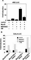
Figure 1
Figure 1
An activated mutant of MKK6EE stimulates the function of MyoD. (A) Activation of the MyoD-dependent promoter 4RE luc (four repeated MyoD-binding E-box sites) was determined by cotransfection of 10T1/2 fibroblasts with the indicated expression plasmids. Cells were transfected in GM containing 20% FBS and harvested 36 hr later for luciferase assay. Where indicated, the p38 MAPK inhibitor SB203580 (5 μm) was added to the transfected cells. The luciferase activity was normalized for the expression levels of MyoD in each sample, and the fold activation was relative to vector-transfected sample. The means and standard deviations from three independent experiments are shown. (B) MKK6EE stimulated the activity of MyoD–Gal4 fusion proteins. Plasmids expressing the indicated MyoD–Gal4 fusion proteins were cotransfected with a gal4–luc reporter gene plus a plasmid expressing MKK6EE (stippled and hatched bars) into 10T1/2 fibroblasts growing in 20% FBS. MKK6EE cotransfected cells were treated with 5 μm SB203580 (hatched bars). Cells were harvested 36 hr after transfection for LUC assay. The luciferase activity was normalized for the expression levels of the transfected Gal4 fusion proteins. The data presented are representative of four independent experiments.
Figure 2
Figure 2
Human RMS cell lines express p38 MAPK but do not modulate its activity under differentiation conditions. The levels of p38 MAPK (A) and phosphorylated p38 MAPK (B) were determined by immunoblotting with the appropriate antibodies (see Materials and Methods) in total lysates of mouse C2C12 (lanes 1,2) and three human RMS cell lines: RD (lanes 3,4), Rh28 (lanes 5,6), and Rh30 (lanes 7,8). (GM) Growth medium; (DM) differentiation medium. (C) p38 MAPK activity was measured by an immunocomplex kinase assay using GST-ATF2 fusion protein as a substrate. The levels of myogenin (D), MHC (E), MyoD (F), and actin (G) were also determined by immunoblotting with the appropriate antibodies. The formation of heterokaryons of RD and 10T/12 fibroblasts (H) was performed as described previously (Tapscott et al. 1993). The heterokaryons were transferred into DM with (+SB) or without (−SB) SB203580 (5 μm) to inhibit p38 MAPK. DMSO and PD98059 were also used as negative controls. After 36 hr in DM, heterokaryons were fixed and stained for the expression of MHC (top) and for the visualization of nuclei with DAPI (bottom). The different pattern of nuclear staining–punctate for mouse nuclei in 10T1/2 cells and uniform staining for human nuclei in RD cells—indicates the formation of heterokaryons. The number of MHC-positive heterokaryons/field was scored and the results of two experiments are summarized. In the case of MKK3KD and MKK6KD overexpression, these dominant-negative forms were expressed by transient transfection in 10T1/2 prior the fusion with RD. A GFP-encoding plasmid was cotransfected to localize successfully transfected cells. The number of GFP/MHC-positive heterokaryons/field was scored and the results of two experiments are summarized.
Figure 3
Figure 3
Introduction of MKK6EE reactivates myogenic differentiation in RMS cells. Two RMS cell lines, Rh30 (A) and RD (B), were infected with adenovirus expressing either the HA–MKK6EE gene or the β-gal gene (Mock) as described previously (Huang et al. 1997). Cells were transferred (12 hr postinfection) into DM with and without SB203580 (5 μm) for 96 hr and then fixed and stained for MHC or for DNA (DAPI). The percentage of MHC-positive multinucleated cells per field was counted and a representative value is reported. Expression of the exogenous MKK6EE proteins in the infected cells was shown by immunoblotting with anti-HA antibody. (C) Induction of CK activity in the indicated cell lines was assayed using a commercial kit (Sigma). (Solid bars) Mock; (stippled bars) MKK6EE; (hatched bars) MKK6EE + SB. The CK activity was calculated as micromoles of creatine formed/min per mg of protein extract. (D) RMS cells were first transfected with a reporter expressing β-gal fused to the nuclear localization signal (nls) and under the control of the MLC promoter (MLC–β-gal). After transfection (24 hr), the cells were infected with MKK6EE-expressing virus in the presence or absence of the p38 inhibitor SB. The percentage of myotubes with β-gal-positive nuclei is a measurement of multinucleation in myogenic cells. As a negative control cells were infected with an adenovirus expressing the β-gal gene (Mock) without the nls. Multinucleation was defined as a single cell with more than two β-gal-positive nuclei. Mock-infected cells were distinguished by the MKK6EE-infected cells as mononucleated cells with exclusively cytoplasmic blue staining. (E) The p21 luc reporter was transfected with the indicated plasmids in Rh30 (left) and RD (right) cells with or without SB (5 μm). At 18 hr post transfection, cells were transferred into DM and they were harvested after 24 hr. Luciferase activity was normalized to the expression levels of the transfected proteins (MyoD and MKK6EE were tagged with Flag and HA epitopes, respectively). The values shown are means and standard deviations from three independent experiments. (F) Plasmids expressing the indicated Gal4 fusion proteins were cotransfected with a gal4–luc reporter gene plus a plasmid expressing MKK6EE into RD and Rh30 cells growing in 20% FBS. After 12 hr, cells were placed in DM. MKK6EE cotransfected cells were also treated with 5 μm SB203580. Cells were harvested after 36 hr of culture in DM for LUC assay. The luciferase activity was normalized for the expression levels of the transfected Gal4 fusion proteins.
Figure 3
Figure 3
Introduction of MKK6EE reactivates myogenic differentiation in RMS cells. Two RMS cell lines, Rh30 (A) and RD (B), were infected with adenovirus expressing either the HA–MKK6EE gene or the β-gal gene (Mock) as described previously (Huang et al. 1997). Cells were transferred (12 hr postinfection) into DM with and without SB203580 (5 μm) for 96 hr and then fixed and stained for MHC or for DNA (DAPI). The percentage of MHC-positive multinucleated cells per field was counted and a representative value is reported. Expression of the exogenous MKK6EE proteins in the infected cells was shown by immunoblotting with anti-HA antibody. (C) Induction of CK activity in the indicated cell lines was assayed using a commercial kit (Sigma). (Solid bars) Mock; (stippled bars) MKK6EE; (hatched bars) MKK6EE + SB. The CK activity was calculated as micromoles of creatine formed/min per mg of protein extract. (D) RMS cells were first transfected with a reporter expressing β-gal fused to the nuclear localization signal (nls) and under the control of the MLC promoter (MLC–β-gal). After transfection (24 hr), the cells were infected with MKK6EE-expressing virus in the presence or absence of the p38 inhibitor SB. The percentage of myotubes with β-gal-positive nuclei is a measurement of multinucleation in myogenic cells. As a negative control cells were infected with an adenovirus expressing the β-gal gene (Mock) without the nls. Multinucleation was defined as a single cell with more than two β-gal-positive nuclei. Mock-infected cells were distinguished by the MKK6EE-infected cells as mononucleated cells with exclusively cytoplasmic blue staining. (E) The p21 luc reporter was transfected with the indicated plasmids in Rh30 (left) and RD (right) cells with or without SB (5 μm). At 18 hr post transfection, cells were transferred into DM and they were harvested after 24 hr. Luciferase activity was normalized to the expression levels of the transfected proteins (MyoD and MKK6EE were tagged with Flag and HA epitopes, respectively). The values shown are means and standard deviations from three independent experiments. (F) Plasmids expressing the indicated Gal4 fusion proteins were cotransfected with a gal4–luc reporter gene plus a plasmid expressing MKK6EE into RD and Rh30 cells growing in 20% FBS. After 12 hr, cells were placed in DM. MKK6EE cotransfected cells were also treated with 5 μm SB203580. Cells were harvested after 36 hr of culture in DM for LUC assay. The luciferase activity was normalized for the expression levels of the transfected Gal4 fusion proteins.
Figure 4
Figure 4
Stable expression of MKK6EE restores terminal differentiation in RMS cells. Polyclonal populations of Rh30 cells stably expressing HA–MKK6EE or vector alone were prepared (see Materials and Methods). (A,B) DNA synthesis was determined following the incubation with BrdU for 12 hr. Nuclei were visualized by staining the DNA with DAPI. Images are presented at the same magnification and show the increased size of nuclei belonging to MKK6EE-expressing Rh30 cells cultured in DM. (GM) Growth media; (DM) differentiation media; (DM > GM) serum restimulation. (C) The expression of HA–MKK6EE was demonstrated by immunoblotting of total cell lysates with anti-HA antibody. (D) Rh30 cells stably expressing MKK6EE (lanes 1,2) or vector control (lanes 3,4). The levels of MyoD, p21Cip1, MHC, troponin T (fast and slow), and actin were determined in total cell lysates by immunoblotting with the appropriate antibodies. The indicated cells were harvested after the indicated hours of culturing in DM. As indicated, SB (5 μm) was added for 48 hr in DM.
Figure 5
Figure 5
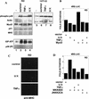
Activation of p38 MAPK in RD cells by stress and cytokines fails to induce myogenic differentiation. (A) The levels of phosphorylated (activated) p38 MAPK were determined by immunoblotting with the appropriate antibodies (see Materials and Methods) in total lysate from RD and 10T1/2 cell lines after exposure to several stress (0.4 m Sorbitol for 30 min; 10 ng/ml TNFα for 15 min; 40 J/m2 UV, for 20 min). The levels of actin, MyoD, p21Cip1, and MHC were also determined by immunoblotting with the appropriate antibodies (see Materials and Methods). The p38 MAPK assay activity was measured by an immunocomplex kinase assay using GST–ATF2 fusion protein as a substrate. (B) Activation of the MyoD-dependent promoter 4RE luc in RD cells exposed or not to UV (40 J/m2, 20 min) and TNFα (10 ng/ml for 15 min) was determined by luciferase assay. Cells were transfected in GM and, at 18 hr post-transfection, transferred into DM, and then exposed to TNFα or UV. After an additional 24 hr, cells were harvested and the luciferase activity calculated. The luciferase activity was normalized to the expression of cotransfected β-gal-encoding plasmid. The data presented is representative of four independent experiments. (C) RD cells were exposed or not to UV (40 J/m2, 20 min) and TNFα (10 ng/ml for 15 min) and kept 36 hr in DM. After fixation, cells were stained for MHC expression (visualized by using a fluorescein-conjugated secondary antibody). (D) Activation of the MyoD-dependent promoter 4RE luc in RD cells exposed or not to UV (40 J/m2, 20 min) and TNFα (10 ng/ml for 15 min) was determined after cotransfection with the indicated plasmids (0.2 μg MKK6EE, 0.4 μg JNKK2CA). Cells were transfected in GM and, at 18 hr post-transfection, transferred into DM. After additional 24 hr cells were harvested and the luciferase activity calculated. The luciferase activity was normalized to the expression of cotransfected β-gal-encoding plasmid. The data presented are representative of four independent experiments.

References

    1. Alemà S, Tatò F. Oncogenes and muscle differentiation: Multiple mechanism of interference. Semin Cancer Biol. 1994;5:147–156. - PubMed
    1. Arndt CAS, Crist WM. Medical progress: Common musculoskeletal tumors of childhood and adolescence. N Engl J Med. 1999;341:342–353. - "V体育官网" PubMed
    1. Arnold H-H, Winter B. Muscle differentiation: More complexity to the network of myogenic regulators. Curr Opin Genet Dev. 1998;8:539–544. - "VSports app下载" PubMed
    1. Bengal E, Ransone L, Scharfmann R, Dwarki VJ, Tapscott SJ, Weintraub H, Verma IM. Functional antagonism between cJun and MyoD proteins: a direct physical association. Cell. 1992;68:507–519. - PubMed
    1. Black BL, Molkentin JD, Olson E. Multiples roles for the MyoD basic region in transmission of transcriptional activation signals and interaction with Mef2. Mol Cell Biol. 1998;18:69–77. - V体育安卓版 - PMC - PubMed

V体育2025版 - Publication types

MeSH terms