<u date-time="GmGpcZpW"></u><sub date-time="eO2SQG"></sub><sub date-time="NrWz9f"><small dropzone="gs4hXTD"></small></sub> Skip to main page content
U.S. flag

An official website of the United States government

Dot gov

The . gov means it’s official. Federal government websites often end in . gov or VSports app下载. mil. Before sharing sensitive information, make sure you’re on a federal government site. .

Https

The site is secure V体育官网. The https:// ensures that you are connecting to the official website and that any information you provide is encrypted and transmitted securely. .

. 1999 Jul 19;190(2):253-65.
doi: 10.1084/jem.190.2.253.

Bcl-2-mediated drug resistance: inhibition of apoptosis by blocking nuclear factor of activated T lymphocytes (NFAT)-induced Fas ligand transcription

Affiliations

Bcl-2-mediated drug resistance: inhibition of apoptosis by blocking nuclear factor of activated T lymphocytes (NFAT)-induced Fas ligand transcription

R K Srivastava et al. J Exp Med. .

Abstract

Bcl-2 inhibits apoptosis induced by a variety of stimuli, including chemotherapy drugs and glucocorticoids. It is generally accepted that Bcl-2 exerts its antiapoptotic effects mainly by dimerizing with proapoptotic members of the Bcl-2 family such as Bax and Bad. However, the mechanism of the antiapoptotic effects is unclear VSports手机版. Paclitaxel and other drugs that disturb microtubule dynamics kill cells in a Fas/Fas ligand (FasL)-dependent manner; antibody to FasL inhibits paclitaxel-induced apoptosis. We have found that Bcl-2 overexpression leads to the prevention of chemotherapy (paclitaxel)-induced expression of FasL and blocks paclitaxel-induced apoptosis. The mechanism of this effect is that Bcl-2 prevents the nuclear translocation of NFAT (nuclear factor of activated T lymphocytes, a transcription factor activated by microtubule damage) by binding and sequestering calcineurin, a calcium-dependent phosphatase that must dephosphorylate NFAT to move to the nucleus. Without NFAT nuclear translocation, the FasL gene is not transcribed. Thus, it appears that paclitaxel and other drugs that disturb microtubule function kill cells at least in part through the induction of FasL. Furthermore, Bcl-2 antagonizes drug-induced apoptosis by inhibiting calcineurin activation, blocking NFAT nuclear translocation, and preventing FasL expression. The effects of Bcl-2 can be overcome, at least partially, through phosphorylation of Bcl-2. Phosphorylated Bcl-2 cannot bind calcineurin, and NFAT activation, FasL expression, and apoptosis can occur after Bcl-2 phosphorylation. .


Figures

Figure 1
Figure 1
Bcl-2 inhibits paclitaxel-induced apoptosis. (A) Jurkat cells were stably transfected with either pSSFV-neo or pSFFV-Bcl-2 plasmid. (B) MDA-MB-231 cells were stably transfected with either pSSFV-neo or pSFFV-Bcl-2 plasmid. (C) Jurkat cells (JT/Neo and JT/Bcl-2) were treated with paclitaxel (0.001, 0.01, and 0.1 μM) with or without anti-FasL neutralizing antibody (NOK-2; 1 μg/ml) for 48 h. Cells were stained with DAPI and visualized under fluorescence microscopy. Cells with fragmented nuclei or condensed chromatin were counted as apoptotic. Data (mean ± SE of quadruplicate determinations) represent one of three separate experiments that gave similar results. (D) MDA/MB/231 (MDA/Neo and MDA/Bcl-2) were treated with paclitaxel (0.001, 0.01, and 0.1 μM) with or without anti-FasL neutralizing antibody (NOK-2; 1 μg/ml) for 48 h. Cells were stained with DAPI and visualized under fluorescence microscopy. Cells with fragmented and condensed chromatin were counted as apoptotic. Data (mean ± SE of quadruplicate determinations) represent one of three separate experiments that gave similar results. (E) JT/Neo and JT/Bcl-2 cells were treated with paclitaxel (0.001, 0.01, and 0.1 μM) with or without anti-Fas blocking antibody (1 μg/ml) for 48 h. Apoptotic nuclei were counted as described for D. (F) JT/Neo and JT/mut CD95 cells were treated with paclitaxel (0.001, 0.01, and 0.1 μM) for 48 h. Apoptotic nuclei were counted as described for D.
Figure 1
Figure 1
Bcl-2 inhibits paclitaxel-induced apoptosis. (A) Jurkat cells were stably transfected with either pSSFV-neo or pSFFV-Bcl-2 plasmid. (B) MDA-MB-231 cells were stably transfected with either pSSFV-neo or pSFFV-Bcl-2 plasmid. (C) Jurkat cells (JT/Neo and JT/Bcl-2) were treated with paclitaxel (0.001, 0.01, and 0.1 μM) with or without anti-FasL neutralizing antibody (NOK-2; 1 μg/ml) for 48 h. Cells were stained with DAPI and visualized under fluorescence microscopy. Cells with fragmented nuclei or condensed chromatin were counted as apoptotic. Data (mean ± SE of quadruplicate determinations) represent one of three separate experiments that gave similar results. (D) MDA/MB/231 (MDA/Neo and MDA/Bcl-2) were treated with paclitaxel (0.001, 0.01, and 0.1 μM) with or without anti-FasL neutralizing antibody (NOK-2; 1 μg/ml) for 48 h. Cells were stained with DAPI and visualized under fluorescence microscopy. Cells with fragmented and condensed chromatin were counted as apoptotic. Data (mean ± SE of quadruplicate determinations) represent one of three separate experiments that gave similar results. (E) JT/Neo and JT/Bcl-2 cells were treated with paclitaxel (0.001, 0.01, and 0.1 μM) with or without anti-Fas blocking antibody (1 μg/ml) for 48 h. Apoptotic nuclei were counted as described for D. (F) JT/Neo and JT/mut CD95 cells were treated with paclitaxel (0.001, 0.01, and 0.1 μM) for 48 h. Apoptotic nuclei were counted as described for D.
Figure 2
Figure 2
Bcl-2 inhibits paclitaxel-, vincristine-, and vinblastine-induced FasL expression. (A) MDA cells were treated with 50 nM of paclitaxel, vincristine, or vinblastine for either 24 or 48 h. At the end of incubation period, cells were harvested and lysed. Equal amounts of protein were resolved on SDS-PAGE. FasL levels were measured by Western blot analysis. The same blot was reprobed with anti–β-actin antibody to check if equal amounts of protein were loaded in each lane. (B) MDA and MCF-7 cells were treated with various concentrations of paclitaxel for 48 h. FasL levels were detected by Western blot analysis. The blot was reprobed with anti–β-actin antibody to check if equal amounts of protein were loaded in each lane. (C) MDA/Neo and MDA/Bcl-2 cells were treated with 50 nM of paclitaxel or vincristine for 48 h. FasL levels were detected by Western blot analysis. The blot was reprobed with anti–β-actin antibody to check if equal amounts of protein were loaded in each lane. (D) JT/Neo and JT/Bcl-2 cells were treated with paclitaxel (50 nM) for 48 h. FasL levels were detected by Western blot analysis. The blot was reprobed with anti–β-actin antibody to check if equal amounts of protein were loaded in each lane.
Figure 2
Figure 2
Bcl-2 inhibits paclitaxel-, vincristine-, and vinblastine-induced FasL expression. (A) MDA cells were treated with 50 nM of paclitaxel, vincristine, or vinblastine for either 24 or 48 h. At the end of incubation period, cells were harvested and lysed. Equal amounts of protein were resolved on SDS-PAGE. FasL levels were measured by Western blot analysis. The same blot was reprobed with anti–β-actin antibody to check if equal amounts of protein were loaded in each lane. (B) MDA and MCF-7 cells were treated with various concentrations of paclitaxel for 48 h. FasL levels were detected by Western blot analysis. The blot was reprobed with anti–β-actin antibody to check if equal amounts of protein were loaded in each lane. (C) MDA/Neo and MDA/Bcl-2 cells were treated with 50 nM of paclitaxel or vincristine for 48 h. FasL levels were detected by Western blot analysis. The blot was reprobed with anti–β-actin antibody to check if equal amounts of protein were loaded in each lane. (D) JT/Neo and JT/Bcl-2 cells were treated with paclitaxel (50 nM) for 48 h. FasL levels were detected by Western blot analysis. The blot was reprobed with anti–β-actin antibody to check if equal amounts of protein were loaded in each lane.
Figure 3
Figure 3
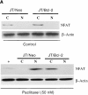
Bcl-2 blocks NFAT translocation to the nucleus. (A) JT/Neo and JT/Bcl-2 cells were either treated with paclitaxel (50 nM) or vehicle (control) for 48 h. Cells were harvested, and cytoplasmic (C) and nuclear (N) fractions were prepared as described in Materials and Methods. Samples were resolved on SDS-PAGE and immunoblotted with anti-NFAT antibody. The same blot was reprobed with anti–β-actin antibody. (B) MDA/Neo and MDA/Bcl-2 cells were either treated with paclitaxel (50 nM) or vehicle for 48 h. Cells were harvested and C and N fractions were prepared as described in Materials and Methods. Samples were resolved on SDS-PAGE and immunoblotted with anti-NFAT antibody. The same blot was reprobed with anti–β-actin antibody.
Figure 3
Figure 3
Bcl-2 blocks NFAT translocation to the nucleus. (A) JT/Neo and JT/Bcl-2 cells were either treated with paclitaxel (50 nM) or vehicle (control) for 48 h. Cells were harvested, and cytoplasmic (C) and nuclear (N) fractions were prepared as described in Materials and Methods. Samples were resolved on SDS-PAGE and immunoblotted with anti-NFAT antibody. The same blot was reprobed with anti–β-actin antibody. (B) MDA/Neo and MDA/Bcl-2 cells were either treated with paclitaxel (50 nM) or vehicle for 48 h. Cells were harvested and C and N fractions were prepared as described in Materials and Methods. Samples were resolved on SDS-PAGE and immunoblotted with anti-NFAT antibody. The same blot was reprobed with anti–β-actin antibody.
Figure 4
Figure 4
Confocal microscopy showing blockage of NFAT translocation by Bcl-2. MDA/Neo and MDA/Bcl-2 cells were treated with paclitaxel (50 nM) for 48 h. Cells were fixed and stained with anti-NFAT antibody along with PI. Cells were washed and restained with secondary antibody conjugated with Alexa-488. Green and red represent cytoplasmic NFAT and nuclear staining, respectively. Yellow, NFAT translocated to the nucleus.
Figure 4
Figure 4
Confocal microscopy showing blockage of NFAT translocation by Bcl-2. MDA/Neo and MDA/Bcl-2 cells were treated with paclitaxel (50 nM) for 48 h. Cells were fixed and stained with anti-NFAT antibody along with PI. Cells were washed and restained with secondary antibody conjugated with Alexa-488. Green and red represent cytoplasmic NFAT and nuclear staining, respectively. Yellow, NFAT translocated to the nucleus.
Figure 5
Figure 5
Bcl-2 binds to calcineurin but not NFAT. (A) JT/Neo and JT/Bcl-2 cells were treated with 50 nM of paclitaxel or vincristine for 48 h. The cell lysates were prepared and immunoprecipitated with 10 μg of anti-NFAT antibody and 20 μl of protein A–Sepharose. The samples were resolved on SDS-PAGE and immunoblotted with either anti–Bcl-2 antibody (top panel) or anti–NF-AT antibody (bottom panel). (B) JT/Neo and JT/Bcl-2 cells were treated with 50 nM of paclitaxel or vincristine for 48 h. The cell lysates were prepared and immunoprecipitated with 10 μg of anti–Bcl-2 antibody and 20 μl of protein A–Sepharose. The samples were resolved on SDS-PAGE and immunoblotted with anticalcineurin. (C) JT/Neo and JT/Bcl-2 cells were treated with 50 nM of paclitaxel or vincristine for 48 h. The cell lysates were prepared and immunoprecipitated with 10 μg of anticalcineurin antibody and 20 μl of protein A–Sepharose. The samples were resolved on SDS-PAGE and immunoblotted with anti–Bcl-2 antibody. (D) Left, JT/Neo and JT/Bcl-2 cells were treated with various concentrations of paclitaxel (0.001, 0.01, and 0.1 μM) with or without FK506 analogue ascomycin (10 μM) for 48 h to measure apoptosis. Cells were stained with DAPI and visualized under fluorescence microscopy. Cells with fragmented nuclei or condensed chromatin were counted as apoptotic. Data (mean ± SE of quadruplicate determinations) represent one of three separate experiments that gave similar results. Right, MDA/Neo and MDA/Bcl-2 cells were treated with various concentrations of paclitaxel (0.001, 0.01, and 0.1 μM) with or without FK506 analogue ascomycin (10 μM) for 48 h to measure apoptosis. Cells were stained with DAPI and visualized under fluorescence microscopy. Data (mean ± SE of quadruplicate determinations) represent one of three separate experiments that gave similar results. (E) JT/Neo and JT/Bcl-2 cells were pretreated with [(Ca2+)i] chelator BAPTA-AM (10 μM) for 45 min and then treated with paclitaxel (50 nM) for 48 h to measure apoptosis. Data (mean ± SE of quadruplicate determinations) represent one of three separate experiments that gave similar results.
Figure 5
Figure 5
Bcl-2 binds to calcineurin but not NFAT. (A) JT/Neo and JT/Bcl-2 cells were treated with 50 nM of paclitaxel or vincristine for 48 h. The cell lysates were prepared and immunoprecipitated with 10 μg of anti-NFAT antibody and 20 μl of protein A–Sepharose. The samples were resolved on SDS-PAGE and immunoblotted with either anti–Bcl-2 antibody (top panel) or anti–NF-AT antibody (bottom panel). (B) JT/Neo and JT/Bcl-2 cells were treated with 50 nM of paclitaxel or vincristine for 48 h. The cell lysates were prepared and immunoprecipitated with 10 μg of anti–Bcl-2 antibody and 20 μl of protein A–Sepharose. The samples were resolved on SDS-PAGE and immunoblotted with anticalcineurin. (C) JT/Neo and JT/Bcl-2 cells were treated with 50 nM of paclitaxel or vincristine for 48 h. The cell lysates were prepared and immunoprecipitated with 10 μg of anticalcineurin antibody and 20 μl of protein A–Sepharose. The samples were resolved on SDS-PAGE and immunoblotted with anti–Bcl-2 antibody. (D) Left, JT/Neo and JT/Bcl-2 cells were treated with various concentrations of paclitaxel (0.001, 0.01, and 0.1 μM) with or without FK506 analogue ascomycin (10 μM) for 48 h to measure apoptosis. Cells were stained with DAPI and visualized under fluorescence microscopy. Cells with fragmented nuclei or condensed chromatin were counted as apoptotic. Data (mean ± SE of quadruplicate determinations) represent one of three separate experiments that gave similar results. Right, MDA/Neo and MDA/Bcl-2 cells were treated with various concentrations of paclitaxel (0.001, 0.01, and 0.1 μM) with or without FK506 analogue ascomycin (10 μM) for 48 h to measure apoptosis. Cells were stained with DAPI and visualized under fluorescence microscopy. Data (mean ± SE of quadruplicate determinations) represent one of three separate experiments that gave similar results. (E) JT/Neo and JT/Bcl-2 cells were pretreated with [(Ca2+)i] chelator BAPTA-AM (10 μM) for 45 min and then treated with paclitaxel (50 nM) for 48 h to measure apoptosis. Data (mean ± SE of quadruplicate determinations) represent one of three separate experiments that gave similar results.
Figure 7
Figure 7
NFAT sites play important roles in FasL promoter activation. (A) Jurkat cells (JT/Neo and JT/Bcl-2) were transfected with 70 μg of the wild-type 486-bp FasL reporter (FasL-486) or a reporter containing mutations in both the distal and proximal NFAT sites (double mutant). Transfectants were left untreated (control) or treated with paclitaxel (100 nM) for 48 h. Cells were lysed and assayed for luciferase activity. Data are expressed as arbitrary luciferase light units and are representative of four independent experiments. Error bars, SE of triplicate samples. (B) MDA/Neo and MDA/Bcl-2 cells were transfected with 70 μg of the wild-type 486-bp FasL reporter (FasL-486) or a reporter containing mutations in both the distal and proximal NFAT sites (double mutant). Transfectants were left untreated (control) or treated with paclitaxel (100 nM) for 48 h. Cells were lysed and assayed for luciferase activity. Data are expressed as arbitrary luciferase light units and are representative of four independent experiments. Error bars, SE of triplicate samples.
Figure 7
Figure 7
NFAT sites play important roles in FasL promoter activation. (A) Jurkat cells (JT/Neo and JT/Bcl-2) were transfected with 70 μg of the wild-type 486-bp FasL reporter (FasL-486) or a reporter containing mutations in both the distal and proximal NFAT sites (double mutant). Transfectants were left untreated (control) or treated with paclitaxel (100 nM) for 48 h. Cells were lysed and assayed for luciferase activity. Data are expressed as arbitrary luciferase light units and are representative of four independent experiments. Error bars, SE of triplicate samples. (B) MDA/Neo and MDA/Bcl-2 cells were transfected with 70 μg of the wild-type 486-bp FasL reporter (FasL-486) or a reporter containing mutations in both the distal and proximal NFAT sites (double mutant). Transfectants were left untreated (control) or treated with paclitaxel (100 nM) for 48 h. Cells were lysed and assayed for luciferase activity. Data are expressed as arbitrary luciferase light units and are representative of four independent experiments. Error bars, SE of triplicate samples.
Figure 8
Figure 8
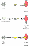
Model for the inhibition of NFAT signaling by Bcl-2. (A) Treatment of cells with paclitaxel results in calcineurin activation, NFAT nuclear translocation, FasL expression, and apoptosis. (B) Unphosphorylated Bcl-2 binds to calcineurin and blocks NFAT nuclear translocation, FasL expression, and apoptosis. (C) Phosphorylation of Bcl-2 results in release of calcineurin from the complex, translocation of NFAT to the nucleus, and induction of FasL expression and apoptosis.
Figure 6
Figure 6
Bcl-2 interacts with calcineurin through its BH4 domain in inhibiting paclitaxel-induced FasL expression and apoptosis. (A) Western blot showing overexpressed wild-type Bcl-2 and ΔBH4 Bcl-2 in MDA-MB-231 cells. (B) MDA/Neo, MDA/ΔBH4 Bcl-2, and MDA/Bcl-2 cells were treated with paclitaxel (50 nM) for 48 h. Cells were harvested and lysed. FasL expression was detected by Western blot analysis. The same blot was reprobed with anti–β-actin antibody. (C) MDA/Neo, MDA/Bcl-2, and MDA/ΔBH4 Bcl-2 cells were treated with paclitaxel (50 nM) for 48 h. The cell lysates were prepared and immunoprecipitated with 10 μg of anticalcineurin antibody and 20 μl of protein A–Sepharose. The samples were resolved on SDS-PAGE and immunoblotted with anti–Bcl-2 antibody. (D) MDA/Neo, MDA/Δloop Bcl-2, and MDA/Bcl-2 cells were treated with paclitaxel (200 nM) for 48 h. Top panel, cell lysates were run on SDS-PAGE and immunoblotted with anti–Bcl-2 antibody. Bottom panel, cell lysates were immunoprecipitated with anti–Bcl-2 antibody, run on SDS-PAGE, and immunoblotted with anticalcineurin antibody. (E) MDA/Neo, MDA/ΔBH4 Bcl-2, MDA/Δloop Bcl-2, and MDA/Bcl-2 cells were treated with paclitaxel (0.001, 0.01, and 0.1 μM) for 48 h to measure apoptosis. Cells were stained with DAPI and visualized under fluorescence microscopy. Data (mean ± SE of quadruplicate determinations) represent one of three separate experiments that gave similar results.
Figure 6
Figure 6
Bcl-2 interacts with calcineurin through its BH4 domain in inhibiting paclitaxel-induced FasL expression and apoptosis. (A) Western blot showing overexpressed wild-type Bcl-2 and ΔBH4 Bcl-2 in MDA-MB-231 cells. (B) MDA/Neo, MDA/ΔBH4 Bcl-2, and MDA/Bcl-2 cells were treated with paclitaxel (50 nM) for 48 h. Cells were harvested and lysed. FasL expression was detected by Western blot analysis. The same blot was reprobed with anti–β-actin antibody. (C) MDA/Neo, MDA/Bcl-2, and MDA/ΔBH4 Bcl-2 cells were treated with paclitaxel (50 nM) for 48 h. The cell lysates were prepared and immunoprecipitated with 10 μg of anticalcineurin antibody and 20 μl of protein A–Sepharose. The samples were resolved on SDS-PAGE and immunoblotted with anti–Bcl-2 antibody. (D) MDA/Neo, MDA/Δloop Bcl-2, and MDA/Bcl-2 cells were treated with paclitaxel (200 nM) for 48 h. Top panel, cell lysates were run on SDS-PAGE and immunoblotted with anti–Bcl-2 antibody. Bottom panel, cell lysates were immunoprecipitated with anti–Bcl-2 antibody, run on SDS-PAGE, and immunoblotted with anticalcineurin antibody. (E) MDA/Neo, MDA/ΔBH4 Bcl-2, MDA/Δloop Bcl-2, and MDA/Bcl-2 cells were treated with paclitaxel (0.001, 0.01, and 0.1 μM) for 48 h to measure apoptosis. Cells were stained with DAPI and visualized under fluorescence microscopy. Data (mean ± SE of quadruplicate determinations) represent one of three separate experiments that gave similar results.

References

    1. Watanabe-Fukunaga R., Brannan C.I., Copeland N.G., Jenkins N.A., Nagata S. Lymphoproliferation disorder in mice explained by defects in Fas antigen that mediates apoptosis. Nature. 1992;356:314–317. - "V体育安卓版" PubMed
    1. Leithauser F., Dhein J., Mechtersheimer G., Koretz K., Bruderlein S., Henne C., Schmidt A., Debatin K.M., Krammer P.H., Moller P. Constitutive and induced expression of APO-1, a new member of the nerve growth factor/tumor necrosis factor receptor superfamily, in normal and neoplastic cells. Lab. Invest. 1993;69:415–429. - PubMed
    1. French L.E., Hahne M., Viard I., Radlgruber G., Zanone R., Becker K., Muller C., Tschopp J. Fas and Fas ligand in embryos and adult miceligand expression in several immune-privileged tissues and coexpression in adult tissues characterized by apoptotic cell turnover. J. Cell Biol. 1996;133:335–343. - PMC - PubMed
    1. Rouvier E., Luciani M.F., Golstein P. Fas involvement in Ca2+-independent T cell–mediated cytotoxicity. J. Exp. Med. 1993;177:195–200. - PMC (V体育官网入口) - PubMed
    1. Arase H., Arase N., Saito T. Fas-mediated cytotoxicity by freshly isolated natural killer cells. J. Exp. Med. 1995;181:1235–1238. - PMC - PubMed

Publication types

MeSH terms

"VSports" LinkOut - more resources