Skip to main page content (V体育2025版)
U.S. flag

An official website of the United States government

Dot gov

The . gov means it’s official. Federal government websites often end in . gov or . mil VSports app下载. Before sharing sensitive information, make sure you’re on a federal government site. .

Https

The site is secure. The https:// ensures that you are connecting to the official website and that any information you provide is encrypted and transmitted securely. V体育官网.

. 1999 Feb 1;13(3):320-33.
doi: 10.1101/gad.13.3.320.

Yeast cohesin complex requires a conserved protein, Eco1p(Ctf7), to establish cohesion between sister chromatids during DNA replication

Affiliations

"V体育官网入口" Yeast cohesin complex requires a conserved protein, Eco1p(Ctf7), to establish cohesion between sister chromatids during DNA replication

A Tóth et al. Genes Dev. .

"VSports最新版本" Abstract

Sister chromatid cohesion is crucial for chromosome segregation during mitosis. Loss of cohesion very possibly triggers sister separation at the metaphase --> anaphase transition. This process depends on the destruction of anaphase inhibitory proteins like Pds1p (Cut2p), which is thought to liberate a sister-separating protein Esp1p (Cut1p). By looking for mutants that separate sister centromeres in the presence of Pds1p, this and a previous study have identified six proteins essential for establishing or maintaining sister chromatid cohesion VSports手机版. Four of these proteins, Scc1p, Scc3p, Smc1p, and Smc3p, are subunits of a 'Cohesin' complex that binds chromosomes from late G1 until the onset of anaphase. The fifth protein, Scc2p, is not a stoichiometric Cohesin subunit but it is required for Cohesin's association with chromosomes. The sixth protein, Eco1p(Ctf7p), is not a Cohesin subunit. It is necessary for the establishment of cohesion during DNA replication but not for its maintenance during G2 and M phases. .


Figures

Figure 1
Figure 1
Scc3p and Eco1p are conserved among eukaryotes. (A) Sequence alignment of the amino-terminal half of S. cerevisiae (Sc) Scc3p and its homologs from mouse (Mm SA-1, Mm SA-2, Mm SA-3), D. melanogaster (Dm SA), C. elegans (Ce F18e2.3), and S. pombe (Sp C17H9.2, Sp Rec11). Identical residues in proteins are shown on a black background. Conservative amino acid substitutions are shown on a gray background. Because the human SA1 and SA2 are almost identical to the corresponding mouse proteins, we did not include the human proteins in this alignment. (B) Phylogenetic tree of the Scc3p homologs based on the above aligned sequences. (C) Sequence alignment of S. cerevisiae Eco1p (Sc) and its homologs from A. thaliana (At F8F16.220), mouse (Mm ESTcons.), fission yeast (Sp SPBC16A3.11).
Figure 2
Figure 2
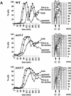
Sister chromatids separate prematurely in scc3-1 and eco1-1 mutants. Small G1 cells of wild-type (K7479), scc3-1 (K7515), and eco1-1 (K7542) strains expressing Pds1–myc18p and with CenV marked by GFP, were isolated by centrifugal elutriation and incubated at 37°C. (A, left) The fraction of budded cells (█), cells that have separated their cenV–GFP ‘dots’ (○) and cells with Pds1p in their nucleus (▵). (A, right) DNA content measured by flow cytometry (FACS). In wild-type cells, degradation of Pds1p always precedes sister chromatid separation. In scc3-1 and eco1-1 mutant cells sister chromatids are separated even in the presence of Pds1p. (B) Immunofluorescence of wild-type (top) and eco1-1 (bottom) cells. DNA was stained by DAPI. Pds1p was detected by antibody to the myc epitope. CenV was visualized by GFP. Bar, 4 μm.
Figure 2
Figure 2
Sister chromatids separate prematurely in scc3-1 and eco1-1 mutants. Small G1 cells of wild-type (K7479), scc3-1 (K7515), and eco1-1 (K7542) strains expressing Pds1–myc18p and with CenV marked by GFP, were isolated by centrifugal elutriation and incubated at 37°C. (A, left) The fraction of budded cells (█), cells that have separated their cenV–GFP ‘dots’ (○) and cells with Pds1p in their nucleus (▵). (A, right) DNA content measured by flow cytometry (FACS). In wild-type cells, degradation of Pds1p always precedes sister chromatid separation. In scc3-1 and eco1-1 mutant cells sister chromatids are separated even in the presence of Pds1p. (B) Immunofluorescence of wild-type (top) and eco1-1 (bottom) cells. DNA was stained by DAPI. Pds1p was detected by antibody to the myc epitope. CenV was visualized by GFP. Bar, 4 μm.
Figure 3
Figure 3
Scc3p and Eco1p are required to prevent sister chromatid separation in esp1-1 mutants. Small unbudded cells of scc3-1 esp1-1 (K7546) and of eco1-1 esp1-1 (K7544) strains containing cenV–GFP were inoculated into YEPD medium at 37°C. Samples were taken every 15 min and the fraction of cells with buds (█) and cells with separated sister chromatids (○) were determined. The control experiment had been published by Ciosk et al. (1998).
Figure 4
Figure 4
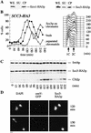
Scc3p and Eco1p are associated with chromatin. (A) Triton X-100 insoluble chromatin pellet was obtained from cycling cultures of ECO1–HA3 and SCC3–HA3 strains as described (Liang and Stillman 1997). Eco1–HA3p and Scc3–HA3p were detected with anti-HA antibody (16B12). (WE) whole cell extract; (SU) Triton X-100 soluble supernatant; (CP) Triton X-100 insoluble chromatin pellet. (B) Small G1 cells expressing Scc3–HA3p (K7461) containing cenV–GFP were incubated at 25°C. DNA content of cells, percentage of budded cells (█), cells with separated sister chromatids (○), and the percentage of cells with Scc3p associated to chromosomes (▵) were determined. (C) Scc3–HA3p, Clb2p, and Swi6p levels were determined by Western blot analysis of protein extracts. (D) Chromosome spreads from 120- and 135-min time points. DNA was stained by DAPI. Scc3–HA3p was detected by antibody to HA epitopes. CenV was visualized by GFP. Scc3p colocalizes with chromatin until the onset of anaphase (top panel). Scc3p association is largely reduced in cells that separated sister chromatids (bottom panel, top left corner). Bar, 4 μm.
Figure 5
Figure 5
Physical associations between proteins involved in sister chromatid cohesion. Protein extracts were prepared from cells of the indicated genotypes. (+) An epitope-tagged gene; (−) a wild-type allele. Myc-tagged proteins were immunoprecipitated with anti-myc mouse mAb 9E10. Myc-tagged and HA-tagged proteins were detected by immunoblotting, using anti-Myc 9E10 or anti-HA mouse mAb 12CA5, respectively. Protein extracts or immunoprecipitates were separated on 7.5% SDS–polyacrylamide gels and either stained with silver (A) or immunobloted with antibodies (B,C). (A) Scc1/3 and Smc1/3 proteins form a soluble complex. Protein extracts of control cells (no tag, lane 1), cells expressing a myc9-tagged version of Cdc23p (lane 2), and cells expressing myc18-tagged versions of either Scc1p or Scc3p (lanes 310), were immunoprecipitated with an antibody specific for the Myc epitope. Proteins that were present in immunoprecipitates were visu alized by staining with silver. Only those proteins that specifically associated with Scc1–myc18p and Scc3–myc18p are shown. Proteins that were specifically present in Scc1–myc18p (lanes 36) and Scc3–myc18p precipitates (lanes 710) were identified by reducing electrophoretic mobility of candidate proteins by their HA tagging. Scc1–myc18p and Scc3–myc18p are indicated with arrows. Scc1p with no tag is not detectable with silver under these conditions. (B) Scc3–myc18p precipitates Scc1–HA6p, Smc1–HA6p, and Smc3–HA6p. (*) A degradation product of Scc1–HA6p. (C) A small fraction of Scc2–HA6p, but not Smc2–HA6p, is present in Scc3–myc18p precipitate.
Figure 5
Figure 5
Physical associations between proteins involved in sister chromatid cohesion. Protein extracts were prepared from cells of the indicated genotypes. (+) An epitope-tagged gene; (−) a wild-type allele. Myc-tagged proteins were immunoprecipitated with anti-myc mouse mAb 9E10. Myc-tagged and HA-tagged proteins were detected by immunoblotting, using anti-Myc 9E10 or anti-HA mouse mAb 12CA5, respectively. Protein extracts or immunoprecipitates were separated on 7.5% SDS–polyacrylamide gels and either stained with silver (A) or immunobloted with antibodies (B,C). (A) Scc1/3 and Smc1/3 proteins form a soluble complex. Protein extracts of control cells (no tag, lane 1), cells expressing a myc9-tagged version of Cdc23p (lane 2), and cells expressing myc18-tagged versions of either Scc1p or Scc3p (lanes 310), were immunoprecipitated with an antibody specific for the Myc epitope. Proteins that were present in immunoprecipitates were visu alized by staining with silver. Only those proteins that specifically associated with Scc1–myc18p and Scc3–myc18p are shown. Proteins that were specifically present in Scc1–myc18p (lanes 36) and Scc3–myc18p precipitates (lanes 710) were identified by reducing electrophoretic mobility of candidate proteins by their HA tagging. Scc1–myc18p and Scc3–myc18p are indicated with arrows. Scc1p with no tag is not detectable with silver under these conditions. (B) Scc3–myc18p precipitates Scc1–HA6p, Smc1–HA6p, and Smc3–HA6p. (*) A degradation product of Scc1–HA6p. (C) A small fraction of Scc2–HA6p, but not Smc2–HA6p, is present in Scc3–myc18p precipitate.
Figure 6
Figure 6
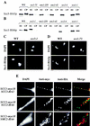
Colocalization of Cohesin subunits and their interdependent chromatin association. Wild-type (K7570), scc1-73 (K7734), scc2-4 (K7689, K7586), scc3-1 (K7548), smc1-259 (K7686, K7589), smc3-42 (K7687, K7583), and eco1-1 (K7586, K7669) strains containing the SCC1 or SCC3 gene tagged with HA epitopes were released from α-factor into nocodazole at 35.5°C. After 2.5 hr, >90% of cells had arrested with large buds and a 2C DNA content. (A,B) The amount of Scc1–HA6p and Scc3–HA3p was detected by Western blot analysis of Triton X-100 insoluble (chromatin pellet, CP) and soluble (supernatant, SU) fractions (Liang et al. 1997). (C) Scc1–HA6p detected in chromosome spreads in wild-type and scc3-1 mutant cells. (D) Scc3–HA3p detected in chromosome spreads in wild-type and scc1-73 mutant cells. Bar, 4 μm. (E) Chromosome spreads were prepared from strains expressing Scc1–myc18p in combination with either Scc2–HA6p, Scc3–HA3p, or Smc2–HA6p. DNA was stained by DAPI. Myc epitopes were detected with anti-myc mouse mAb 9E10 and anti-mouse cy3-conjugated goat antibody (Amersham); HA-tagged proteins were visualized with anti-HA rat mAb 3F10 and anti-rat cy2-conjugated goat antibody (Amersham).
Figure 7
Figure 7
Regulation of Scc1p and Scc3p association with the chromatin is normal in eco1-1 mutant cells. Small unbudded G1 eco1-1 mutant cells expressing Pds1–myc18p and either Scc1–HA6p (K7752) or Scc3–HA3p (K7669) were incubated at 37°C. Samples were taken every 15 min. (A) DNA content of cells and the percentage of budded cells (█), cells with separated sister chromatids (○), cells with Pds1p in their nucleus (▵), and chromosome spreads associated with high amounts of Scc1–HA6p or Scc3–HA3p (⋄). (B) Chromosome spreads of cells taken at 135 min (eco1-1 SCC3–HA3) or at 165 min (eco1-1 SCC1–HA6). DNA was stained by DAPI. CenV was visualized by GFP. Chromatin-associated Scc1–HA6p and Scc3–HA3p were detected by indirect immunofluorescence. Bar, 4 μm.
Figure 7
Figure 7
Regulation of Scc1p and Scc3p association with the chromatin is normal in eco1-1 mutant cells. Small unbudded G1 eco1-1 mutant cells expressing Pds1–myc18p and either Scc1–HA6p (K7752) or Scc3–HA3p (K7669) were incubated at 37°C. Samples were taken every 15 min. (A) DNA content of cells and the percentage of budded cells (█), cells with separated sister chromatids (○), cells with Pds1p in their nucleus (▵), and chromosome spreads associated with high amounts of Scc1–HA6p or Scc3–HA3p (⋄). (B) Chromosome spreads of cells taken at 135 min (eco1-1 SCC3–HA3) or at 165 min (eco1-1 SCC1–HA6). DNA was stained by DAPI. CenV was visualized by GFP. Chromatin-associated Scc1–HA6p and Scc3–HA3p were detected by indirect immunofluorescence. Bar, 4 μm.
Figure 8
Figure 8
Eco1p is essential during S phase but not during G2 and M phases. (A,B) Small G1 cells of eco1-1 (K7542) and scc1-73 (K6800) strains expressing Pds1–myc18p were inoculated into YEPD medium at 25°C and 37°C (⋄). Aliquots from cultures grown at 25°C (█) were shifted to 37°C at 120 min (scc1-73; ▵) and at 140 min (eco1-1; ▵). DNA contents and the percentages of viable cells were determined in each of these cultures every 20 min. The presence of Pds1p and short bipolar spindles within nuclei were determined by immunofluorescence at each time point from the cultures grown at 25°C. (C) Wild-type (K7101; ⋄), eco1-1 (K7542; ▴), and scc1-73 (K6800; □) strains were released from α-factor arrest into nocodazole-containing medium at 22.5°C. Cultures were shifted to 37°C when >85% of cells had budded (time point 0 min). We followed the separation of CenV–GFP dots for 180 min after the temperature shift. Cells were arrested with 2C DNA content during the entire experiment.
Figure 8
Figure 8
Eco1p is essential during S phase but not during G2 and M phases. (A,B) Small G1 cells of eco1-1 (K7542) and scc1-73 (K6800) strains expressing Pds1–myc18p were inoculated into YEPD medium at 25°C and 37°C (⋄). Aliquots from cultures grown at 25°C (█) were shifted to 37°C at 120 min (scc1-73; ▵) and at 140 min (eco1-1; ▵). DNA contents and the percentages of viable cells were determined in each of these cultures every 20 min. The presence of Pds1p and short bipolar spindles within nuclei were determined by immunofluorescence at each time point from the cultures grown at 25°C. (C) Wild-type (K7101; ⋄), eco1-1 (K7542; ▴), and scc1-73 (K6800; □) strains were released from α-factor arrest into nocodazole-containing medium at 22.5°C. Cultures were shifted to 37°C when >85% of cells had budded (time point 0 min). We followed the separation of CenV–GFP dots for 180 min after the temperature shift. Cells were arrested with 2C DNA content during the entire experiment.
Figure 9
Figure 9
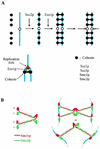
Models for the function of different cohesion proteins. (A) A Cohesin complex composed of Scc1p, Scc3p, Smc1p, and Smc3p is loaded onto chromosomes at the end of G1. Association of Cohesin with chromosomes depends on Scc2p. During S phase, Eco1p, acting at replication forks, catalyzes the formation of links between sister chromatids. These links are portrayed as being composed of Cohesin. It is possible, however, that the links merely need Cohesin for their formation. (B) Two models for how Smc1/3 heterodimers might join sister chromatids.

VSports app下载 - References

    1. Berben G, Dumont J, Gilliquet V, Bolle P, Hilger F. The YDp plasmids: A uniform set of vectors bearing versatile gene disruption cassettes for Saccharomyces cerevisiae. Yeast. 1991;7:475–477. - PubMed
    1. Birkenbihl RP, Subramani S. The rad21 gene product of Schizosacharomyces pombe is a nuclear, cell cycle regulated phosphoprotein. J Cell Biol. 1995;270:7703–7711. - PubMed
    1. Carramolino L, Lee B, Zaballos A, Peled A, Barthelemy I, Shav-Tal Y, Prieto I, Carmi P, Gothelf Y, Gonzalez de Buitrago G, Aracil M, Marquez G, Barbero J, D Z. SA-1, a nuclear protein encoded by one member of a novel gene family: Molecular cloning and detection in hemopoietic organs. Gene. 1997;195:151–159. - "V体育官网" PubMed
    1. Ciosk R, Zachariae W, Michaelis C, Shevchenko A, Mann M, Nasmyth K. An Esp1/Pds1 complex regulates loss of sister chromatid cohesion at the metaphase to anaphase transition in yeast. Cell. 1998;93:1067–1076. - PubMed
    1. Cohen-Fix O, Peters J-M, Kirschner MW, Koshland D. Anaphase initiation in Saccharomyces cerevisiae is controlled by the APC-dependent degradation of the anaphase inhibitor Pds1p. Genes & Dev. 1996;10:3081–3093. - "VSports注册入口" PubMed

"VSports在线直播" Publication types

MeSH terms (VSports)